Removing a tooth is the last thing a dentist will suggest. Due to a number of subjective and objective reasons, it is impossible to maintain the dentition in ideal condition until old age.
Dental defects are the loss of one or more dental units, leading to a violation of the integrity of the dentition, deviation in the development of physiological occlusion, and an abnormal arrangement of individual teeth.
Reasons contributing to the development of dental defects
Dental defects arise due to:
- untimely treatment of caries;
- inflammatory processes in the oral cavity;
- presence of malocclusion;
- genetic conditioning;
- diseases leading to changes in metabolic processes;
- damage to the jaw apparatus;
- various neoplasms.
Classification of dentition defects
There are several types of classification. Dentists mainly use three of them:
- Kennedy classification:
- Group 1 - bilateral absence of chewing teeth;
- Group 2 - unilateral absence of a molar;
- Group 3 - unilateral absence of lateral teeth with preservation of distal support;
- Group 4 - defect of the anterior jaw.
- Beltman classification:
- Class 1 - the defect includes edentulous chewing teeth.
a) dentition with unilateral terminal defects;
b) dentition with bilateral terminal defects.
- Class 2 - there is one or more included defects (if the terminal units are present, there may be no teeth in other parts of the row).
a) the number of missing units does not exceed three;
b) the number of missing units is three or more.
- Classification according to the Gavrilov system:
- Group 1 - dentition with terminal one- and two-sided defects;
- Group 2 - dentition with anterior and lateral one- and two-sided defects;
- Group 3 - combined defects;
- Group 4 - single preserved dental units.
Phenomena occurring before teething
- hypoplasia, enamel hyperplasia
- endemic fluorosis;
- anomalies of tooth formation;
- color anomalies;
- genetic disorders.
Enamel hypoplasia is a disorder that is caused by changes in the cells from which enamel is formed. In these cells - ameloblasts, a change in mineral metabolism occurs and the trophism of hard tissues is disrupted. It develops in the fetal state or in childhood. It entails deformation of the pulp, dentin, and provokes malocclusion. Enamel hypoplasia affects up to 14% of all children.
Enamel hyperplasia involves excessive development of tooth tissue. Most often observed on the neck of the tooth, it may affect the contact surface of the teeth. Enamel hyperplasia does not cause functional impairment, but the orthopedist will have to take this feature into account when creating metal-ceramic and porcelain prostheses.
Dental fluorosis is considered a chronic disease that is caused by excess fluoride intake. As a rule, it occurs when drinking water containing a large amount of this element. Fluoride removes calcium from the body, as a result of which the mineralization of teeth is disrupted, they become fragile, and various associated anomalies appear.
Anomalies of hard dental tissues can be hereditary. This is due to diseases affecting the development of enamel and dentin. Often accompanied by changes in the color and shape of teeth.
Treatment of hypoplasia
Treatment for hypoplasia may vary depending on the degree of the disease and consist of bleaching and other measures, as well as remineralization therapy and subsequent prevention. Hyperplasia is the excessive formation of tooth tissue, in which so-called enamel drops of different sizes are formed, often located at the border of enamel and root cement in the neck area, less often in another place. Treatment is most often not required, but if the pathology has affected the front teeth, grinding and thorough polishing of the tooth surface can be used.
Endemic fluorosis
Endemic fluorosis is a lesion of hard tooth tissue due to the consumption of water containing more than 2 mg/l of fluoride compounds. In this case, treatment is prescribed depending on the period of residence of the patient in the area in which such water is used, as well as on the diet and social situation. It can consist of either remineralization of teeth in mild cases of disease, or restoration using composite materials or the use of orthopedic structures.
Anomalies of tooth formation
Anomalies in the formation and pathological processes during tooth eruption occur with developmental disorders in general, as well as diseases of the endocrine and nervous systems, and require complex treatment. Changes in tooth color depend on many factors - taking medications of a certain group, including by the mother during pregnancy, as well as other phenomena.
Symptoms and consequences of dental defects
Dental defects appear:
- chewing function disorder;
- incorrect bite;
- articulation disorder;
- single displacement of teeth;
- violation of the integrity of the dentition;
- violation of the aesthetics of a smile.
Diagnosis of dental defects
The removal of one or more dental units requires consultation with a dentist and an orthodontist, especially when it comes to the anterior part of the dentition. Untimely correction of the defect does not have the best effect on the condition of adjacent teeth.
As a rule, a visual inspection is sufficient to determine how to correct the defect. In some cases, x-rays may be needed.
Orthopedic treatment of tooth crown defects using inlays
For several decades, the search for optimal ways to replace defects in the crown of the tooth has been ongoing. The development of technology constantly tilts the scales, which tilts towards either therapeutic or orthopedic techniques. Our goal was not to conduct a comparative analysis of various methods for restoring crown defects. This publication is devoted only to illustrating certain recovery methods that, in our opinion, deserve attention.
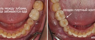
The main method of eliminating defects in the crown of a tooth, especially in the initial and middle forms, is undoubtedly filling. Dental filling compared to prosthetics is a less labor-intensive process, which consists of surgical removal of the affected tissue, medicinal treatment of the cavity and filling the defect with filling material. However, this method of treatment, even at the modern level of development of dentistry, cannot always provide a solution to the problem of restoring the shape and function of teeth, especially with carious defects of classes II, IV of the Black classification, systemic hypoplasia, increased abrasion, hereditary disorders and malformations of hard tissues, etc.
The significant and most characteristic disadvantages of fillings include, first of all, secondary caries and loss of fillings (Abolmasov N. G. et al., 2003). M. B. Bushan and V. N. Kopeikin note that filling materials have a lower coefficient of wear resistance compared to metal and porcelain. They may be porous and not strong enough. Therefore, interest in alternative types of treatment and, in particular, in replacing tooth crown defects with inlays is quite justified.
Inlays are called microprostheses used to restore defects in the coronal part destroyed as a result of carious and non-carious lesions. Unlike a filling, the tab is inserted into the prepared cavity not in a plastic state, but in a solid state. This avoids shrinkage that occurs when the filling material hardens, and therefore improves the marginal fit and reduces the frequency of caries relapses.
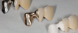
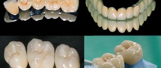
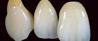
The design of the inlay is selected taking into account the topography, shape, size of the defect, anatomical and topographic relationships of hard and soft tissues, type of bite, direction of loads, tooth inclination, radiographic results, presence or absence of pulp. Depending on the material from which the inlays are made, they are divided into: a) metal (gold 900, 750, cobalt-chrome alloy, silver-palladium alloy, titanium alloys, for example VT5L); b) non-metallic (porcelain, composite); c) combined metal-ceramic and metal-composite (Fig. 1).
Rice. 1a. Inlay made of 900-carat gold alloy.
Rice. 1b. Inlay made of 900-carat gold alloy.
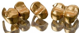
For a number of years, the main structural material for inlays has been gold alloys. The method of manufacturing a cast inlay using a wax model was described by Taggart in 1907. Since then, the technology has undergone certain changes, but the high strength characteristics and accuracy of the fit of the gold inlays to the walls of the cavity, maintaining a constant volume, protecting the edges of the enamel from chipping, inertness, non-oxidation in the oral cavity, the ability to unravel during chewing and better adapt to the walls of the cavity.
In connection with the increasing requirements for the aesthetics of restorations and constantly improving technologies for the manufacture of dentures, one of the most promising areas is the prosthetics of partial defects in the coronal part of the tooth with ceramic inlays. The current level of development of dentistry makes it possible to produce these structures in the following ways:
- firing on foil;
- firing on a fireproof model;
- production of a ceramic frame by firing on a refractory model, followed by cladding;
- production of ceramic inlays by pressing;
- manufacturing a frame from aluminum oxide and zirconium dioxide by electrophoretic deposition with subsequent lining;
- manufacturing the inlay frame by ultrasonic condensation of aluminum oxide with the addition of zirconium dioxide, followed by layer-by-layer application and firing of porcelain;
- firing of the aluminum oxide frame followed by cladding;
- milling using a computer program.
The technology for manufacturing ceramic dentures by hot pressing is implemented using the IPS-Empress I, II systems. One of the advantages of these systems is the use of fluorapatite as a structural material, which, compared to other ceramic materials, causes less abrasion of natural antagonist teeth (Fig. 2).
Rice. 2a. Porcelain inlay made by pressing (painting technique).
Rice. 2b. Porcelain inlay made by pressing (painting technique).
Initially, the IPS-Empress I system provided for the complete reconstruction of the anatomical shape of the coronal part of the tooth from wax and the subsequent simultaneous hot pressing of the ceramic mass. Thus, the finished restoration was monochrome, with a high degree of transparency, since porcelain of the same color was used. Color correction was carried out by applying dyes before glazing, which did not always make it possible to optimize the color rendition of the restoration with the individual coloring characteristics of natural teeth (coloring technique).
Ivoclar specialists continued to work on this issue, which was reflected in their later developments. When working with the IPS-Empress II system, you can create a wax model of the base of the inlay with subsequent pressing. Subsequently, dentin and enamel masses are applied to the model on the frame, ceramics are modeled and fired (layer-by-layer porcelain application technique).
The manufacture of the inlay frame by ultrasonic condensation of aluminum oxide with the addition of zirconium dioxide, followed by layer-by-layer application and firing of porcelain involves the introduction of a suspension of the VITA In-Ceram ZIRCONYA material into the cavity of the double model of the prepared tooth, compacted in a Vita Sonic II type apparatus, drying, and correction of the shape of the inlay base , glass infiltration and firing. After trying on the base in the oral cavity, it is veneered with ceramic mass (Fig. 3).
Rice. 3a. Ceramic inlay made using ultrasonic condensation technology of VITA In-Ceram ZIRCONYA material, followed by firing and layer-by-layer application and firing of ceramics.
Rice. 3b. Ceramic inlay made using ultrasonic condensation technology of VITA In-Ceram ZIRCONYA material, followed by firing and layer-by-layer application and firing of ceramics.
Rice. 3c. Ceramic inlay made using ultrasonic condensation technology of VITA In-Ceram ZIRCONYA material, followed by firing and layer-by-layer application and firing of ceramics.
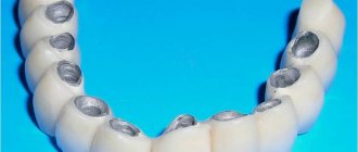
The most interesting direction may be related to milling a ceramic block using a computer program. Since the early 90s, zirconium oxide has been increasingly used in dentistry. According to in-vitro studies, zirconium oxide denture frames have high tensile strength comparable to that of metal alloys. The introduction of CAD/CAM technology in dental prosthetics makes it possible to produce structures with high accuracy and predictably reproducible quality. Based on the general user algorithm and hardware layout, CAD/CAM systems can be divided into 3 main groups:
- Centralized macrosystems (Procera, Decim).
- Individual mini-systems (Hint-Els, Precident).
- Custom microsystems (Cerec).
The use of modern computer technologies can significantly increase the accuracy of restoration manufacturing and minimize the influence of the human factor. However, most of the well-known CAD/CAM systems are expensive, which limits their distribution in the dental market. A number of inventors set out to create compact and inexpensive milling equipment that makes it possible to produce extended structures from zirconium oxide (Fig. 4).
Rice. 4a. Manufacturing of inlays using copy-milling technology and a zirkonzahn machine.
Rice. 4b. Manufacturing of inlays using copy-milling technology and a zirkonzahn machine.
Rice. 4c. Manufacturing of inlays using copy-milling technology and a zirkonzahn machine.
Rice. 4g. Manufacturing of inlays using copy-milling technology and a zirkonzahn machine.
One of them was Enrico Steger, inventor, owner and CEO of Zirkonzahn. The operation of his system is based on copy-milling technology. First, a prototype of the product is created from a light-curing composite material, then it is milled from a zirconium oxide blank in a copy-milling machine.
To facilitate this process and reduce wear on grinding tools, the Zirkonzahn machine works with an unsintered (pre-agglomerated) form of zirconium dioxide. The zirconium oxide inlay blank is 25% larger than the composite counterpart, since further firing of the product will lead to significant shrinkage of the material. The author managed to calculate these volumetric changes and transfer them with high accuracy into the basis of the operation of his copy-milling machine.
Along with the above-described methods for making inlays, restoration of defects in the coronal part of the tooth is also currently used using inlays made of light-curing composite materials. Compared to porcelain and metal-ceramic inlays, composite inlays have a less abrasive effect on natural teeth; due to the elasticity of the composite, they absorb part of the occlusal load, are less labor-intensive to manufacture and do not require expensive bulky equipment and specially trained dental technicians, and can be easily restored in the oral cavity. , much cheaper.
The disadvantages of composite inlays include insufficient color stability, the possibility of allergic reactions to the composite, and the abrasion of modern composite materials, although close to the abrasion of tooth enamel, is somewhat higher, which can lead over time to a decrease in the height of the lower third of the face in the position of central occlusion with multiple restoration of lateral teeth, with single restorations - to the development of deformation of the occlusal curves. Composite inlays have lower strength, greater porosity, and, compared to porcelain and metal ones, are characterized by less tolerance to the gingival margin, but despite this, they can be a serious alternative to direct restorations (Fig. 5).
Rice. 5a. Stages of tooth restoration using a composite inlay.
Rice. 5 B. Stages of tooth restoration using a composite inlay.
Rice. 5th century Stages of tooth restoration using a composite inlay.
Rice. 5g. Stages of tooth restoration using a composite inlay.
Rice. 5d. Stages of tooth restoration using a composite inlay.
Rice. 5e. Stages of tooth restoration using a composite inlay.
The development of dentistry at the present stage is characterized by the rapid growth of new technologies for the manufacture of dentures. And it’s quite difficult for a dentist to navigate this diversity. We hope that this publication expands the horizons of choosing restoration techniques for defects in the coronal part of the tooth.
LITERATURE
- Abolmasov N. G. Replacement of defects of teeth and dentition with fixed dentures N. G. Abolmasov, N. N. Abolmasov, V. A. Bychkov, V. R. Shashmurina. - Smolensk, 1995. - 175 p.
- Bragin E. A. Fundamentals of microprosthetics. Pin structures of dentures, inlays, veneers, artificial crowns, decorative dental onlays. / E. A. Bragin, A. V. Skryl. - M.: Medical press, 2009. - 508 p.: ill.
- Volvach S.I. CAD/CAM technologies in the dental laboratory - myth or reality? / S. I. Volvach // New in dentistry for dental technicians. - 2000, No. 4 (12). - P. 3-13.
- Dyakonenko E. E. Orthopedic treatment with metal-free ceramics as an alternative method of tooth restoration / E. E. Dyakonenko // New in dentistry for dental technicians. - 2000, No. 1 (9). - P. 3-14.
- Prosthetic restoration of teeth. CEREC system. Textbook, ed. prof. V. N. Trezubova, S. D. Arutyunova. - St. Petersburg: Spetslit, 2003. - 64 p.
- Skryl A.V. Restoration of defects in tooth crowns with ceramic inlays / A.V. Skryl // Modern orthopedic dentistry. - 2008, No. 10. - P. 46-48.
- Steger E. ZIRKON milling technology / E. Steger // Dental technician. - 2007, No. 4. - P. 33-41.
Treatment of localized pathological abrasion
Localized pathological abrasion in most cases is compensated by local hypertrophy of the alveolar process and dentoalveolar elongation, therefore the first stage of treatment of localized pathological abrasion is aimed at eliminating the deformation of the dentition. Elimination of dentoalveolar elongation is carried out using a mouth guard or bite block on a group of protruded teeth. If it is impossible to use the hardware method, they can resort to a surgical method to lengthen the crown of the tooth and resection of part of the alveolar process.
If teeth are severely worn out, they are depulped and the core part is restored using pin structures. Depulpation also allows you to eliminate hyperesthesia when drug treatment is ineffective.
After eliminating the protrusion of teeth, prosthetics begins. For prosthetics, metal, metal-plastic and metal-ceramic inlays and crowns are used, as well as covering the occlusal and palatal surfaces of the teeth with cast metal overlays to eliminate further abrasion. In the area of the frontal group of teeth, restoration with composite restorations is also used.
The article was written by N.A. Sokolov. especially for the OHI-S.COM website. Please, when copying material, do not forget to provide a link to the current page
What happens if the crown is not restored in time?
Ignoring crown defects leads to worsening pathological changes, up to complete destruction of the upper part and damage to the root.
The loss of even one crown leads to the following consequences:
- disturbances in the chewing process;
- overload and wear of other sectors of the dentition;
- displacement of neighboring units;
- destabilization of antagonist teeth;
- diction disorders;
- expansion of defects;
- cosmetic defect (especially if the crowns in the smile area are damaged).
Timely restoration of the crown part is much simpler, cheaper and more physiological than replacing your own tooth with an artificial one.