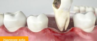
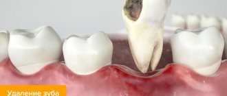
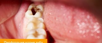
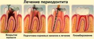
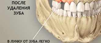
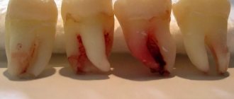
Tooth extraction is a process that traumatizes the gums. After healing a diseased tooth, nerves, ligaments and small blood vessels are torn, which causes bleeding. This is a natural process - the blood does not allow pathogenic organisms to enter the circulatory system, washing them out. And then, curled up, it forms a barrier between unprotected tissues and bacteria. After tooth extraction, white plaque on the gum is not pus, but another stage of protection and restoration of the body.
When removing a tooth, the patient must be prepared:
➢
to moderate bleeding that may last for some time;
➢
the appearance of a dense blood clot in the gum;
➢
unpleasant odor 10-12 hours after surgery;
➢
the formation of a white coating on the blood clot;
➢
pain after surgery;
➢
temperature rise.
The listed symptoms are not a sign of an unsuccessful operation or infection in the body - this is the body’s natural response to tooth extraction.
Content:
- When white plaque is the norm
- When white plaque is a harbinger of disease
- Why do complications occur after tooth extraction?
- How to recognize the problem
- Diagnostic measures
- Treatment of alveolitis
After tooth extraction, it is important to monitor the condition of the mucous membrane.
Very often a white coating appears in the extraction area. It frightens patients because it seems something strange to them, and to some it resembles an accumulation of purulent masses. It should be noted that white plaque in the socket after tooth extraction can be either normal or a complication. In the first case we are talking about natural regeneration, in the second - about alveolitis.
Dentists' recommendations
After surgery, it is important to monitor the condition of the wound and follow the doctor's instructions. The hole filled with blood gradually overgrows and is completely covered with granulation tissue 14-20 days after the operation. The first three days after tooth extraction you cannot:
- rinse the mouth,
- eat solid food
- drink through a straw
- smoking and drinking alcohol,
- use a brush with stiff bristles,
- take a hot bath and go to the sauna.
If possible, you should avoid physical activity and stop taking medications that prevent blood clotting. Compliance with the listed recommendations contributes to the speedy healing of the wound.
When white plaque is the norm
Nature intended it so that damaged tissues should be protected from the penetration of infectious agents. This is why, after a tooth is removed, a clot and white plaque forms in the socket. The latter is nothing more than fibrin, a protein needed for blood clotting. It protects the wound surface from infection and does not allow the inflammatory process to spread.
At first, the white film seems very thin, but gradually it turns into bone tissue. Some even mistake plaque for food debris and try to remove it with their tongue or using foreign objects. Under no circumstances should this be done. If the wound heals according to plan, there are no severe pain symptoms, there is no need to worry or touch it. This can lead to complications.
So, fibrin in the form of a whitish coating is needed to:
- the wound healed quickly;
- no infection penetrated into the socket;
- soft tissues were not inflamed.
If the patient has questions regarding the white clot on the surface of the damaged gum, he should consult with his dentist. You don't need to do anything yourself.
General overview
Extraction of an element of the dentition is a process during which the gums receive extensive trauma. Tooth extraction leads to rupture of nerves and ligaments, as well as destruction of small blood vessels, resulting in local bleeding. This process is natural in nature and is a protective mechanism - the blood limits the possibility of pathogenic bacteria entering the socket that can cause infection. During the folding process, a reliable barrier is formed that protects the internal tissues until complete recovery. The formation of white plaque is a normal phenomenon, since it is not a purulent structure, but an element necessary for the regeneration of the jaw region.
When undergoing tooth extraction surgery, you should prepare in advance for possible consequences, the list of which includes:
- Moderate bleeding that persists for several hours;
- Formation of a dense blood clot over the socket;
- Unpleasant odor that occurs 10-12 hours after the procedure;
- Formation of a white coating on the surface of the blood clot;
- Painful manifestations in the area of intervention;
- Increased body temperature.
These symptoms are not pathological in nature and are a normal reaction of the body to radical intervention.
When white plaque is a harbinger of disease
It also happens that a whitish coating indicates the development of alveolitis after tooth extraction. This means that the tooth socket has become infected. In this condition, the patient is concerned about:
- severe pain in the area of the removed unit;
- increased body temperature;
- weakness, decreased performance;
- enlargement of the submandibular lymph nodes;
- headache;
- unpleasant putrid odor from the mouth and the same taste in the mouth.
If you experience these symptoms, you should make an appointment at the dental clinic as soon as possible.
Exostosis, fistula or beneficial plaque: types of growths after removal
Tooth extraction is always traumatic. The doctor extracts the tooth, damaging soft and bone tissues and breaking blood vessels. After the tooth is extracted, the wound fills with blood, which coagulates into a clot. The naturally formed blood clot closes the hole, protecting it from bacteria and stimulating regeneration processes.
With the correct course of events, the hole gradually overgrows, closes, and ceases to disturb the patient. But sometimes a formation forms at the site of removal - on the hole itself or nearby. Possible options:
- white dense coating;
- cyst;
- fistula;
- exostosis.
Each formation has its own nature and reasons for its appearance. Some require treatment, others are beneficial and go away on their own.
White plaque or growth after tooth extraction
During the first hours after removal, bleeding stops in the hole and a dark blood clot appears. After a couple of days, a white coating is noticeable on the clot. Externally similar to purulent discharge, it frightens many patients. But this is the next stage of healing - fibrin protein fibers.
Fibrin performs several functions at once:
- accelerates tissue regeneration, including bone;
- helps reduce the size of the hole;
- protects the wound from bacteria and pathogenic microflora;
- helps the blood clot dissolve.
The plaque often acquires a dense, hard structure, and patients mistake the fibrinous film for a growth. White growth on the tooth socket after extraction is a natural healing process and should not be a cause for concern.
Cyst or fistula after tooth extraction
The cyst and fistula only indirectly resemble a white growth. But often patients describe the pathological condition this way. The reason is the filling of the formation with liquid, pus, which gives the lump a whitish tint.
A fistula is a channel through which purulent contents are removed from the hole. It is located near the site of the extracted tooth. The tissues around the formation are inflamed.
A cyst is a cavity or lump that occurs when tissue is damaged during surgery. It has walls and contents, and stands out strongly above the surface of the gum. Causes pain.
Both a cyst and a fistula are pathological formations that require removal. Externally, patients define them as growths in the area of tooth extraction.
If there are no visible manifestations, indirect signs of ill health will be:
- a feeling of heaviness and pressure in the area of the hole;
- soft tissue swelling;
- jaw pain;
- heat;
- discomfort when eating;
- smell from the mouth;
- burning gums;
- individual signs of intoxication of the body.
If you have unpleasant symptoms, you should consult a doctor to determine the type of pathology and its treatment. The main methods are drug therapy or surgical removal of the formation. The dentist chooses the method after assessing the clinical situation.
Exostosis - bone growth after tooth extraction
Exostosis - bumps, spines, growths on the gums that arise due to hereditary factors or after complex tooth extraction. The cause of protruding ridges is improperly fused bone, the consequences of displacement of bone tissue due to careless work of the surgeon.
The pathology looks unaesthetic, but does not cause much discomfort. If the formation is significant in size, it can put pressure on the bone and nearby teeth and become a contraindication for dental implantation.
Removal of exostosis is carried out surgically. After anesthesia, the doctor makes an incision into the soft tissue, saws off the growth, straightens the bone, and applies sutures. Healing occurs quickly and the formation does not reappear.
Other causes of gum growth
Lumps and growths on the gums appear after removal and for other reasons:
- infectious processes that began due to non-compliance with the surgeon’s recommendations after removal;
- allergic reaction to anesthetic drugs;
- mechanical damage - due to careless removal.
Sometimes a growth occurs at the site where the anesthetic injection was given. Such a hematoma resolves on its own in 2-3 days.
Other lumps require consultation with a dentist.
Why do complications occur after tooth extraction?
The most common reason for the development of complications is the patient’s failure to comply with medical prescriptions. Normally, the hole should heal within a few days. She shouldn't be sick for a long time. As a result of the fact that the blood clot closes it, it is reliably protected from viruses and food debris.
It happens that a person actively rinses the wound or constantly touches it with his tongue, fingers, and tries to remove the light coating. Then the clot displaces or does not form completely, the tissue becomes infected and alveolitis develops.
Untreated neighboring teeth can also lead to disease (which is why all carious “cavities” need to be treated before removal), and failure to comply with the rules of oral hygiene. If you properly prepare for extraction and follow all the doctor’s instructions, the risk of developing alveolitis after removal will be minimal.
Diagnostics
Such a diagnosis is made only by a doctor when he examines the oral cavity. You can't diagnose yourself. After all, alveolitis of the socket is a serious disease.
If it is not treated promptly, serious complications are possible, including blood poisoning. If there is no blood clot at the place where the tooth was pulled out, then this indicates alveolitis.
How to recognize the problem
How to understand that white plaque signals a problem?
If the gums heal well, then within 2-3 days after surgery the pain becomes very weak and barely noticeable. The wound closes with a dense clot and practically does not bother the person. With the development of alveolitis on days 3-5, the pain, on the contrary, intensifies. It's pulsating. It can become unbearable. Sometimes it radiates to the entire half of the face. Body temperature rises, lymph nodes enlarge, appetite decreases, salivation becomes stronger. The face may also become swollen.
In such a situation, staying at home is dangerous - inflammation can spread to the deep tissues of the jaw. You need to immediately go to the dental clinic.
Prices
| Name of service | Price, rub.) * |
| Initial consultation with a dentist-therapist | 1,500 rub. |
| Repeated consultation with a dentist-therapist | 800 rub. |
| Treatment of caries (imaging, anesthesia, isolation of the oral cavity, installation of an insulating lining and filling made of composite light-curing imported material) | from 4,350 rub. |
| Treatment of alveolitis with socket revision | 3,000 rub. |
We accept VISA, MASTERCARD, MAESTRO bank cards for payment.
Would you like us to call you back?
Treatment of alveolitis
If alveolitis occurs after tooth extraction, you need to undergo dental treatment aimed at:
- elimination of the infectious focus;
- preventing possible complications;
- maintaining the integrity of the rest of the dentition.
First, the dentist cleans the socket and rinses it. It is important to wash away all purulent masses and dead tissue from it. Antiseptics and hydrogen peroxide work well for this purpose.
To reduce painful symptoms, which deprive the patient of the opportunity to fully rest and work, analgesics are used. It is better to use them by applying applications. Compresses are changed every half hour. It is also possible to take drugs orally, that is, in the form of painkillers and anti-inflammatory tablets.
If the patient has concomitant diseases or reduced immunity, the doctor may decide to prescribe antibiotics. They quickly relieve inflammation and speed up the healing of damaged tissue.
If you seek qualified dental care in a timely manner, the prognosis for alveolitis is favorable. After 3-5 days, the symptoms begin to subside, and the person’s well-being improves. Residual socket pain may persist for another 2-3 weeks, but it does not pose a threat.
Prevention of alveolitis at SM-Dentistry
If the operation is not an emergency, then dentists first treat caries and other inflammatory diseases in the oral cavity, which can become a source of infection.
After the operation, you will receive detailed recommendations for caring for the socket so that the blood clot forms correctly and does not break down prematurely. To do this, you must follow a diet (do not eat hard or hot foods), do not use intensive rinsing and cleaning in the area of the injured hole, and use antiseptic and painkillers.
Sign up for a consultation with a dental surgeon by calling 24/7 in Moscow +7 (495) 777-48-06.
Dentist consultation Treatment of caries
Sharp edge of hole
Healthy wound healing involves the presence of two parallel processes: mucosal formation and bone formation. The latter must be protected by a gum or a blood clot. If any of the walls of the hole has an excessively sharp (not rounded) edge, it injures (cuts through) the surrounding mucosa and protrudes, remaining unprotected, in the oral cavity.
A blood clot is a protective barrier that protects the empty socket from infection
This phenomenon can lead to alveolitis or the formation of a so-called sharp edge. After extraction, more than one week may pass, and something white still remains in the hole. On palpation, the depression is compacted and sharp.
What to do if a sharp bone edge has formed: in some cases the problem resolves on its own and does not require medical intervention, in others a minor operation may be necessary. So, under local anesthesia, the dentist will move the edge of the gum in the area of the protruding fragment, remove it with forceps or a drill, and, if necessary, apply a postoperative suture.
