A sinus cyst is a fluid-filled formation with elastic walls; the location of the cyst affects the symptoms of the disease. Most often, cysts form in the maxillary sinuses. The reasons for the development of cysts in the nasal sinuses are varied; there are a number of reasons for the appearance of a neoplasm:
- Blockage of the ducts of the mucus-secreting glands.
- Dental diseases, tooth roots protruding into the paranasal sinuses.
- Allergic diseases of the nose.
- Frequent inflammatory processes.
- Anatomical features of the structure of the nose.
- Mechanical injuries of the nose.
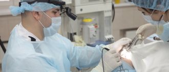
In the oncology department of the Yusupov Hospital, you can undergo examination using MRI, CT, ultrasound, diagnosis of diseases of the paranasal sinuses if a nasal tumor is suspected. The patient will be able to undergo a full examination at the diagnostic center, take tests in the hospital laboratory, and receive advice from an otolaryngologist, oncologist and other specialists.
What is a sinus cyst?
This is a large formation in which mucus or pus accumulates. The cyst is not dangerous, it does not degenerate into cancer, but over time it can grow and put pressure on the walls of the sinus. Because of this, breathing problems appear, the nasal mucosa swells, a runny nose almost never goes away, and recurrent bronchitis and pneumonia may occur. The cyst is sometimes located in such a way that there are no symptoms, and it is discovered by chance during another examination - for example, before dental prosthetics.
What causes cysts to form in the sinuses?
To ensure that the mucous membrane of the respiratory organs does not dry out, is not overcooled and is protected from dust, it contains glands that constantly secrete moisturizing mucus. This mucus flows to the surface through the ducts of the glands. If at least one of them is blocked, mucus continues to be produced, but will accumulate inside the gland, filling it and stretching the walls. As a result, a cyst appears.
The formation of cysts can occur due to:
- frequent infections;
- severe allergies;
- structural features of the nasopharynx;
- injuries and curvature of the nasal septum;
- inflammatory processes in the upper jaw (usually dental diseases).
The most common symptoms of a cyst are headaches, difficulty in nasal breathing due to swelling of the mucous membrane, discomfort or pain over the upper jaw.
General information
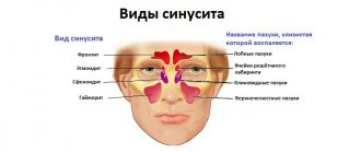
In the structure of diseases of the ENT organs, the pathology of the paranasal sinuses has occupied a leading place in recent years. cyst is a pathological formation in the form of a benign formation in the shape of a sphere/ball, filled with liquid contents.
The cyst shell is two-layered, in which the inner layer has an epithelial lining containing glands that produce fluid. Cysts gradually increase in size and never disappear on their own. The cyst is attached directly to the wall of the sinus, completely/partially filling it from the inside. Most often, the fluid that accumulates in a cyst is mucous in nature; purulent cysts are less likely to form, and serous cysts are extremely rare. Most often, cysts of the maxillary (maxillary) sinuses are formed (75-80%), somewhat less often - cysts of the ethmoidal labyrinth (about 15%) and extremely rarely - cysts of the frontal/sphenoid sinuses (about 5%). Most often, a cyst is discovered accidentally during an X-ray examination. There were no differences in the incidence of cysts in the left sinus and the right sinus. According to the classification of diseases (ICD-10), cystic pathologies of the paranasal sinuses belong to chronic forms of sinusitis.
You can suspect the presence of a cyst in the maxillary sinuses during periods of local hypothermia , acute respiratory viral infections , and exacerbations of chronic sinusitis . What can a maxillary sinus cyst lead to? An enlarged, untreated cyst is dangerous because it puts pressure on the nerve endings of the mucous membrane of the maxillary sinuses, provoking migraines . Also, an overgrown cyst in the maxillary sinuses often leads to blocking of the ducts between the nasal cavity and the maxillary sinuses, provoking the development of purulent sinusitis , which occurs in a severe form.
Symptoms of a sinus cyst
The cysts and polyps themselves are soft and lack sensitivity, so if they are small, many people may not notice them at all. However, multiple growths or a large cyst can block the nasal passages and sinuses.
Common signs and symptoms of nasal cysts include:
- runny nose
- reduced or absent sense of smell
- loss of sense of taste
- facial or headache
- pain in upper teeth
- feeling of pressure on the forehead and face
- snore
- frequent nosebleeds
Is it always necessary to remove a cyst?
There are a number of absolute indications for removal. Firstly, patients who are preparing to enter flight and military schools. Secondly, persons who are undergoing dental implantation. Thirdly, for large cysts that cause severe pain in the area where they are located. However, in 60% of cases, identified cysts of the paranasal sinuses do not manifest themselves clinically (that is, they do not cause discomfort to the patient) and therefore we do not recommend removing them.
You can safely live your whole life with a sinus cyst without even knowing it!
Indications for sinus cyst removal
It will not be possible to remove the cyst simply by relieving the inflammation; surgery is not necessary. Surgery is necessary if:
- pus accumulates inside the cyst;
- it is growing rapidly or its diameter is already more than 6 cm;
- pressure on the lower wall of the orbit increases (because of this, vision may deteriorate and double vision may appear);
- worried about frequent or constant runny nose and nasal congestion.
Important! Surgery to remove a sinus cyst is also recommended if the cyst is causing any discomfort. However, the size of the cyst in this case does not matter.
Classification
The classification of maxillary sinus cysts is based on the factor of their origin and secondary changes in the sinus , which is due to the specific treatment of each form and the need for surgery. Based on the mechanism of occurrence and morphological characteristics, they distinguish:
- True (retention) cysts. They are formed as a result of complete/partial obstruction of the excretory ducts of mucus-producing glands. They have an epithelial lining with columnar ciliated epithelium. The reasons for their obstruction are: swelling , blockage , scarring or hyperplasia . The gland continues to function and produce secretions. Over time, the walls stretch, it overflows and closes the lumen of the sinus.
- False cysts (cyst-like formations). They do not have an epithelial lining and are formed in the thickness of the mucous membrane.
- Odontogenic cyst (radicular/follicular). Filled with pus. Radicular cysts form around the root of the inflamed upper tooth, penetrating into the sinus by growing through the atrophied bone tissue of the upper jaw. Follicular cysts form directly from the follicle of a baby tooth.
- Congenital cysts. Caused by developmental defects (anomalies of the mucous membranes of the maxillary sinuses/deformations of the upper jaw), contributing to the formation of cysts.
Contraindications
The operations for removing sinus cysts are quite simple. Patients recover quickly after the intervention, and complications are rare. Contraindications are only general for any surgical interventions:
- acute infection;
- diabetes mellitus (severe);
- increased bleeding (blood does not clot well);
- pregnancy;
- epilepsy;
- oncology.
Before surgery to remove a cyst from the sinus, you need to take tests (for HIV, syphilis, hepatitis B and C, general blood test).
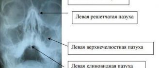
Computer 3D diagnostics - for reliable results
Ultra-modern SIRONA-SIEMENS tomograph with ENT settings - ultra-precise and safe search for hidden odontogenic and ENT pathologies
- Highly accurate assessment of the condition of teeth and maxillary sinuses
- Detection of the extent of the inflammatory process, location and size of the cyst
- Analysis of efficiency and forecast of ongoing operations
Diagnostics
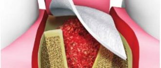
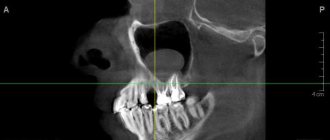
Figure 1. Cyst in the maxillary sinus.
Source: Contemporary clinical dentistry / Open-i (Attribution-NonCommercial-ShareAlike 3.0 Unported) Before surgery to remove a sinus cyst, you need to take tests (for HIV, syphilis, hepatitis B and C, general blood test).
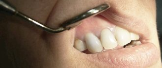
Usually, to make a diagnosis, the doctor only needs to collect an anamnesis and a general physical examination, including the nose.
Other diagnostic tests include:
Endoscopy of the nose. The doctor performs a detailed examination of the nose and sinuses using a narrow tube with a lighted magnifying lens or small camera.
Visualization methods. Images obtained from a computed tomography (CT) scan can help the doctor accurately determine the size and location of cysts in the deeper areas of the sinuses, and assess the degree of swelling and inflammation.
Test for cystic fibrosis. If a child needs diagnosis, the doctor may suggest testing for cystic fibrosis. This is an inherited disease that affects the glands that produce nasal mucus, tears, sweat, saliva and digestive juices. This is usually a non-invasive sweat test that can determine whether your sweat is more or less salty than most people.
Why you should entrust treatment to the ENT department of dentistry
ENT dentistry combines two areas of medicine - dentistry and otolaryngology. This is a comprehensive approach to the treatment of combined inflammatory diseases of the paranasal sinuses of odontogenic origin.
The main difficulty in diagnosing and treating ENT-combined situations is that 100% of patients, when symptoms appear or cysts are accidentally discovered, turn only to an otolaryngologist or dentist. Narrow specialization does not allow us to determine the cause-and-effect relationship and create a thoughtful, comprehensive treatment plan.
- The ENT doctor prescribes a standardized conservative treatment, which does not bring results without eliminating the cause, namely the dental cyst.
- The dentist, by persuasion of the patient, tries to save the tooth, but in case of extensive neoplasms, therapeutic treatment of cysts is ineffective.
A person travels from one doctor to another in search of an adequate specialist and a solution to the problem; money and time are wasted on unsuccessful treatment, and the situation does not change.
70% of patients turned to our Center after unsuccessful treatment of cysts , many painful “punctures” of the sinus by an otolaryngologist, or attempts to persuade dentists not to remove, but to cure a tooth cyst without removal.
It is better to entrust the situation to a maxillofacial surgeon with ENT training together with an otolaryngologist. A multidisciplinary approach to patient problems in our Center, extensive experience and practice in two related areas, the necessary diagnostic and operating equipment greatly expand our overall chances of success and increase the overall quality and comfort of treatment.
KolchinSergey Alexandrovich
Maxillofacial surgeon, 7 years of experience
Surgeon with ENT training. He specializes in ENT dentistry, including performing endoscopic operations and carefully removing tumors in the sinuses.
More about the doctor
Types of operations to remove maxillary sinus cysts
The method of removing a cyst in the nose is chosen by the doctor. He takes into account its location and size. There are several types of operations.
Source: Symbolbild/Pixabay
Radical operation
This is a maxillary sinusotomy, in which an incision is made in the soft tissue (behind the upper lip) to gain access to the inside of the sinus in the oral cavity. They are peeled off, exposing the bone - the front wall of the sinus. Part of the bone fragment of the wall is removed to gain access to the sinus cavity. At the main stage, the surgeon cleans the sinus cavity from cysts, polyps and accumulated fluid, and rinses the sinus. The anastomosis with the nasal passage is widened, a drain is installed and taken out. The incision on the gum is sutured. The tampon is left in place for two days and then removed under anesthesia.
Figure 2. Stages of radical maxillary sinusotomy. Source: CC0 Public Domain
Important! Radical surgery is used relatively rarely, only if it is necessary to remove a large cyst or there are complications (separation of pus, inflammation, proliferation of polyps). Such an intervention allows you to completely scrape out and wash out the sinus, but it is dangerous due to complications (neuritis, the appearance of a fistula, bleeding). It is relatively difficult to tolerate and may require anesthesia and hospitalization.
Such an intervention allows you to completely clean and rinse the sinus, but is dangerous due to complications (neuritis, the appearance of a fistula, bleeding). It is relatively difficult to tolerate and requires hospitalization and anesthesia.
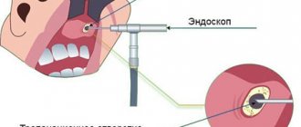
Endoscopic surgery
This is a minimally invasive procedure in which an endoscope is used to remove the cyst. The entire procedure takes about half an hour and is performed under local anesthesia.
To remove the cyst, an endoscope is inserted into the sinus through the nasal passage. Sometimes it is inserted through the tubercle on the upper jaw or the alveolar socket (through an additional channel with a diameter of 4–5 mm). The technique of access to the sinus depends on the location, shape and size of the cyst, as well as the individual characteristics of the patient’s nasopharynx. The path to the sinus through the nose is less traumatic and does not require an additional puncture. Access through the alveolar socket is used if the patient has had at least one molar removed (in adults, these are the sixth, seventh and eighth teeth on the left and right sides) on the upper jaw.
After entering the sinus under video control, the doctor pierces the cystic cavity and excises its walls, performs sanitation of the maxillary sinus, cleans it and rinses it with an antiseptic solution.
Endoscopic surgery has several advantages:
- there is no need to make an incision, the tissue is not injured;
- a video endoscope allows you to combine diagnostics with surgery (examine the sinus and immediately remove the tumor);
- The tissues of the mucous membrane are not damaged and scarring does not occur.
- Complications rarely occur;
- patients tolerate the intervention more easily and recover from it faster.
Therefore, the endoscopic technique is used more often than the radical one.
Figure 3. Endonasal removal of nasal cysts. Source: CC0 Public Domain
Important! Endoscopic removal is used for certain anatomical features of the structure of the nasopharynx, which can be diagnosed by a doctor. The technique is safe, almost does not injure the nasopharynx, and long-term rehabilitation is not required after its use.
Microsinusrotomy
In this case, a trocar (something like a surgical awl) and an endoscope are used. The procedure is also considered gentle. It is performed if the tumor can be removed through an opening in the anterior wall of the maxillary sinus.
At the first stage of the operation, access to the sinus is provided. To do this, a puncture with a diameter of 7 mm is made in the upper jaw. The puncture is performed with a trocar and an endoscope is inserted into the sinus cavity through it. Under video control, the cyst is punctured, its walls are excised, and the contents are removed.
In almost a third of cases, after maxillary sinusotomy, trigeminal neuropathy develops, which usually regresses without special treatment. Advantages: quick recovery, low swelling. Healthy tissues are almost not damaged.
Laser therapy
The laser is rarely used and only in combination with other surgical methods, provided that the cyst is small (otherwise it will take too long to remove it).
After surgery to remove a cyst, swelling of the mucous membrane, a feeling of dryness or profuse discharge from the nose, and pain in the area through which the sinus was accessed may appear. These symptoms are not complications of surgery and usually go away within a few days. If they persist and intensify, you should consult a doctor.
The nature of complications after surgery depends on the technique used:
- if access to the sinus was carried out through the alveolus of the tooth, an incision or puncture in the upper jaw, a fistula tract may form;
- with micromaxillary and radical maxillary sinus there is a risk of damage to the infraorbital or trigeminal nerve with the development of neuropathy. In this case, areas of the skin, nasal mucosa and mouth become numb, which may require treatment;
- inflammation and/or suppuration of the wound develops if it becomes infected (with poor quality oral care, in cases where the patient does not follow the doctor’s orders).
Pathogenesis
The pathogenesis of paranasal sinus cysts varies depending on the type of cyst. Thus, true cysts are formed when the lumen of the ducts of the glands of the sinus mucosa is obliterated due to squamous metaplasia of their epithelium and disruption of the process of outflow of their secretions. They develop against the background of inflammatory reactions caused by blockage of the lumen of the excretory duct with necrotic masses and stretching of the tissues of the gland and the proximal part of the duct. The leading mechanism of cystic transformation of the glands is a change in the composition of the secretory epithelium and hyperplasia of the glands. It is based on a sharp mucoidization of the secretory epithelium. At the same time, serous elements undergo pronounced changes, which, against the background of high mucoidization, are completely reduced. The corresponding transformation of the structure of the glands, together with goblet cell hyperplasia, leads to disruption of the transport function of the mucosa, excessive accumulation of secretions in the terminal sections of the glands, followed by cystic expansion, gradual enlargement and fusion into one common cavity. Also, the formation of cysts may be preceded by compression of the gland and duct by connective tissue.
False cystic formations develop primarily as a result of progressive disorganization of connective tissue (edema, degenerative changes in the mucosa and morphologically confirmed separation of connective tissue). At the same time, the leading role in the development of false cysts belongs to immunopathological mechanisms, as evidenced by the cellular composition of inflammatory infiltrates (eosinophils/mature plasma cells) and the predominant damage to blood vessels.
Odontogenic radicular cysts of the maxillary sinuses develop as a result of epithelial granulomas/necrotic changes in the apical part of a tooth affected by caries in combination with atrophic processes in the bone of the upper jaw. The development of follicular cysts is based on inflammatory lesions of the impacted tooth germ of primary teeth. Congenital cysts develop against the background of various developmental anomalies of glandular tissue/ducts of the mucous glands.
Possible complications
After surgery to remove a cyst from the maxillary sinus, swelling of the mucous membrane, a feeling of dryness or profuse nasal discharge, and pain in the area through which the sinus was accessed may appear. These symptoms are not complications of surgery and usually resolve within a few days. If they persist and intensify, you should consult a doctor.
The nature of complications after surgery depends on the technique used:
- if access to the sinus was carried out through the alveolus of the tooth, an incision or puncture in the upper jaw, a fistulous tract may appear;
- with micromaxillary and radical maxillary sinus there is a risk of damage to the infraorbital or trigeminal nerve with the development of neuritis. In this case, areas of the skin, nasal mucosa and mouth become numb, which requires additional treatment;
- inflammation and/or suppuration of the wound develops if it becomes infected (with poor quality oral care, in cases where the patient does not follow the doctor’s orders).
Stages of treatment
Treatment at our Center is comprehensive; we try to combine preparatory, surgical and rehabilitation procedures in one visit
- Preparation Surgeries are performed only in a sanitized oral cavity to avoid the risk of infection. We perform hygienic cleaning and dental treatment of teeth with inflammation in preparation for sterile surgical work.
- The operation is carried out in a sterile operating room of the ENT department. Ultrasound is used to create access to the sinus, under the control of a microscope, the causative tooth is removed, and bone inflammation is removed. Using special instruments, the cyst is removed and the sinus is inspected.
- Prosthetics In case of tooth extraction, which provoked inflammation and the growth of a cyst in the sinus, we fix temporary crowns or immediate dentures that hide the work performed. We make camouflage structures so as not to send the patient home with a missing tooth complex.
Rehabilitation period
Figure 4. A cyst removed from the maxillary sinus with a tooth in it.
Source: Indian journal of ophthalmology / Open-i (Attribution 2.0 Generic) After surgery to remove a sinus cyst, no special recovery or treatment is usually required. Full recovery takes only a few days. With a small amount of intervention, patients feel well within a few hours after the intervention.
For the first time after surgery, it is advisable to avoid physical activity. You cannot visit the pool and sauna, you must not allow injuries. To avoid inflammation and other complications, you need to ensure high-quality oral hygiene and follow your doctor’s prescriptions.
Our services in otorhinolaryngology
The administration of CELT JSC regularly updates the price list posted on the clinic’s website. However, in order to avoid possible misunderstandings, we ask you to clarify the cost of services by phone: +7
| Service name | Price in rubles |
| X-ray of the paranasal sinuses | 2 200 |
| MSCT of the paranasal sinuses | 5 000 |
| Endonasal maxillary sinusotomy | 40 000 — 75 000 |
All services
Make an appointment through the application or by calling +7 +7 We work every day:
- Monday—Friday: 8.00—20.00
- Saturday: 8.00–18.00
- Sunday is a day off
The nearest metro and MCC stations to the clinic:
- Highway of Enthusiasts or Perovo
- Partisan
- Enthusiast Highway
Driving directions