Classification of non-carious enamel lesions
The concept of “non-carious lesions” combines many pathologies, each of which is designated in the International Classification of Diseases (ICD 10). Such diseases are usually divided into 2 groups.
- Lesions that occurred before teething (during the period of follicular development of hard tissues, occur in 5-14% of the population):
- hypoplasia – underdevelopment of dental tissues;
- hyperplasia – excessive formation of dental tissue structures;
- endemic fluorosis, or “speckled teeth” - destruction of enamel due to excess fluoride in the body;
- hereditary diseases.
- Lesions that occur after teething (the overall prevalence of such diseases is 50-75%):
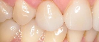
- wedge-shaped defect - the appearance of V-shaped defects in the cervical area of the crown;
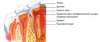
- erosion - in addition to the enamel, the dentinal layer is destroyed, as a rule, it occurs on the upper incisors and canines;
- pathological abrasion of enamel - intense loss of hard tissue on all or some teeth;
- mechanical injuries to dental crowns – chips, cracks, fractures;
- necrosis (death) of hard dental tissues;
- enamel pigmentation – tetracycline stains, etc.
Dental injuries
With any injury to the dental system, you should immediately contact a dentist (if you suspect a fracture of the jaw bone, contact a surgeon). The specialist will conduct an examination, study the X-ray data and give an opinion on the possibility of restoring the tooth or the need for its removal. There are different types of dental injuries, both adults and children suffer from them, and in most cases the problems could have been avoided if the patient had shown more attention and caution.
Causes of dental injuries:
- mechanical damage received during a fall, fight, accident;
- a foreign object that has entered the oral cavity with food;
- habit of biting nails and biting threads when sewing;
- using teeth for purposes other than their intended purpose, for example, to crack nut shells;
- installation of a metal pin of the wrong size or errors made during the manufacture or installation of an orthopedic inlay into the tooth root;
- untimely treatment of caries, periodontitis, hypoplasia, fluorosis and other dental diseases.
Symptoms of dental injuries
The most common tooth injury is a partial or complete fracture of the crown (supra-gingival part); bruises and dislocations of teeth are less common. The loss of the crown is visible to the naked eye; the root may also suffer from the injury, receiving a longitudinal or diagonal fracture, but only a doctor can determine this. If the matter is limited to a bruise and the tooth visually retains its integrity, its trouble, in particular the rupture of the pulp (neurovascular bundle), is indicated by aching pain that intensifies when the jaws are closed. If the injury causes hemorrhage, the supragingival part of the affected tooth sometimes takes on a red tint. Dislocation of the latter relative to the socket indicates dislocation. If a tooth falls out of its socket, the dislocation is called complete (typical for the frontal group, especially the central incisors).
Treatment of dental injuries
The most important question that arises during an injury is whether the tooth can be restored. In case of partial loss of the supragingival part, but maintaining the integrity of the root, artistic restoration with filling material is possible (if at least 50% of its own tissues are preserved). If the supragingival part is completely destroyed or little remains of it, then the tooth is restored using an inlay, onlay or crown. It is worse if the root could not withstand the load and cracked - in this case, the tooth can only be removed and an implant installed in its place, and this can be done (in the absence of inflammation) at the same time, combining the removal operation with implantation and prosthetics. The same treatment is relevant for complete dislocation, but if the tooth has simply moved, it is returned to the socket and wait for healing. During the treatment of injured teeth, it is recommended to exclude too hard foods from the diet.
Why is tooth enamel destroyed?
The etymology of non-carious enamel lesions is still controversial. Since bacterial infection and inflammation are absent, then the cause of tooth decay is a combination of external and internal unfavorable factors.
External reasons:
- bruxism (involuntary grinding of teeth at night);
- eating too hard food;
- using a toothbrush with hard bristles, as well as excessively intense movements when brushing your teeth;
- malocclusion;
- consumption of foods high in acids (citrus fruits, berries, carbonated drinks, wine).
Internal reasons:
- diseases of the kidneys, gastrointestinal tract, cardiovascular system;
- endocrine disorders;
- deficiency of calcium and other important elements in the body.
Prevention
If the cause is not eliminated, tooth enamel defects will return after treatment. To avoid this, it is important to take preventive measures:
- Do not use whitening toothpastes containing abrasive particles. Give preference to enamel strengthening products;
- if you can’t give up carbonated sweet drinks, reduce their quantity and drink through a straw;
- For those suffering from bruxism, dentists often recommend using a protective mouth guard while sleeping;
- balance your diet: include foods containing calcium and phosphorus in your diet. They strengthen the enamel;
- after eating sour and sweet foods, be sure to drink water;
- Some medications, such as Aspirin, can weaken enamel. If it is not possible to stop taking the drug, consult your doctor, maybe he will reduce the dosage or prescribe additional calcium intake.
As we can see, even if the enamel is weakened and destroyed, it is restored through remineralization, filling, restoration or prosthetics. Simple preventive measures will help strengthen your teeth at home. If you see enamel damage, consult a doctor immediately. It may be possible to restore it without resorting to radical treatment methods.
Non-carious lesions of teeth in children
Non-carious lesions in children most often belong to group I, that is, tooth germs are destroyed or completely die at the development stage (the period of intrauterine life of the child).
This may occur due to genetic disorders, toxic effects of medications taken by the expectant mother, as well as intrauterine infections. Excess or deficiency of vitamin D, as well as excessive fluoride concentrations, also have a detrimental effect.
Early toxicosis of a woman during pregnancy can also disrupt the mineralization of a child’s teeth.
Pathogenesis
The reasons for the development of necrosis are most often the negative effects of chemical vapors saturated with acid solutions, improper diet or taking certain medications. In this case, decalcification is observed, that is, the loss of calcium salts by tissues, which reduces the strength of enamel and dental dentin, the formation of cracks, chips or destruction of a large area. Excessive sensitivity and increased abrasion appear. If treatment is not started, the lesion gradually affects the entire coronal part and pulp.
Symptoms:
- pigmentation, the appearance of dark spots on the surface;
- sticking of the lower and upper teeth when closing, which is caused by softening;
- the appearance of uneven contours, breaks and microcracks;
- the enamel surface becomes matte, slightly rough;
- the bite decreases, the gaps between individual units increase;
- dentin acquires a dark, pronounced shade;
- over time, increased sensitivity decreases and disappears;
- destruction gradually begins from the cutting edge to the neck;
- the root cavities are closed, the formation of a volume of replacement dentin is diagnosed.
Diagnosis of non-carious lesions
The main signs of destruction of dental tissues:
- change in enamel color;
- change in the structure of the tooth surface, which becomes wavy, dotted or grooved;
- visual reduction of the coronal part, formation of round defects with a diameter of up to 5 mm.
Also, one of the main symptoms is increased sensitivity of the enamel (hyperesthesia). In the first stages, a reaction to hot and cold food appears; over time, pain arises from chemical irritants (sour, sweet, salty) and the slightest tactile influence.
Causes
People whose work is associated with radiation or some kind of harmful production are at greatest risk of developing necrosis of hard tissues of the oral cavity. Main reasons:
- problems with the nervous system;
- hormonal surges (usually during adolescence or pregnancy);
- hypothyroidism – impaired functioning of the thyroid gland;
- regular intoxication of the body;
- genetics;
- regular exposure to acids in the oral cavity (work in hazardous industries, frequent vomiting, impaired acid-base balance in the body);
- high dose of radiation (for example, chemotherapy);
- taking certain medications that negatively affect tooth enamel.
Symptoms:
- excessive sensitivity, manifested by a painful reaction to cold and hot;
- causeless teeth set on edge, which usually occurs when sour fruits are consumed in large quantities;
- the enamel stops shining and becomes dull and pale;
- formation of chalk stains on enamel;
- the surface of the affected areas becomes rough;
- when using a probe to diagnose pathology, peeling of some areas of tooth enamel may be observed;
- sometimes the pathology is accompanied by constant aching toothaches;
- the cutting edges of the dental units are gradually destroyed, which leads to severe abrasion of the dentition and even malocclusion;
- at an advanced stage of the disease, the teeth are worn down so much that the distance from the edge of the tooth to the gum becomes completely insignificant.
Treatment and prevention of non-carious diseases
Prevention of non-carious enamel lesions includes increasing the body's protective functions and strengthening the dental structure. For this, vitamin therapy (vitamin A, C, E, B6) and medications with microelements (magnesium, zinc, etc.) are prescribed. It is important to include foods rich in calcium and protein in your diet.
An integral part of the therapy is enamel remineralization. In the initial stages of the disease, you can use homemade gels and pastes containing fluoride - “ROCS”, “Splat” and others. At later stages, deep fluoridation in the dentist's office is recommended.
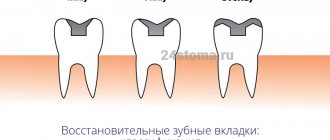
In the presence of significant deformations, filling is performed with ionomer cements with minimal preparation or photopolymer composites. You can restore the anatomical shape of your teeth and prevent their further destruction with the help of ceramic veneers and crowns.
The sooner you see a dentist, the easier it will be to stop the disease. You can find a list of specialized dentists on our website.
Types of pathology
Depending on the cause of formation and location, necrosis is classified into several types:
- Cervical necrosis , affecting the area in the gum area or even extending under it. The pathology is manifested by the formation of a chalky spot, which quickly turns black. Cervical necrosis can quickly spread to adjacent dental units.
- Acid necrosis , as the name suggests, is caused by exposure to dangerous acids. As a rule, this type of disease is detected in people working in hazardous industries. It can also be observed in people suffering from gastritis. This necrosis begins with demineralization of a small area of tooth enamel, which gradually leads to thinning of the enamel and tooth destruction.
- The radiation type is formed as a result of harmful radiation. It is observed in people whose work involves hazardous radiation, as well as in patients with cancer undergoing chemotherapy. In addition to the fact that the tooth is destroyed, the general condition of the mucous membrane worsens, and the periodontium may become inflamed.
- Computer necrosis is a relatively new type of pathology that affects people who spend a lot of time at the computer. Such necrosis can develop after several years of sustained, constant exposure to a computer on the human body. The pathology practically does not manifest itself at all; it can only be visualized by darkening of the tooth enamel.
After diagnosing the disease and making a diagnosis, the specialist selects the most suitable treatment option. If cervical necrosis is diagnosed, the tooth surface is treated with a composition that reduces the sensitivity of the enamel. Then the tooth is filled. If a tooth has darkened due to exposure to a computer, the necrotic tissue is removed, the resulting cavity is filled several times with a special compound, and only then a filling is placed. If necrosis has developed under the influence of dangerous acids, it is first necessary to eliminate their exposure, and only then carry out remineralizing therapy. Treatment of necrosis is carried out comprehensively, in addition to filling, medications and vitamins are prescribed, as well as applications with special pastes.
Advantages of the GrandMed clinic
- Dentistry has created an excellent diagnostic base that allows identifying non-carious dental pathologies in the early stages. Doctors have modern equipment at their disposal (radiovisiograph, orthopantomograph, computed tomograph), and all necessary clarifying tests can be done in the clinic’s biochemical laboratory.
- We employ professionals of the highest level, whose experience and qualifications allow us to treat complex non-carious pathologies, which often require the use of special techniques and non-standard approaches. Clients are provided with high-quality therapeutic and orthopedic care, including prosthetics on implants. So even in the most advanced cases, our patients can count on restoration of the beauty and functionality of their dentition.
- This disease is treated using local anesthesia. Patients of our clinic do not experience pain and feel comfortable during the procedure after injection of one of the modern anesthetic drugs that do not have side effects (Ultracaine, Ubestezin, Scandonest). You should not be afraid of the injection itself - it is performed quickly and accurately, and therefore is absolutely painless.
- Our clinic adheres to the highest sanitary standards, which is confirmed by the use of the latest sterilization equipment, which completely eliminates infection during treatment; Dentists have all the necessary consumables and disposable dental instruments at their disposal.
Note: computer necrosis of teeth
Experts started talking about this non-carious lesion of teeth relatively recently; it definitely did not exist in the 20th century, however, like people who spent most of their working and free time at the computer. The death of dental tissue threatens patients who are ready to sit at a computer for more than 8 hours a day, without days off and a full night’s rest. After 3–5 years of living in this mode, a person (and these are mainly young people under 35 years old) are guaranteed to have problems with the front teeth located in the path of the electrostatic radiation of the monitor.
Symptoms of computer necrosis of teeth
Experts, comparing computer and post-radiation lesions, find much in common between them: dying areas in both cases spread over the entire surface of the supragingival part of the teeth, which acquires a dark, almost black color. The focus of necrosis is filled with softened brown tooth tissues; the dentist easily removes them with special instruments, and the patient does not feel pain. As for the teeth of the chewing group, which are exposed to relatively less computer radiation, they also do not look the best: the enamel loses its shine and acquires a gray tint. Studies have shown that not only hard tissues undergo negative changes, but also the pulp. Thus, during electroodontometry, the neurovascular bundle does not respond to an electric current of 25-30 μA. In addition, changes in the functioning of the salivary glands are observed.
Treatment of computer necrosis of teeth
The “newness” of the disease complicates diagnosis, since the dentist may never have encountered it. An X-ray examination is required (sight image, orthopantomogram, computed tomogram), in the images the teeth look translucent and have unclear contours, which indicates a deficiency of mineral substances.
Treatment of computer necrosis is complicated by the fact that during the removal of damaged tissue with a bur, the tooth falls apart; in such conditions, it is impossible to preserve the vitality of the pulp, and often the tooth itself. If recovery is possible, it will take a lot of time, as it will be carried out in several stages, including local and general therapy, preceding filling or prosthetics. At the first stage of treatment, dead tissue is removed and healthy tissue is remineralized, and the patient is recommended to take mineral-vitamin complexes containing calcium, antioxidants, and vitamin A. After the condition improves, therapeutic linings are placed in the dental cavity and a temporary filling is performed. The result of this stage of treatment, which takes up to two months, is the restoration of dentin, after which the tooth is finally filled.
Prevention of computer necrosis of teeth
Computer necrosis of teeth can be prevented by a rational approach to working at the computer. Firstly, comply with sanitary standards, which require organizing a work area of at least 6 sq.m., located in a room with an area of 20 - 24 sq.m. The monitor should be located no closer than 60 - 70 cm from the face; if there are several computers installed in a room, the distance between them should be at least 2 m. Every two hours you should take a break of 15–20 minutes, during which the room is ventilated. Secondly, monitor your diet; if you don’t get enough antioxidants, vitamins and microelements with food, then take vitamin-mineral complexes. Thirdly, use new models of monitors that have minimal electrostatic effect on the body.