N. A. Yudina Doctor of Medicine, Professor, Head of the Department of General Dentistry of the Belarusian Medical Academy of Postgraduate Education (Minsk)
Dental patients present with various problems, among which non-carious lesions of hard dental tissues occupy a significant place. For a dentist, knowledge about etiological factors and features of the clinical course of various types of non-carious lesions remains relevant.
Foreign authors have proposed the term “tooth wear”, which combines various non-carious lesions that develop after teething [1].
Tooth wear increases from 3% at age 20 to 17% at age 70 [2, 3].
The purpose of this work is to present information from modern literary sources on the issues of terminology, etiology, clinic, differential diagnosis, prevention and treatment of wear of hard dental tissues.
Increased abrasion - “attrition”, abrasion (grinding) - “abrasion”, erosion - “erosion”, abfraction - “abfraction”, erosion, foreign scientists combine into broader terms: “tooth wear” - tooth wear - or “tooth surface loss (TSL)” - loss of hard tooth tissue. Tooth wear is the loss of hard tooth tissue due to a process not related to caries [4].
Wear or loss of hard tissue occurs due to tooth contact, stretching and compression during chewing, mechanical, chemical, acidic action and other factors [5]. In the etiology of wear, an important role, along with the combination of abrasive and erosive factors, is played by the failure of compensatory-protective mechanisms.
Risk factors predisposing to the development of non-carious lesions affect not only dentistry, but also other areas of medicine. The incidence is affected by changes in eating habits, psycho-emotional stress, an increase in the number of various social stressful events and much more.
Erosion is the loss of hard tissue due to the action of an acid or chelating agent [6]. In the dental literature, the term “erosion” more often refers to chemical wear as a result of external or internal acids or enterosorbents acting on the tooth surface without the involvement of a bacterial factor [7, 8].
However, the definition of “erosion” does not take into account proteolytic and piezoelectric effects, which are also involved in the biochemical and electrochemical wear of dental hard tissues. According to foreign authors, erosive wear is, rather, the abrasive destruction of a material, which occurs as a result of the movement of a liquid or gas, containing or not containing solid particles, over its surface. Therefore, the term "biocorrosion" is a more accurate definition for this condition. “Biocorrosion” is wear (degradation) of a material (including teeth) due to chemical, biochemical or electrochemical influences [9, 10].
Abfraction of teeth (literally - “breaking off”, from the Latin “ab” - from, “fractio” - destruction; microcrack, microchip) is a non-carious lesion of the enamel, having the shape of a notch or wedge, resulting from repeated exposure to lateral occlusal loads (J. Grippo, 1991) [11]. The term stress corrosion is used to refer to this phenomenon. The main cause of abfraction is considered to be tooth bending caused by excessive occlusal load on it. A special role is assigned to frequent and extreme lateral loads during chewing or in the case of parafunctions of the dental system and tongue. Lateral occlusal forces cause the appearance of stress concentration nodes in the enamel in the cervical region, which leads to the breaking of chemical bonds in the crystalline structure of enamel and dentin.
Grinding of teeth (syn. abrasion, abrasive wear, abrasion, wedge-shaped defect) is a progressive loss of hard dental tissues due to repeated mechanical contacts with any objects other than teeth [12]. As a rule, the cause of abrasion is the mechanical impact of a foreign body: a hard brush, tooth powder, seeds, which manifests itself on the cervical and occlusal surfaces. One of the most common names for this type of pathology - wedge-shaped defect - is due to the shape of the defect.
In the development of V-shaped abrasion, the role of diseases of internal organs, especially the gastrointestinal tract, endocrine and nervous systems, has been established. According to literature sources, in patients with gastrointestinal pathology, a wedge-shaped defect occurs in 23.6% of cases. Most often it is detected in chronic colitis, gastric ulcer and duodenal ulcer.
Increased tooth abrasion is the loss of hard dental tissues due to occlusal contacts during chewing. This is the progressive loss of hard tissue on the occlusal surfaces of the teeth, exceeding normal wear. The condition may be caused by defects in the dentition, improper design of dentures, or the habit of chewing on one side. There are many other factors that contribute to erasure. These include bruxism, clenching of teeth, eating habits, and malocclusions.
Clinic, diagnosis and differential diagnosis of wear of hard dental tissues
Only a third of practicing dentists in Europe report erosive or other tooth wear in their medical records [13]. The codes and criteria used in medical documentation do not allow recording of non-carious lesions.
The classic description of erosion in the literature is “a saucer-shaped defect with clear boundaries and a smooth bottom, located on the vestibular surface between the neck of the tooth and its equator” [14].
| Table No. 1. Variants of clinical manifestations of erosive lesions of teeth depending on the etiological factor | |||
| Clinical picture | Saucer-shaped defects in the cervical region of the anterior group of teeth of the upper jaw, unilateral or bilateral | Defects on the oral surface of the chewing group of teeth in the upper jaw | Defects on the oral surface of the anterior group of teeth or all teeth of the upper jaw |
| Cause of erosion | Diet, medications | Gastrointestinal diseases gastroesophageal reflux | Vomiting in eating disorders |
However, today the clinical manifestations of erosion are more varied and depend on the etiological factors of this disease (Table No. 1).
In the early stages, erosive tooth wear appears as a loss of physiological surface gloss. In later stages, changes occur in the original morphology of the tooth.
With frequent consumption of foods and drinks with low pH, concavities appear on the vestibular surface of the front teeth, the width of which clearly exceeds the depth.
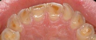
In patients with eating disorders [15], due to frequent vomiting, enamel loss first occurs on the palatal surfaces of the central and lateral incisors of the maxilla. The enamel loses its characteristic pattern and looks “glazed.” Next, in descending order, the palatal surface of the molars, the palatal surface of the canines, the occlusal surface of the molars, the palatal surface of the premolars, and the occlusal surface of the premolars are affected in the upper jaw. The cutting edge of the incisors becomes thinner, the chewing surfaces become flattened. In severe cases, the occlusal morphology disappears. The rate of decline depends on episodes of vomiting and usually becomes noticeable within 3 years.
In patients with gastroesophageal reflux, enamel is lost from the palatal and occlusal surfaces of the upper molars [16].
The clinical picture of an abfraction defect differs significantly from that of an erosive one: a defect in the form of a step or ledge with an acute angle at the base, occurs in the area of the tooth neck, and sometimes extends into the subgingival region (Fig. 1).
Rice. 1a. Abfraction defects.
Rice. 1b. Abfraction defects.
Gingival recession may complement the clinical picture, but its presence is not always observed.
To diagnose abfractions, a thorough history and diagnosis of occlusal relationships are very important. An abfraction defect, unlike erosion, necessarily has sharp edges. A wedge-shaped defect is always combined with gum recession, and its depth always exceeds its width (Fig. 2a, Table No. 3).
Rice. 2a. Wedge-shaped defects.
The wedge-shaped defect is localized in the cervical region on the vestibular surfaces of the teeth that protrude most strongly from the dental arch, most often the first premolars and canines. It may be more pronounced on one side depending on which hand the patient uses to brush their teeth. The shape resembles a triangle, the apex of which faces the tooth cavity.
For the purpose of differential diagnosis of wedge-shaped and abfraction defects, occlusal relationships are diagnosed. And if supracontacts are detected on teeth with V-shaped defects and overload of individual teeth and groups of teeth is recorded, then the diagnosis of “abfraction defect” is more justified. Sometimes in the same patient we can observe a combination of wedge-shaped and abfraction defects.
With increased abrasion, the teeth of the upper and lower jaws are affected. Tissue loss is observed on the chewing surfaces of molars and premolars, the cutting edges of the frontal teeth (Fig. 2b), the palatal surfaces of the anterior teeth of the upper jaw and the vestibular surfaces of the anterior teeth of the lower jaw. The affected surfaces are hard, smooth and shiny. Sharp edges and chips are observed in patients suffering from bruxism.
Rice. 2b. Increased erasure.
Differential diagnosis according to clinical manifestations is given in Table No. 2.
| Table No. 2. Clinical picture of various forms of tooth wear | ||
| Diagnosis | Characteristics of the lesion | Location of the lesion |
| K03.2 Erosion | An oval or saucer-shaped defect with clear boundaries, the bottom is smooth, shiny, loss of occlusal pattern, “vitrified” enamel |
|
| K03.1 Grinding (abrasive wear) of teeth - wedge-shaped defect | Defect in the shape of a triangle, the apex faces the oral cavity, the depth exceeds the width |
|
| K03.18 Other specified diseases of dental hard tissues (abfraction) | Angular defect with sharp edges |
|
| K03.0 Increased abrasion (wear) | Horizontal or vertical loss of hard tooth tissues |
|
We should not forget that in the clinic we can observe a combination of various lesions in one patient (Fig. 3-8).
Rice. 3. Patient I., 48 years old, erosion, abfraction, abrasion, bruxism + caries.
Rice. 4. Patient K., 42 years old, erosion (covered by restorations), abfraction, abrasion, bruxism.
Rice. 5. Patient Shch., 53 years old, erosion, abfraction.
Rice. 6. Patient L., 63 years old, crossbite, erosion (covered by restorations), abrasion, bruxism.
Rice. 7. Patient K., 43 years old, tetracycline teeth, abrasion, bruxism.
Rice. 8. Patient S., 44 years old, erosions (covered by restorations), wedge-shaped defects, abrasion, bruxism.
The stress concentration due to bruxism can act synergistically as a cofactor with acids as well as abrasives to produce various manifestations of non-carious lesions. Signs of bruxism in the clinic include increased abrasion, abfraction defects, chipping, tension in the masticatory muscles, and much more.
Causes of cervical caries
To date, more than 400 theories have been developed regarding the causes of caries.
- Meanwhile, most of them are based on the assertion that the main factor contributing to the development of the disease is non-compliance with hygiene rules, which entails the formation of soft or hard plaque on the surface of the tooth enamel.
- Plaque plaques provide a favorable environment for the growth, reproduction and development of pathogenic bacteria. In the process of life, pathogenic microflora produces lactic acid, as a result of which demineralization of tooth enamel occurs.
- The increased risk of developing caries in the cervical area is due to the fact that at the base of the tooth the layer of enamel is thinner than on other parts of its surface.
- In addition, the cervical area is considered difficult to access for care.
- It should be noted that caries rarely occurs indirectly. The most important pathogenetic link in its development are gastrointestinal pathologies, decreased local and general immunity, errors in diet and other malfunctions in the body, and external factors.
Prevention of wear of hard dental tissues
For the purpose of prevention, it is necessary to identify and eliminate risk factors that are important in the occurrence of tooth wear. These include the patient's use of highly abrasive hygiene products, frequent consumption of drinks and foods with low pH, eating disorders, bruxism, disorders of occlusal relationships, and somatic pathology.
Basic strategies for preventing wear of hard dental tissues, based on the main risk factors:
1. A balanced diet with minimizing the consumption of foods and drinks with low pH.
Careful assessment of dietary habits is important in assessing the erosive potential of a diet. The patient should be familiarized with a list of foods with low pH (salad dressings, vinegar, alcoholic beverages, carbonated soft drinks, herbal teas, fruit juices). The patient is advised to keep a food diary for at least 4 days, including weekends. Identification of consumption of foods and drinks with low pH in any combination more than 4 times [17] per day allows the patient to be classified as at risk of dental erosion.
2. Control of hygiene skills and informed choice of hygiene products.
Scientists assign the leading role in the erosive and abrasive wear of tooth tissue to mechanical factors: the active use of certain abrasive hygiene products, irrational brushing of teeth [18-21].
Patients at risk (gastrointestinal diseases, eating disorders, hazardous occupations, bruxism, etc.) should use toothpastes with a low degree of abrasiveness (RDA index = 30-50) and a soft toothbrush. It is not recommended to cleanse immediately after consuming fruits and drinks with low pH, vomiting, regurgitation or reflux.
To prevent erosion, at least 1 hour should pass between meals or drinks.
3. Strengthening hard tissues and normalizing saliva function. Strengthening hard tissues is possible due to fluorides and calcium phosphate technologies.
Scientists have recognized effective technologies for increasing the acid resistance of hard dental tissues using fluoride-containing preparations [22]. When exposed to erosion, there is no need for remineralization of the subsurface layer. The main thing is to strengthen the thin surface layer. Therefore, when treating erosions, high concentration fluoride applications are a more appropriate application. Fluoride gels provide good protection for eroded enamel from the abrasive action of a toothbrush.
The use of bicarbonate-containing toothpastes is one of the ways to introduce buffering agents into the oral cavity [23]. It is recommended to apply alkaline pastes or gels before bed to protect teeth from erosion due to reflux that occurs during sleep.
Adding calcium and phosphate to sour drinks can significantly reduce their erosive potential. Juices and carbonated drinks containing calcium and phosphates have appeared on sale.
Measures to increase the secretion of stimulated saliva, such as chewing sugar-free gum, are accordingly anti-erosive and support remineralization.
4. Referral to related specialists for timely detection and treatment of somatic pathology (endocrine pathology, hormonal disorders, gastrointestinal diseases, serious eating disorders: anorexia, bulimia, bruxism).
The appearance of tooth wear may be associated with endocrine pathology, disorders of hormonal and mineral homeostasis. Yu. M. Maksimovsky (1981) assigns an important role in the development of erosions to hyperfunction of the thyroid gland. Dental erosions in patients with thyrotoxicosis were detected twice as often as in persons with normal thyroid function; a direct connection was established between the intensity of dental damage and the duration of thyrotoxicosis {24}. In women, dental erosions are detected in a higher percentage of cases {25}. Examination of patients with non-carious lesions of teeth should be carried out jointly with a gynecologist and endocrinologist. The examination algorithm by related specialists includes hormonal indicators (estradiol, TSH, prolactin, cortisol).
If gastroesophageal reflux is suspected, patients need a referral to a physician or gastroenterologist. The dentist should be alerted to erosions on the oral surface of the chewing group of the teeth of the upper jaw and the patient’s specific complaints (sour taste in the mouth, halitosis, increased salivation, permanent cough, stomach pain, feeling of a “lump” in the throat, burning sensation, hoarseness, belching, heartburn) . Patients with a history of asymptomatic gastroesophageal reflux report wet spots on the pillow and a sour taste in the mouth after sleep.
Eating disorders include three main diagnoses [26]: anorexia, bulimia, and other unspecified eating disorders (a combination of several disorders). Anorexia is characterized by a conscious refusal to eat, a prolonged lack of appetite, and the disappearance of the basic instinct - hunger; bulimia - uncontrolled eating of large portions of food, followed by spontaneous vomiting.
Early detection of eating disorders is important for the outcome of the disease, secondary prevention, prevention of complications and reduction of harmful consequences in relation to the dental health and general somatic status of the patient. Patients with eating disorders often avoid medical professionals, and if they do, they hide the true origin of the problem. Therefore, knowledge of the manifestations of these disorders in the oral cavity is extremely important for the dentist.
Hypoplasia and hyperplasia
These are rare congenital pathologies. With hypoplasia, the enamel is underdeveloped or absent altogether. With hyperplasia, its quantity, on the contrary, is excessive. Such disorders arise due to metabolic failure, general pathologies of fetal development, diseases of the gastrointestinal tract, as well as health problems that arise in women during pregnancy. Usually the problem of hyper- and hypoplasia is solved in childhood by removing “extra” or replenishing missing hard tissues. Sometimes those who had congenital hypoplasia may require more frequent remodeling therapy in adulthood.
Treatment of wear of hard dental tissues
Normalization of occlusal relationships is the first and most important stage. Measures to correct occlusion include restoration of the occlusal surfaces of teeth, orthodontic treatment, and more.
Treatment of bruxism requires a comprehensive interdisciplinary approach [27]. Depending on the severity of manifestations and causes, psychiatrists, neurologists and physiotherapists take part in treatment. There is a need to manufacture an occlusal splint or mouth guard and use intraoral devices.
Therapeutic and orthopedic treatment methods are used to eliminate hard tissue defects. When restoring the vestibular surfaces of teeth, the leading requirement is aesthetics; for the occlusal surfaces, mechanical strength is the main requirement.
With increased abrasion, preference is given to orthopedic treatment methods - crowns and onlays.
In the case of cervical defects, it is recommended to prepare hard tissues to a depth of 0.5 mm, form a bevel, and restore with composites or GIC [28]. The enamel is ground around the defect using fine-grained diamond burs coated with 80 microns (with a red marking strip); some authors recommend reducing the enamel etching time to 5 seconds. [29].
An innovative method for closing defects is the use of direct composite onlays, manufactured in a factory. For example, COMPONEER manufactured by Coltene/Whaledent (Switzerland) - thin composite onlays (0.3-0.7 mm) with a uniform surface - allows you to close defects in the hard tissues of teeth [30] (Fig. 9a - d).
Rice. 9a. Patient 1 before treatment.
Rice. 9b. Patient 1 after treatment: metal-ceramic bridges in the area of the lateral group of teeth, Enamel White Opalescent composites on teeth 14,13,12,11, base A1/B1; lower incisors - restorations from Synergy D6.
Rice. 9th century Patient 2 before treatment.
Rice. 9y. Patient 2 after treatment: Enamel White Opalescent compositors on teeth 12, 11, 21, 22, S0 Miris base and W paint on teeth 12 and 21.
Erosion
Looks like areas of damaged enamel with a changed shade. In the affected areas, during erosion, hard tissues soften, gradually collapse, and a cavity is formed that reaches the dentin. At this stage, the tooth begins to hurt, react to any irritants, and the defect itself becomes clearly visible. Erosion has several possible causes:
- frequent consumption of sour or sweet foods;
- brushing teeth with hard brushes or pastes with an abrasive effect;
- low calcium content in hard tissues (due to metabolic disorders, pregnancy, certain diseases).
Erosion looks like a spot with a smooth, non-shiny surface. It is treated by performing remineralization and tooth restoration. It is important to find and eliminate the cause of the defect. If this is not done, erosion will appear in new places.
Injury
The tooth may become damaged due to trauma, and this requires urgent treatment. Usually these are chips, fractures or dislocations that occur due to impacts, accidental falls, or gnawing on hard objects. If the defect as a result of injury is superficial (it is a small chip), it is enough to restore the hard tissues by performing a restoration. If it is a severe bruise, crack, or fracture, more serious recovery will be required. If you quickly consult a dentist, the tooth will most likely be saved (there are such chances even if it was knocked out). In the most severe cases, removal is required, and then implantation and prosthetics are performed.
Necrosis
This is the death of hard tissue cells, due to which multiple defects appear on the surface of the crowns, as well as on the tooth root. The structure of the cells changes, and destruction can affect not only dentin, but also the pulp. Necrosis is rare and is usually not directly related to the condition of the teeth. It can be caused by severe toxin poisoning, hormonal imbalance, or severe systemic diseases. Treatment at the dentist is symptomatic: restoration of hard tissues, remineralization, prosthetics for complete tooth destruction. The main therapy must be related to the disease that caused the necrosis, and dental treatment only complements it.
You have questions?
We will call you back within 30 seconds
+7