Anomalies of teeth - various types of deviations of teeth from the norm (in number, size, shape, structure, etc.), as a rule, congenital or acquired disorders of the functioning of the dental system. According to dental practice, various dental anomalies are observed in 40-50% of children and adolescents and 30-40% of adults. Despite the fact that dental anomalies sometimes require long-term and multi-stage treatment (orthodontic, orthopedic, therapeutic, surgical, etc.), many prefer to turn a blind eye to the presence of problems, some even try to make a feature out of a problem (as in the case of diastema, despite the charm and charm of some public representatives of the fair sex, this is in any case an anomaly).
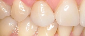
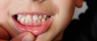
Examples of dental anomalies from the YuliSTOM dental practice
Anomalies of the dental system can be divided into the following groups:
- anomalies of individual teeth (size, shape, number, position);
- dental anomalies;
- malocclusion;
Below we will look at each group separately.
Description and causes of the disease
Both children and adults are susceptible to pathology. Depending on the etiological reasons, acquired and congenital anomalies of dental development are distinguished. The first appear throughout life as a result of various external and internal factors affecting the dental system. Congenital are formed due to the presence of unfavorable heredity, or a violation of the embryonic development of the fetus.
Main reasons:
- genetic predisposition;
- teratogenic effects of various substances on the embryo during the formation of organs and systems;
- hormonal imbalance (hypothyroidism, hypocortisolism), vitamin deficiency during pregnancy;
- infections that cause genetic mutations in the fetus (herpes, rubella, HIV, syphilis, toxoplasmosis, cytomegalovirus, etc.);
- fetal hypoxia;
- toxicosis of a pregnant woman;
- bad habits of childhood: prolonged use of a pacifier, bottle feeding, finger sucking, etc.;
- difficult childbirth, birth injuries of the jaw;
- infectious diseases, rickets, hypovitaminosis in the first three years of life, leading to impaired development of segments.
Various factors can provoke abnormalities in the development of teeth in children: alcohol, drugs used by the expectant mother before and during pregnancy, unbalanced nutrition, sudden climate change, radiation, lack of fluoride, calcium, etc.
Preventive measures
Specific methods for the prevention of tortoanomalies have not yet been developed. This is quite understandable, given their polyetiological nature and the impossibility of accurately predicting how teeth will behave during the process of eruption and growth.
You can help reduce the risk and consequences of an anomaly only by regular preventive visits to a doctor , which allow you to identify the development of disorders at the initial stage and take timely measures to prevent them.
The recommended frequency of visiting a doctor depends on the age of the child. In the period from the second to the fifth year (the period of growth and development of baby teeth), it is recommended to visit the dentist 3 times a year; in the future, you can reduce visits to 2 times a year.
It is also necessary to take into account the possibility of a hereditary predisposition to tortoanomaly; it should be the reason for genetic analysis.
Types of dental anomalies
| View | Characteristic |
| Size pathology | Macrodentia. The crown is significantly enlarged. The developmental anomaly is associated with the fusion of several rudiments into one due to hormonal imbalance. Defective units are more often located in the upper row of the smile zone. Microdentia. Excessively small units. Pathogenesis is associated with genetic predisposition. The violation affects the incisors, mainly the upper ones. |
| Anomalies of dental structure | Hyperplasia. “Pearls” are formed on the crown - formations of enamel of various shapes, 1.5 - 3.5 mm in diameter. Droplets are most often localized near the neck of the segment, or in the bifurcation zone of the root. Dysplasia. The surface of the unit is heterogeneous, thinned in places, and gray spots are present. There are many serrations on the incisors. Chipping and increased sensitivity may occur. Hypoplasia. The enamel layer is sharply thinned, sometimes absent (aplasia). Leads to premature development of inflammatory diseases of hard tissues (caries, pulpitis). Dentinogenesis imperfecta. Units are amber in color, opalescent. Enamel and dentin are fragile, susceptible to abrasion and destruction. Insufficient amelogenesis. It manifests itself as thinning of the enamel, the appearance of brown spots, and hyperesthesia to stress factors. |
| Form defect | Spine-shaped (in the form of a thorn or awl). The segments are wide at the base and sharply taper towards the periphery. Possible combination with microdentia. The surface is uneven and spotty. The pathology is characteristic of segments of the smile zone. Barrel-shaped (Hutchinson's teeth). The neck is thickened, the cutting surface has a recess in the center of which there is no enamel layer. Molar segments with a shortened surface and enamel hypoplasia, etc. |
| Growth disorder | Anomalies in tooth growth are associated with their abnormal spatial arrangement: vestibular displacement (outward); oral (inside); high or low localization; trema, diastema (gaps between units); tortoanomaly (rotation of a segment around an axis); crowding; transposition (change of place). |
| Pathology of numbers | Supernumerary (hyperdentia). Extra units. Hypodentia (insufficient quantity). Adentia (complete absence of units in the mouth). |
| Impaired eruption | Retention (retention of a unit in the jaw or alveolar process). Late eruption of incisors. Impaired eruption of paired segments. Premature eruption. |
| Abnormalities of tooth roots | Characterized by a change in the number of roots (downwards or upwards), their curvature |
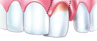
Common symptoms that occur with most developmental anomalies are: discomfort in the mouth (sometimes soreness), problems with chewing food, and speech defects. Such pathologies are often accompanied by other genetic defects (cleft palate, lips, etc.).
Types and classification of malocclusions
Bite is the position of the segments at the moment of tight closure of the jaw bones. Occlusion is assessed in 3 planes:
- by crown height (vertical);
- lateral arrangement of segments relative to the jaws (transvesial);
- ratio of the length of the row and the jaw (sagittal plane).
Malocclusion can be of 2 types: skeletal and dental. In the first case, the cause of pathological closure of the elements is the non-standard size of the jaws or their position in the mouth. Dental abnormal bite is formed due to dental defects (number, size, shape, etc.).
Classification of dental malocclusions:
- Open: the segments of the top and bottom rows do not meet each other vertically. The mouth remains slightly open. Due to the defect, speech, breathing, and chewing are impaired.
- Deep. The lower crowns are significantly (more than ½ covered by the upper ones). The face takes on a flattened shape. Often leads to injuries to the mucous membrane, rapid abrasion of teeth, and defects of the temporomandibular joint.
- Distal (prognathic). Occurs due to disproportionately developed jaws (overdevelopment of the upper or underdevelopment of the lower).
- Mesial: the lower row overlaps the upper segments.
- Crossed: on one side of the jaw, the teeth are located without pathology, and on the other, the lower crowns protrude above the upper ones.
Diagnostics
An obvious defect is noticeable even upon visual inspection. To identify hidden anomalies (unerupted teeth), as well as to clarify the diagnosis and find treatment methods, additional X-ray diagnostic methods are used.
To obtain a general picture of the condition of the dentition, an orthopantomogram is prescribed - a detailed x-ray of both jaws, which demonstrates the position of erupted and unerupted teeth on both jaws. To visualize local disorders, targeted intraoral photographs are taken.
Possible consequences
Improper development of teeth and abnormal bite cause uneven distribution of chewing load. This leads to the development of various complications:
- rapid abrasion of closing teeth;
- caries, pulpitis;
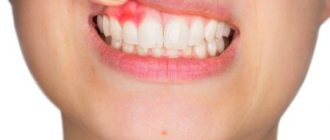
- gingivitis, periodontitis;
- bleeding of the mucous membrane;
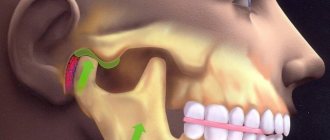
- displacement of the articular head of the temporomandibular joint.
When bite deviations occur, problems arise with oral hygiene and microcirculation in the periodontal tissues is disrupted. Ultimately, cellular nutrition, gas exchange, and metabolism are disrupted. A complex of pathological processes leads to early destruction of dentin, periodontal ligaments and tooth loss. Timely consultation with a doctor will help prevent adverse consequences. In Moscow, jaw bite correction is carried out by the Center for Aesthetic Dentistry near the Otradnoye metro station.
General overview
Tortoanomaly is the most common violation of the position of individual teeth. According to various sources, it occurs in 10-20% of all diagnosed disorders.
The anomaly is a rotation of the tooth around a vertical axis, the magnitude of which can vary from several to 180°, that is, the position when the lingual (palatal) surface becomes vestibular.
The most common case is a rotation of approximately 45°. The maxillary lateral incisors are most often affected by tortoanomalies, and the lateral incisors of the mandible are somewhat less common.
But in principle, any element can, for one reason or another, be rotated relative to its longitudinal axis.
The most common reason why patients consult a doctor with a thoracic anomaly is dissatisfaction with the aesthetics of their smile. But when the anomaly is significant, functional problems arise along with aesthetic ones.
People begin to experience difficulty biting and chewing food, the tongue and mucous membrane of the cheeks are injured by the abnormally located unit, and they lose clarity of speech.
Methods for correction and treatment of anomalies
Treatment depends on the clinical situation and is determined by the doctor after a comprehensive dental examination. Correction of dental abnormalities may include:
- surgical removal of wisdom teeth or supernumerary units;
- prosthetics: for adentia, hypodentia;
- orthopedic correction with a crown, veneer, lumineer: for size anomalies;
- restoration with crowns, composite materials, veneers: for non-standard shapes;
- if the structure of enamel and dentin is damaged, complex therapeutic, orthodontic, orthopedic treatment, enamel mineralization, and drug therapy are prescribed;
- eruption disorders: physiotherapy, massage. Sometimes orthopedic treatment (plates, aligners), removal of a baby tooth (if a permanent tooth has formed underneath it), surgical care (for impacted segments), or orthodontic preparation (system braces) may be required in order to create conditions for stretching the segment retained in the gum.
Malocclusions are most often corrected with fixed orthodontic structures (braces, aligners) in adults. Children are mostly fitted with removable devices.
Retention period
Tortoanomalies corrected with orthodontic appliances require a long period of retention as the periodontal and interdental ligaments strive to return the tooth to its previous abnormal position.
Retention methods are divided into orthodontic, surgical and physiotherapeutic:
- Orthodontic retention primarily involves the use of lingual retainers bonded to the inner surface of the teeth. The device used to correct the anomaly can also be used as a retainer.
- The surgical method of retention involves dissection of the interdental papillae with ligaments that tend to return the displaced unit to its previous position.
- The physiotherapeutic method consists of vibration stimulation of the rotated tooth with a frequency of up to 50 Hz. Vibrations are accompanied by repair of the bone tissue of the alveolar sockets, which helps maintain the tooth in a rotated position. Vibration stimulation sessions are carried out several times a week.
The retention time by orthodontic methods depends on the severity of the tortoanomaly and the correction method used, and can be several months.