The dentofacial system is an important component of the human masticatory apparatus. It meets morphological and functional characteristics, such as number, size, shape, color, structure of dental units. There are a number of genetically determined or external factors that lead to disruption of these characteristics. Dental anomalies can have various consequences: some lead to periodontal pathologies, various forms of caries, others seriously limit chewing function, cause injury to the mucous membrane, and others cause aesthetic problems.
Improper growth of teeth results in speech and bite defects and breathing problems. As a result, the patient’s general health seriously suffers, and psychological problems arise.
Reasons causing anomalies
Anomalies of the dental system are very diverse and include both anomalies of dental units and incorrect formation of the dentition, as well as abnormalities in the development of the jaws. The most frequently diagnosed abnormalities of dental units. They can be caused by a wide variety of reasons, including both exogenous (caused by external factors) and endogenous (formed as a result of the physiological state of the patient).
Endogenous causes
Endogenous anomalies are formed as a result of genetic and endocrine disorders.
- Many structural features of the dental system, leading to improper development of teeth, are genetically determined. This can occur through direct inheritance (abnormal number and shape of teeth, edentia, diastema), through inheritance of a mismatch in the size of the jaw bones, through inheritance of a mismatch in the size of the jaw and teeth (crowding of teeth or sparse arrangement of teeth). Dental anomalies of this type include disorders associated with hereditary and congenital pathologies (clefts of the soft and hard palate, cleft lip, Down syndrome, Vaanderburg syndrome, Seckel syndrome, Shershevsky-Turner syndrome).
- Another part of the endogenous causes of dental anomalies are problems of the endocrine system, such as hypothyroidism (in later stages), hyperparathyroidism, hypocortisolism. Their frequent symptoms may be delays in the eruption and replacement of teeth, and disturbances in the formation of the enamel layer.
Exogenous causes
Exogenous (external) causes of dental anomalies are, for the most part, external unfavorable factors that affect the baby’s body during the formation of tooth germs. There are prenatal (prenatal), postnatal (postpartum) and intranatal (associated with childbirth) periods.
- An example of a prenatal factor is a pregnant woman living in poor environmental conditions. Various developmental disorders of the fetus can also occur as a result of the mother’s poor lifestyle.
- Factors in the intranatal period may include complicated labor, birth injuries, consequences of asphyxia and oligohydramnios.
- In the postnatal period, dental anomalies develop as a complication of pathologies in early childhood. These include childhood infections, rickets, hypovitaminosis, and micronutrient deficiency.
All these reasons are of a common nature. Local factors that affect dental development include everything related to improper oral care, feeding errors or bad habits. Among them are feeding too soft food, prolonged sucking of a pacifier or finger in early childhood.
Childhood injuries and caries with complications lead to dental anomalies.
The death of tooth germs or the development of supernumerary teeth can occur due to osteomyelitis.
Possible consequences
Improper development of teeth and abnormal bite cause uneven distribution of chewing load. This leads to the development of various complications:
- rapid abrasion of closing teeth;
- caries, pulpitis;
- gingivitis, periodontitis;
- bleeding of the mucous membrane;
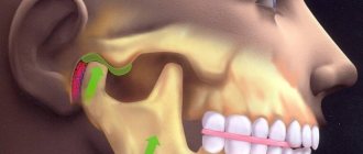
- displacement of the articular head of the temporomandibular joint.
When bite deviations occur, problems arise with oral hygiene and microcirculation in the periodontal tissues is disrupted. Ultimately, cellular nutrition, gas exchange, and metabolism are disrupted. A complex of pathological processes leads to early destruction of dentin, periodontal ligaments and tooth loss. Timely consultation with a doctor will help prevent adverse consequences. In Moscow, jaw bite correction is carried out by the Center for Aesthetic Dentistry near the Otradnoye metro station.
Anomalies in the number of teeth
All anomalies in the number of teeth are divided into three types:
- hyperodontia (excess of dental units above normal);
- edentia (complete absence of teeth);
- hypodontia (insufficient number of teeth).
Hyperodontia
Hyperodontia is a disorder of the formation of the dentition, which is expressed in the presence of supernumerary teeth (over 32 dental units in an adult). The cause of the disease may be a violation of the mechanism of formation of tooth germs in childhood or infancy. Hyperodontia mainly affects the incisors of the upper jaw. Other teeth are involved in the pathological process less frequently. Extra dental units grow small and have a cone shape.
The disease can be true or false.
- With true hyperodontia, the formation of extra dental units occurs in the human jaw.
- False hyperodontia occurs when the loss of primary teeth is delayed, which interferes with the normal growth of permanent teeth.
With polyodontia, teeth may erupt away from the dentition or next to a correctly positioned dental unit, which leads to its gradual displacement. The dentition can be seriously deformed, which leads to serious diction problems.
If supernumerary teeth interfere with the development of normal teeth, they must be removed. Broken teeth are corrected by installing braces.
In those rare cases when a row violation does not occur, the supernumerary tooth is preserved. Prosthetics are used to correct the shape.
Hypodentia
Hypodentia is a deficiency of dental units. This disorder occurs most often due to the death of tooth germs in the fetus. A small number of teeth causes a shift in the midline, the formation of wide gaps between teeth (diastema and three), and shortening of the dentition. All this adversely affects the bite. Possible ways of correction are prosthetics.
Edentia
Adentia is a complete or partial absence of teeth. With edentia, the continuity of the dentition is disrupted, which leads to difficulties in chewing and speaking. The reason is a violation of the development or death of tooth germs under the influence of genetic factors or under the influence of unfavorable external factors during the formation of the dental plate in the fetus (the formation of the germs of permanent teeth in the fetus occurs after the 17th week of intrauterine development). The disorder is corrected using prosthetics or dental implantation.
Methods for correction and treatment of anomalies
Treatment depends on the clinical situation and is determined by the doctor after a comprehensive dental examination. Correction of dental abnormalities may include:
- surgical removal of wisdom teeth or supernumerary units;
- prosthetics: for adentia, hypodentia;
- orthopedic correction with a crown, veneer, lumineer: for size anomalies;
- restoration with crowns, composite materials, veneers: for non-standard shapes;
- if the structure of enamel and dentin is damaged, complex therapeutic, orthodontic, orthopedic treatment, enamel mineralization, and drug therapy are prescribed;
- eruption disorders: physiotherapy, massage. Sometimes orthopedic treatment (plates, aligners), removal of a baby tooth (if a permanent tooth has formed underneath it), surgical care (for impacted segments), or orthodontic preparation (system braces) may be required in order to create conditions for stretching the segment retained in the gum.
Malocclusions are most often corrected with fixed orthodontic structures (braces, aligners) in adults. Children are mostly fitted with removable devices.
Anomalies of magnitude
There are two forms of this type of deviation, including macrodentia (increase in the size of dental units) and microdentia (small teeth).
Macrodentia
With macrodentia, the size of dental crowns is significantly increased. The cause of the disorder is, in most cases, dysfunction of the endocrine system, which is characterized by the fusion of several rudiments together. The disease is localized mainly in the area of the upper incisors. Large teeth interfere with the process of eruption and growth of neighboring dental units, which leads to their abnormal arrangement and crowding. Giant teeth are found on or outside the dentition line. This is a serious cosmetic defect that causes serious functional impairment and psychological disorders. Pathologically enlarged teeth are removed, and the growth of adjacent teeth is corrected using prosthetics or braces.
Microdentia
Microdentia – teeth that are too small. The disorder mainly affects the upper lateral incisors, but may affect the incisors of the lower or both jaws. The cause of the anomaly has not been studied, but it is reliably known that it develops in genetically predisposed patients. With the problem of small teeth, patients develop too large interdental spaces, which significantly disrupts the aesthetics of the dentition. To correct the pathology, incorrectly growing teeth are removed or covered with crowns.
Anomalies of the dentition
Anomalies of the dentition are characterized by a change in the shape of the typical dentition of the upper or lower jaw, which is caused by their narrowing or expansion in various areas and is expressed by crowding of the teeth, their rotation along the axis, vestibular or oral teething, partial edentia, the presence of supernumerary teeth, diastemas and etc. The following irregular forms of dentition are distinguished when they are narrowed:
- acute-angled, when the narrowing is localized in the canine area;
- saddle-shaped, when the narrowing is more pronounced in the premolar area;
- V-shaped, when the dentition is narrowed in the lateral areas, the frontal area acts as an acute angle;
- trapezoidal, when the frontal area is narrowed and flattened;
- common, when all the teeth - frontal and lateral - stand closely together;
- asymmetrical, in which the narrowing is more pronounced on one side of the dentition of the upper or lower jaw, resulting in a crossbite.
The main etiological factors of anomalies in the shape of the dental arches are underdevelopment of the jaws and their deformations caused by diseases of early childhood.
Shape anomalies
There are pathologies in the development of dental units, due to which they acquire an unnatural shape. These disorders are named after the scientists who first described them. The following types of dental shape anomalies are found: spiny teeth, Pflueger teeth, Fournier teeth, Hutchinson teeth.
- Spiked or awl-shaped teeth. With this pathology, the teeth acquire a cone-shaped shape. Wide at the base, they gradually narrow and, towards the cutting edge, become sharpened into a spike shape. This problem is combined with microdentia. There are irregularities and stains on the surface of the teeth. The disease affects the front and lateral incisors. The disease occurs in childhood and is caused by genetic factors in combination with external factors. Among them are vitamin D deficiency and endocrine system problems.
- Hutchinson's teeth are characterized by a modified crown shape of the incisors. Externally, they have the shape of a barrel, since the neck is significantly thickened. The cutting edge of the teeth acquires an arched notch. The enamel layer also suffers, which is present only on the sides and absent in the center.
- Fournier's teeth are a form of systemic hypoplasia of dental units associated with metabolic disorders at the stage of intrauterine development of the fetus. The barrel-shaped shape of the teeth is preserved, but the notch of the incisal edge is absent. The color of the enamel is not disturbed, but the enamel layer is underdeveloped, which can be seen during microscopic examination.
- Pflueger's teeth are a disorder that affects permanent dental units. Their crowns take on a conical shape. Thickening develops in the cervical region, and the chewing surface is significantly underdeveloped. The chewing function of the teeth is completely preserved.
- Turner anomaly. With this pathology, there is no enamel on the teeth. They have an abnormally lumpy surface, and replacement tissue, dentin, forms in the exposed areas.
Systemic hypoplasia with a violation of the shape of the teeth has three degrees of development. The third degree is the most dangerous, in which the crown is severely deformed and the enamel layer decreases. In this case, the defective teeth are removed and replaced with dentures. It is also possible to restore affected teeth using reflective components.
Publications in the media
Dental developmental disorders - structural anomalies and malformations of teeth - can appear in isolation or in combination with structural anomalies and malformations of organs and systems of the entire child's body.
Etiology. Dental tissue defects are caused by hereditarily fixed changes in the genetic code, but can be caused by environmental factors. Pathogenesis. Teeth during the period of their follicular development, i.e. Before eruption, several stages pass: laying, formation and calcification of crown enamel, formation and calcification of dentin. At each stage of tooth development, conditions can be created that lead to disruption of the formation of its shape, mineralization and color.
Classification • All disorders are divided into: •• anomalies of the structure of dental tissues, transmitted by inheritance •• anomalies in the number, size and shape of teeth, due to the hereditary transmission of the sample •• anomalies of the structure and malformations of dental tissues, which arose as patterns of the pathogenesis of systemic pathology in the child’s body (hereditary, congenital, acquired) •• structural anomalies and malformations of dental tissues caused by the influence of external factors. • Each group has individual clinical and radiological features, determined by the cause of occurrence and the mechanism of development.
Hereditary disorders of the development of hard dental tissues. Men and women suffer equally often. Both temporary and permanent teeth can be affected. • Enamel defects (most often dysplasia) are an isolated symptom (130900, Â). Combinations with cataracts and stenosis of the Sylvian aqueduct (600907, r), nail defects and hypohidrosis (ameloonychohypohidrotic syndrome, *104570, Â), also a sign of a number of hereditary diseases, for example: •• Renal tubular acidosis type II •• Albright’s disease •• Lacrimal duct defect (149700) •• Citrate transport protein defect (190315) •• Dysosteosclerosis (224300) •• Cranioectodermal dysplasia (218330) •• Tumor calcification (114120) •• Mucopolysaccharidosis, type IVB (253010) •• Neurosensory hearing loss, hypoplasia enamel, nail defect (234580) •• Goltz-Gorlin syndrome (305600) •• Menkes syndrome (261900) •• Naegeli syndrome (161000) •• Polyglandular autoimmune syndrome, type I (*240300) •• Stimmler syndrome (202900) • • Tuberous sclerosis (191092) •• Epidermolysis bullosa (226440). Oculodental-digital dysplasia (164200, genes ODDD, SDTY3, ODOD, 6q22 q24) •• Amelogenesis imperfecta (impairment of the process of enamel formation) is the most common pathology. Usually manifested by grooved (wrinkled) enamel or its sharp thinning. Erupting teeth are small, cylindrical, yellowish or brown in color. Amelogenesis imperfecta is inherited independently and is part of a number of hereditary syndromes ••• Amelogenesis imperfecta, type 1 (301200, genes AMELX, AMG, AIH1, AMGX, Xp22.3 p22.1, À) ••• type 2 (104500, gene AIH2 , 4q11 q21 À) ••• type 3 (301201, AIH3 gene, Xq22 q28 À) • with taurodontia (104510, À) ••• with nephrocalcinosis (204690, r) •• Enamel abnormalities are also observed in the following phenotypes: •• • platyspondylia with amelogenesis imperfecta (601216) ••• Kohlschutter syndrome (*226750, r): epilepsy, dementia and amelogenesis imperfecta ••• photoreceptor dystrophy and amelogenesis imperfecta (217080, r).
• Defects in dentin formation are numerous ; traditionally, the groups of dentinogenesis imperfecta and dentin dysplasia are distinguished. In addition, dentin defects occur in a variety of other osteogenesis disorder syndromes •• Dentinogenesis imperfecta. The structure of the enamel is not changed, but due to the imperfection of dentin, its connection with the enamel is fragile, which leads to enamel chipping ••• type I (*125490, 4q13–q21, DGI1 gene, Â). Synonyms: opalescent dentin, opalescent teeth without osteogenesis imperfecta ••• dentinogenesis imperfecta, type II, Capdepont teeth ••• type III (125500, Â). Clinically: Rapidly abraded dental crowns, smooth amber-colored dentin, open bite, very large pulp chambers and root canals of primary teeth, small or absent pulp chambers of permanent teeth •• Dentin dysplasia is a dental anomaly in which the teeth are clinically, morphologically and color-wise normal , but x-ray examination reveals short roots, obliteration of the pulp cavity and canals, increased mobility and premature tooth loss ••• type I (*125400, Â) •• type II (*125420, Â) - occlusion of the pulp cavity, the structure of the pulp in the form straws ••• Dentin dysplasia and cortical osteosclerosis (*125440) - compacted long bones, compaction of the alveoli of the teeth, narrow or sometimes closed medullary canals, thickening of the cortical layer.
• Mesoectodermal odontopathy (Stainton–Capdepont disease; simultaneous disruption of the formation of enamel, dentin and pulp). The teeth erupt at the usual time, have a normal size and shape, but their color is changed (from gray to blue-gray and yellowish-brown). The length and shape of the roots are changed. The tooth cavity and root canals are obliterated. Characterized by rapid wear of teeth (especially temporary ones). Their surface is shiny, flat and smooth (“polished”). At the same time, teeth practically do not react to mechanical, chemical and temperature stimuli.
• Oculodental-digital dysplasia (ODDD gene, 6q22–6q24, Â - *164200, r -*257850) - microphthalmos, coloboma or iris abnormalities in combination with congenital dental defects and finger abnormalities. Clinically: microphthalmos, microcornea, glaucoma, dental anomalies (enamel hypoplasia, small teeth), camptodactyly of the little fingers, syndactyly of the fourth and fifth fingers, absence of the middle phalanges of the toes, hypotrichosis, small nose, hypoplastic wings of the nose, hyperostosis of the skull bones, orbital (bone) hypotelorism, jaw overgrowth, spinal cord compression, spastic tetraparesis, progressive spastic paraplegia, the effect of paternal age on the frequency, pathology of the white matter of the brain on MRI, calcification of the basal ganglia. Synonyms: oculo-dental-digital syndrome, ODD (oculo-dento-digitalis) syndrome, Meyer-Schwickerath and Weyers syndrome. Note: a mutation at this same locus is the cause of syndactyly type III.
• ADULT syndrome (from Acro-Dermato-Ungual-Lacrimal-Tooth, 103285, Â). Clinically: hypodontia, early eruption of permanent teeth, ectrodactyly, obstruction of the lacrimal ducts, onychodysplasia, freckles, hypoplasia of the mammary glands. • Treatment depends on the degree of tooth tissue loss due to increased abrasion. In case of minor loss, it is recommended to regularly (2 times a year) conduct courses of remineralization therapy; in case of severe loss, orthopedic treatment is recommended. Enamel hyperplasia is the excessive formation of tooth enamel during its development. Typically, enamel hyperplasia is observed in the neck of a permanent tooth in the form of drop-shaped formations. After teething, these formations do not progress and are not affected by caries; do not cause any unpleasant sensations. Treatment is indicated only for noticeable cosmetic defects; Grinding is carried out followed by remineralization.
Enamel hypoplasia is a developmental defect that manifests itself in a violation of the structure and mineralization of tooth tissue during the period of their formation. Usually, only the formation of enamel is disrupted, and in severe cases, dentin as well. Most often, enamel hypoplasia is observed on permanent teeth, very rarely on temporary teeth, which is explained by their formation in the prenatal period. The localization of areas of hypoplasia depends on the age at which the child suffered from a general disease, and its severity depends on the severity of the disease.
• Causes: severe infectious diseases, hypo- and avitaminosis, disorders of the gastrointestinal tract (dyspepsia), endocrine organs (especially the parathyroid glands), hemolytic jaundice, hereditary syphilis, etc. • Classification : •• systemic hypoplasia - a violation of the structure of the tissues of all or a group of teeth formed in the same period of time •• focal hypoplasia - a malformation of the tissues of one or more teeth that formed in the focus of inflammation of the tissues surrounding the follicle, in the area of tumor growth or exposed to trauma. • Manifestations. Hypoplasia of dental enamel manifests itself in the form of whitish spots or depigmentation of various sizes with a smooth shiny surface. They can be combined with point or cup-shaped depressions, grooves, and constrictions of the enamel of the crowns of the teeth. In these lesions, the enamel loses its inherent color, but retains its density. Sometimes a complete absence of enamel is observed in a limited area of the tooth, which looks like a cup-shaped depression. Rarely, enamel is absent on almost the entire surface of the tooth crown and then the tooth takes on a bizarre, ugly shape (Turner's, Hutchinson's, etc. teeth).
• Treatment and prevention . Treatment of dental enamel hypoplasia depends on the form and severity of the disease. For spotted hypoplasia, bleaching with medications or grinding is carried out. To prevent the occurrence of caries, remineralizing therapy is carried out. Small enamel defects are filled after preparation. Teeth significantly affected by hypoplasia are covered with artificial crowns. Prevention of dental enamel hypoplasia - harmonious physical development of the child, balanced nutrition, timely treatment of diseases of primary teeth. Fluorosis is an acquired malformation of dental tissues caused by excessive intake of fluoride during tooth formation (a kind of enamel hypoplasia). Most often it develops with an excessive fluoride content in drinking water (more than 1.5 mg/l), but it is also possible with an optimal fluoride content (0.8–1.2 mg/l) in children with a reduced immune status.
• Pathogenesis. Fluoride salts have a toxic effect on ameloblasts, which leads to impaired calcification of tooth tissue. Subsequently, the content of fluorine and calcium in the surface layers of the enamel increases, and, accordingly, the acid solubility of the enamel decreases. The severity of fluorosis depends on the dose of fluoride entering the body during amelogenesis. These teeth are not affected by caries.
• Manifestations: •• chalky spots and stripes on the vestibular surface of the teeth •• chalky and pigmented (to brown and black) spots •• erosive lesions of the enamel •• disturbances in the shape of the crowns of the teeth in combination with stains, erosions and fragility of the enamel (fractures of areas of enamel during mechanical influence).
• Treatment is prescribed taking into account the severity of the process. If the form is spotty, teeth are whitened. Enamel defects are filled or orthopedic treatment is performed (crowns, pinned teeth). • Prevention •• Public prevention: reducing the intake of fluoride from drinking water into the child’s body during the period of teeth formation (up to replacing the water source) •• Individual prevention: partial defluoridation of drinking water by boiling, settling •• Additionally, milk and dairy products are recommended throughout the year , careful hygienic care of the oral cavity.
ICD-10 • K00 Disorders of development and eruption of teeth •• K03.7 Discoloration of hard tissues of teeth after eruption
Anomalies of hard tissue structure
An altered shape, abnormal size or color of enamel is formed against the background of an abnormality in the structure of the hard tissues of the dental unit. Among such anomalies are:
- Hypoplasia (underdevelopment of tissue). The initial degree of the disorder is manifested by the presence of chalky spots on the enamel and areas where tissue deficiency is observed. Subsequently, all kinds of pits, grooves, and recesses appear on the surface of the enamel. The defect affects all teeth in the dentition.
- Hyperplasia (excessive tissue formation). Pathology also affects all teeth at the same time. It is characterized by areas where tissue growth is observed - tubercles, sagging enamel. The consequence of the pathology is a change in the shape and size of the teeth, a violation of the occlusion (the line of contact of the upper and lower jaws).
- Anomalies of amelogenesis (enamel formation) are expressed in the presence of brown or yellow spots on the surface of the teeth. Areas where the natural composition of the enamel is disrupted become especially sensitive. Microdentia develops against the background of pathology. The cause of the development of pathology is a deficiency of microelements that take part in the formation of dental tissue. Treatment consists of replenishing this deficiency. Additionally, local remineralizing therapy and physiotherapy are performed.
- Disturbance of dentinogenesis consists of dysfunction of the mechanism of dentin formation. The main symptoms of the disease are as follows: teeth become yellow-brown or grayish in color. The fragile dentin in these areas quickly wears off, causing tooth decay and then tooth loss. The disease occurs in genetically predisposed patients. The problem can only be solved by replacing the units destroyed by the disease with prosthetics.
What it is?
Microdentia refers to diseases characterized by the eruption of permanent teeth with abnormal development of their size.
With this pathology, they are smaller not only in diameter, but also in height , although the height, more often than not, is only slightly different from the average statistical norm.
Basically, the pathology affects the teeth of the anterior or lateral sections, but can also affect the entire dentition. The lateral and central incisors are especially .
As a rule, microdentia is characterized by asymmetry, but there are exceptions here too.
Color anomalies
Healthy, young teeth look snow-white due to a thick dentin layer and high-quality enamel, which has sufficient characteristics of whiteness, shine and transparency. Lifestyle, age, genetic factors, and ecology can slightly change the color of teeth from bluish to yellow. Small deviations are not considered pathology, although they indicate a change in the quality of the enamel.
Various pathological processes occurring in the body have a more significant impact on the condition of the enamel. The cause may be carious lesions, the use of medications, or a lack of certain microelements in the body. Teeth can take on a gray, pinkish, brown, purple and even black tint.
When starting to treat a patient, the dentist excludes the development of chronic pathologies. After a course of therapy and stable remission is achieved, other causes of discoloration of the enamel are eliminated. The final stage of treatment is a course of teeth whitening.
Specialists at the Consilium Dent choose the enamel lightening technique together with the patient. The selection criteria are age, condition of the enamel, and the wishes of the patient.
Diagnosis of microdentia
According to statistics, about 5% of people face the problem of microdentia – abnormally small teeth in an adult. There are several signs that allow you to distinguish microdentia from other dental pathologies.
Symptoms of microdentia
- teeth of regular shape, but with a significantly reduced crown;
- distances between teeth visible to the naked eye;
- damage mainly to the front teeth;
- wavy or jagged shape of the cutting part of the affected tooth.
In most cases, the presence of microdentia can be determined by visual examination of the patient, but for a more accurate diagnosis, hardware diagnostics are performed. For example, the doctor can measure the overall width of the incisors of both jaws using a special instrument, as well as resort to x-rays. A deviation is considered to be a reduction of teeth by 1.5 mm or more relative to the norm. Based on the results of the examination, one of three types of microdentia is determined.
Isolated
If the patient has one or two small teeth, then we are talking about isolated microdentia. Most often, the front incisors (mainly the lateral incisors) are underdeveloped.
Relative
Relative microdentia is said to occur when the patient has an abnormally enlarged jaw. On such a jaw, even normal-sized crowns can look disproportionately small. Thus, relative microdentia is an overly large gum and small teeth on it.
Generalized
In the most severe cases, when small teeth are located throughout the jaw, the patient is diagnosed with “generalized microdentia.”
Position anomalies
Incorrect position of teeth always develops as a consequence of another pathology and forms an incorrect bite. May affect one or both jaws. Incorrect position of teeth makes chewing food and oral hygiene difficult. This can lead to digestive problems and the development of cavities. There are several options for the pathological position of teeth within or outside the dentition:
- External or vestibular position means that the tooth is located outside the dentition, closer to the vestibule of the mouth. This position is typical for canines and incisors and develops as a serious cosmetic defect.
- Oral or internal position. The teeth are deviated inward closer to the tongue. The disorder is typical for canines, incisors and premolars. May cause tongue injuries and dysfunction of the temporomandibular joint.
- Mesial and distal position. Dental units are displaced forward or backward along the dentition, which leads to its shortening.
- Supra and infraocclusion are high or low positions of teeth relative to the occlusal curve. The cause of the disorder is underdevelopment of the alveolar process. It appears due to the presence of some obstacle that interferes with the normal formation of the tooth germ. Effective therapy is surgery.
- Tortoanomaly. The tooth is rotated around a vertical axis. The disorder is typical for incisors, canines, and premolars. The anomaly can cause injury to adjacent teeth. Dental units are rotated into the correct position using orthodontic structures.
- Transposition. The teeth change their location with each other. The disease affects canines that exchange places with premolars or lateral incisors. The disorder develops at the stage of tooth germ formation. The therapy is as follows: problematic teeth are removed followed by prosthetics.
- Crowding of teeth occurs due to lack of space when tooth germs are located too closely. Crowded teeth are closely adjacent to each other and rotate around their axis. Occurs in combination with macrodentia or with an undeveloped basal part. To eliminate the violation, some dental units are removed.
- Diastemas and tremas are wide spaces between teeth. The disorder develops as a consequence of an abnormal shape and size. The problem is treated with orthodontic methods or by installing veneers, which helps restore the aesthetics of the smile.
Misaligned teeth cannot always be cured using braces. Severe pathology, which is characterized by a skeletal disorder of the maxillofacial region, is treated with surgical reconstruction. The pathological fragment is transferred to the desired position and then secured. A fragment containing part of the dentition or the entire dentition can be used.
Types and classification of malocclusions
Bite is the position of the segments at the moment of tight closure of the jaw bones. Occlusion is assessed in 3 planes:
- by crown height (vertical);
- lateral arrangement of segments relative to the jaws (transvesial);
- ratio of the length of the row and the jaw (sagittal plane).
Malocclusion can be of 2 types: skeletal and dental. In the first case, the cause of pathological closure of the elements is the non-standard size of the jaws or their position in the mouth. Dental abnormal bite is formed due to dental defects (number, size, shape, etc.).
Classification of dental malocclusions:
- Open: the segments of the top and bottom rows do not meet each other vertically. The mouth remains slightly open. Due to the defect, speech, breathing, and chewing are impaired.
- Deep. The lower crowns are significantly (more than ½ covered by the upper ones). The face takes on a flattened shape. Often leads to injuries to the mucous membrane, rapid abrasion of teeth, and defects of the temporomandibular joint.
- Distal (prognathic). Occurs due to disproportionately developed jaws (overdevelopment of the upper or underdevelopment of the lower).
- Mesial: the lower row overlaps the upper segments.
- Crossed: on one side of the jaw, the teeth are located without pathology, and on the other, the lower crowns protrude above the upper ones.
Diagnostics
An experienced dentist diagnoses dental abnormalities after an external examination and questioning of the patient. To clarify the diagnosis and identify the causes of the disease, a more detailed examination of the dental system is carried out:
- The construction of diagnostic plaster models and odontometric measurements make it possible to accurately identify changes in the size of dental units and their characteristics, which indicate the presence of pathologies.
- The color and transparency of the enamel are determined using a photographic method.
- Computed tomography or teleradiography makes it possible to obtain information about the condition of abnormal dental units in order to determine further treatment tactics.
- Panoramic radiography of the jaws is an important diagnostic method
- Electromyography of the jaws helps to assess the functional state of the facial muscular system.
To carry out differential diagnosis, the patient is referred for consultation to specialized specialists: endocrinologist, otolaryngologist, geneticist. Based on the diagnostic results, the attending physician determines treatment tactics. Depending on the type of disorder and the complexity of the disease, the patient may need the help of a dentist, surgeon, orthopedist, or implantologist.
Treatment
Treatment of abnormalities in the position, size and shape of teeth is carried out mainly surgically. When reducing the size of a tooth, prosthetics, implantation or restoration are used.
You can change the shape of a tooth using veneers and laminates made from composite filling materials or ceramics. If the teeth are in an abnormal position, they are moved or removed and then replaced with a prosthesis. The first is implemented using special devices or plates that cause compression of the teeth and their displacement. For this purpose, removable and non-removable orthodontic devices, braces, and a face bow are used.
Complex dental anomalies require an integrated approach to treatment, which will correct the bite and completely eliminate the defect. During a consultation at the Mositalmed clinic network, the dentist will select the necessary treatment regimen, taking into account the individual characteristics of the patient and the clinical picture. The production of implants in our clinic is carried out using reliable technology and high-strength materials.
Prognosis and prevention
Consilium Dent clinic uses advanced methods of therapeutic, surgical, and orthopedic treatment of dental anomalies in patients. By skillfully combining them, experienced specialists are able to restore the aesthetics of a smile and ensure the normal functioning of the dental system, even if the degree of deviation is significant.
Prevention of anomalies in the formation and development of dental units begins during the period of intrauterine development of the fetus and is carried out throughout a person’s life. The main stages are as follows: monitoring the successful course of the prenatal period, caring for harmonious postnatal development, organizing a correct lifestyle while the baby is growing up, caring for dental health and general health throughout life. By conducting an annual preventive examination, the dentist identifies dental anomalies at an early stage, when the pathological process is still reversible.
Expert of the article you are reading:
Peculiarities
Dental crowding is a pathology characterized by:
- incorrect angle of rotation of some teeth relative to the rest (tortoanomaly),
- vestibular (outward) or palatal (inward) inclination of the teeth,
- “pile up” of teeth, their position is too close,
- eruption of teeth outside the dental arch due to lack of space in the dentition.
Depending on the severity, crowding of teeth can be:
- mild - crowding on one jaw is about 2-3 mm, no more than 2-3 teeth are crooked;
- medium - the displacement reaches 4-65mm;
- severe - more than 6 mm, many teeth are displaced, which is clearly visible to others;
- very severe - the lack of space in the dental arch reaches 7-8 mm, the position of all or almost all teeth is disturbed.
Prevention of the development of micrognathia and macrognathia
Let's talk about possible measures to prevent the development of maxillofacial anomalies. Since the influence of negative factors on the health of the child’s future dental system is especially active during the mother’s pregnancy and in the first months of the baby’s life, preventive measures must be followed even before the birth of the child and in the first year of life . To promptly prevent the development of dentofacial deformities, you should adhere to the following:
- carefully monitor the course of pregnancy, try, if possible, to avoid the risk of infectious diseases of the expectant mother;
- try to avoid long stays of a pregnant woman, mother and baby in an unfavorable ecological environment;
- eat a nutritious and varied diet and lead a healthy lifestyle during pregnancy;
- give up bad habits in the process of planning pregnancy, during pregnancy and breastfeeding;
- adhere to proper feeding of the baby (use an anatomically correct nipple and discard it in a timely manner; when artificially feeding, make sure that the bottle with the mixture does not put pressure on the alveolar processes);
- if possible, stop bad habits (sucking and biting fingers and cheeks, tongue, various objects) that provoke deformations of the dentition, disturbances in the growth of the jaw bones;
- observe the timing and sequence of teething, monitor the development of speech;
- promptly identify and eliminate congenital pathologies in the form of a shortened frenulum, cleft lip, palate, etc.;
- wean the child from sucking fingers, objects, etc.;
- conduct myogymnastics with the child to prevent disorders of respiratory, swallowing, and speech functions; ensure that the child does not breathe through his mouth;
- timely treatment of baby teeth and periodontal diseases;
- adhere to preventive measures and promptly treat common diseases (rickets, diseases of the ENT organs, endocrine pathologies, etc.)
Thus, in the prevention and timely identification of risk factors that contribute to the occurrence of various dental anomalies, the most important thing is the active and careful participation of parents at all stages of the child’s growth and development.