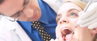
Medical treatment of carious cavity
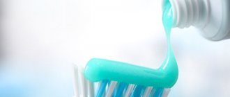
The next stage of caries treatment is drug treatment, the main purpose of which is to clean the carious cavity, have an antibacterial effect on the cavity and dry it. Previously, for these purposes they used a 3% solution of hydrogen peroxide, medical alcohol, and medical ether for drying. For deeper cavities, a special solution was prepared, consisting of 1% hydrogen peroxide, 1% chloramine solution, 0.1% furatsilin solution, and such cavities were dried with warm air. However, with the advent of composite materials, the medicinal treatment of carious cavities has changed significantly - they stopped using alcohol and ether for drying, since it was found that they are toxic and do not dry the cavity well, and also reduce the adhesive properties of composite materials and destroy the so-called polymer matrix. Therefore, in our time, for medicinal treatment of a carious cavity, they began to irrigate with warm antiseptics (sodium hypochlorite, chlorhexidine, hydrogen peroxide, furatsilin), using a syringe for these purposes, and dry the cavity with a stream of air or sterile cotton balls. However, dentists recognized that the effectiveness of cavity treatment with such methods is far from high, and is also technologically complex. At the same time, the drugs used are not very pleasant to the patient in taste and smell, and the safety of hydrogen peroxide and sodium hypochlorite for future fillings is questionable. Therefore, the following antiseptic treatment of a carious cavity is considered the most appropriate:
— to begin with, the cavity is washed generously with clean distilled water or a water-air spray, then dried with a “gun”; - medicinal treatment is carried out with a 2% aqueous solution of chlorhexidine by applying it to the walls and bottom of the cavity, as well as to the tooth tissue and gums, with a cannula brush (for about one minute). The drug is not washed off. — after the antiseptic treatment, the doctor etches the enamel and dentin, applies an adhesive system and fills the cavity with the selected filling material.
Modern filling materials are biologically compatible with human tissue, have high strength, low thermal conductivity, are plastic, and airtight. They are also resistant to chemical compounds that are present in the human mouth and have an anti-caries effect. The filling will be selected exactly according to the transparency, color and shade of the tooth.
If everything is done correctly – and at the Apollonia clinic everything is done professionally – then the dentin surface is sealed for a long time. Therefore, reintroduction of infection into dentin and the occurrence of relapse of caries, as well as complications in the pulp, is impossible. The molding mass is prepared in front of the patient and applied while it is still in a plastic state. The room temperature should not be higher than + 20 degrees Celsius.
You can be sure that caries treatment at the Apollonia private dental clinic will be carried out painlessly using the most modern pain-relieving materials, and the medicinal treatment itself will be carried out professionally and will guarantee that the treated tooth will serve you for a long time. The cost of the procedure for medicinal treatment of the cavity can be found in our price list or by calling the numbers listed on the clinic’s website.
Adhesive systems: what does a practicing dentist need to know?
In modern dentistry, the use of adhesive agents is considered a prerequisite for filling with composite materials. Failure to implement or violation of the technology for using the adhesive system leads to disruption of adhesion to tooth tissues, which can manifest itself in the form of postoperative sensitivity, the appearance of a marginal gap, microbial invasion, staining of the “hard dental tissues - restoration” interface, and the development of recurrent caries. Any dentist involved in aesthetic dental restoration is faced with the problem of choosing an easy-to-use and clinically effective adhesive system.
In this article we will try to provide in an accessible form recommendations on the choice of adhesive systems and the basic rules for working with them based on an analysis of foreign publications in recent years, the authors’ own clinical experience, and the results of our original clinical and laboratory studies and experiments. The choice of such a topic is explained by the lack of objective scientific data on this topic in modern Russian dental literature.
Despite the emergence of easier-to-use self-etching adhesives and self-adhesive composites, 5th generation adhesive systems remain the most popular among Russian dentists. Over the course of two years (July 2011 - May 2013), we conducted a survey of dentists in a number of Russian cities. One of the questions in the questionnaire concerned the choice of generation of adhesive system. The results of the survey are shown in the diagram (Fig. 1).
Rice. 1. What adhesive systems do you use in your practice? (Questionnaire results).
We explain the popularity of 5th generation adhesives for several reasons. On the one hand, adhesive systems that require total etching, with the correct application technique, demonstrate excellent results both after the restoration and in the long term.
On the other hand, the acidity level of self-etching systems is not high enough; when they are used, insufficient etching of the enamel occurs, which increases the risk of the formation of a “white” line after filling and leads to a violation of the marginal seal in the near future (Fig. 2).
Rice. 2. Violation of the marginal fit of restorations of teeth 15 and 16 one year after filling with a nanohybrid self-adhesive composite.
Other disadvantages of self-etching systems include sensitivity to storage conditions, extremely pronounced activation of matrix metalloproteinases (MMP) in dentin and endogenous enzymes responsible for the degradation of the hybrid layer (“rejection” of the restoration by the human body) [1], insufficient stability of these adhesives even during shelf life [2]. Many clinicians note psychological discomfort due to the feeling of a “missed step” (total etching) during adhesive preparation of the cavity.
Having studied and compared the properties of different generations of adhesive systems in sufficient detail, analyzed literature data and performed a number of experiments on our own, we have almost completely abandoned self-etching systems in our work. But 5th generation adhesive systems are also far from ideal. Their use involves multi-stage preparation of the cavity in compliance with all technological nuances at each stage. Errors and errors in work have a significant impact on the result and lead to complications such as postoperative sensitivity and the appearance of a “white” line along the edge of the restoration. The scrupulous attitude of the dentist to each stage of the adhesive preparation of the cavity indirectly affects the rate of degradation of the hybrid layer, and, consequently, the service life of the restoration. It should be remembered that a number of rules for operating these adhesive systems, unfortunately, are not specified in the manufacturer’s instructions and the pictogram diagrams for use included in the packaging.
Without comparing 5th generation adhesive systems from different manufacturers and without delving into theoretical subtleties, we want to discuss in detail the basic rules for working with them.
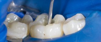
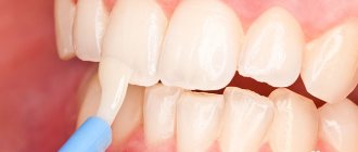
When working with modern light-curing materials, the dentist should take into account that the process of their polymerization is significantly influenced by active oxygen and chlorine compounds. Therefore, hydrogen peroxide and sodium hypochlorite should not be used for medicinal treatment of the cavity. The optimal preparation for working with modern light-curing materials is an aqueous solution of chlorhexidine. It is most convenient, in our opinion, to use a 2% aqueous solution of chlorhexidine bigluconate for these purposes [3]. The drug is applied to all the walls and bottom of the carious cavity with a cannula brush (Fig. 3).
Rice. 3. Drug treatment of the carious cavity before filling.
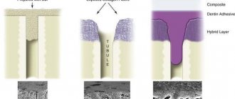
After exposure 30-60 sec. it is carefully inflated and dried with air. It is not recommended to wash off chlorhexidine. After drug treatment, the enamel and dentin are etched. Recommended exposure of the etching composition: on enamel - 15-30 seconds. [4], on dentin - no more than 15 seconds. [5]. We recommend rubbing the acid applied to the tooth tissue into the enamel. It was recently discovered that simply applying acid to the enamel (the so-called static etching of the enamel) may not be enough for high-quality etching [6]. On the one hand, this may be due to the structure of the enamel. Applying acid for 15-30 seconds. guarantees high-quality etching of only internal areas consisting of enamel prisms. In this case, the etching of the outer aprismatic areas of the enamel occurs unevenly. As a result, islands of unetched enamel remain on the surface, with which the adhesive does not interact (Fig. 4).
Rice. 4. Formation of islands of unetched enamel during static etching (diagram).
This leads to the formation of microspaces, the appearance of a “white” line, and marginal staining of the restoration. This problem is quite relevant during aesthetic fillings and is critical when etching enamel that has not undergone preparation, since in this situation such islands make up the majority of the bonding surface. On the other hand, insufficient etching of enamel with acid may be due to uneven distribution of the etching gel and its insufficient adaptation to the enamel. Rubbing acid into the enamel will also solve this problem (Fig. 5).
Rice. 5. Uniform etching of enamel as a result of dynamic etching (diagram).
The dynamic etching technique involves constantly rubbing an etching gel into the enamel surface using a stiff applicator brush. With this etching technique, regardless of the initial structure of the enamel, a uniform micro-roughness of its surface is achieved (Fig. 6) [7].
Rice. 6. Applying etching gel and rubbing it into the enamel (dynamic etching).
After etching, the cavity is washed for 30 seconds. water and slightly dried with air. In this case, the enamel should become matte white, and the dentin should remain slightly moist, “sparkling” (Fig. 7).
Rice. 7. View of etched and dried tooth tissue.
As a result of a correctly performed total etching technique, the enamel surface becomes micro-roughened, the smear layer on the dentin surface dissolves and is completely removed, the surface layers of dentin are demineralized, collagen fibers are exposed, and dentinal tubules open.
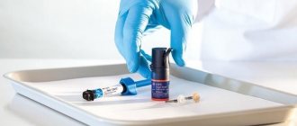
In recent years, a recommendation has appeared in the literature to apply a 2% solution of chlorhexidine to the etched dentin for one minute before applying the adhesive (Fig.  [8].
[8].
Rice. 8. Reapply a 2% aqueous solution of chlorhexidine bigluconate to the etched dentin.
After which the drug is not washed off, but dried with air. It was found that it is acid etching of dentin that activates matrix metalloproteinases (MMPs), responsible for the degradation of the hybrid layer [9], and chlorhexidine is their inhibitor [10]. In vivo experiments have proven that performing this step can stop clinically significant degradation of the hybrid layer for at least 14 months [11].
After drying the chlorhexidine, adhesive is applied to all etched fabrics (Fig. 9).
Rice. 9. Scheme of formation of nanoleakages: a - the depth of demineralization exceeds the depth of penetration of the adhesive into the tooth tissue; b - penetration of the adhesive occurred to the depth of demineralization.
It is important to remember that the number of layers of adhesive is determined by the manufacturer’s instructions, and not by the personal preferences of the dentist or the “general” recommendations of consultants or dental product managers. There are adhesive systems that are applied in one, two or three layers. Changing the recommended number of applications, both downward and upward, is fraught with complications such as the appearance of a “white” line and postoperative sensitivity. The applied adhesive should be lightly rubbed into the etched fabrics, and before starting to dry it, be sure to pause for 15-20 seconds. to prevent nanoleakage. During this time, the adhesive permeates the tissue to the entire depth of demineralization (Fig. 10).
Rice. 10. Pressure of dentinal fluid on the hybrid layer (diagram).
Drying the adhesive is the most important step in adhesive cavity preparation, the purpose of which is to completely remove the solvent. Technologically, the stage is performed quite simply: the dentist dries the adhesive from a distance of 15-20 cm with a weak stream of air, gradually reducing the distance to the tooth.
Do not start drying the adhesive from close distance or do it with a strong stream of air. This can lead to splashing of the adhesive, or to rapid evaporation of the solvent, which will lead to a difference in osmotic pressure in the dentinal tubules and injury to odontoblasts. Proper drying of the adhesive takes approximately 30 seconds, as a result of which the cavity walls should be covered with a thin, shiny film. This film should not move under the influence of a stream of air. The adhesive is then polymerized by light from an activating lamp.
It should be remembered that after polymerization of the adhesive, the flow of fluid in the dentinal tubules does not stop. The dentinal fluid continues to exert constant pressure on the formed hybrid layer (Fig. 11).
Rice. 11. Deformation of the hybrid layer resulting from the pressure of the dentinal fluid (diagram).
Moreover, this pressure (25-30 mm Hg) is enough to deform and even break through the hybrid layer over time (Fig. 12, 13)
Rice. 12. Breakthrough of the hybrid layer resulting from the pressure of the dentinal fluid (diagram).
Rice. 13. Prime & Bond® NT™ and XP Bond™ (DENTSPLY) are 5th generation filled adhesives.
[eleven]. There are several ways to stabilize the hybrid layer: sequential rather than parallel adhesive preparation of several prepared teeth; applying a thin layer of flowable composite to all cavity walls immediately after polymerization of the adhesive, etc. The most reliable and effective method is the use of filled adhesive systems, such as Prime & Bond® NT™ and XP Bond™ (Fig. 14).
Rice. 14. Results of our own in vitro experiment: a, b - numerous defects and heterogeneity of the adhesive film; c, d — the film of the filled adhesive is preserved on the tooth surface, no defects were found.
Filler particles included in the adhesive form a stronger hybrid layer that can resist the pressure of dentinal fluid.
In cases where the dentist, for one reason or another, prefers to use self-etching systems, we can make the following recommendations:
- choose those adhesive systems that do not require special storage conditions and remain stable throughout their shelf life at room temperature;
- It is imperative to carry out medicinal treatment of the cavity with a 2% aqueous solution of chlorhexidine bigluconate in order to inhibit matrix metalloproteinases in dentin;
- carry out selective etching of the enamel in doubtful cases or in an aesthetically significant area to remove the smear layer from the enamel and improve the adhesion of the adhesive to the tooth tissues - preventing the formation of a “white line” in the short term and “seam leakage” in the long term.
Thus, adhesive preparation of a cavity is a complex process that requires maximum concentration of attention from the doctor, compliance with all technological nuances, theoretical training, as well as the use of an adequate adhesive system.
In our opinion, the most current scientific trends and technologies are implemented in the DENTSPLY line of adhesive systems. All of them form a strong, stable hybrid layer of minimal thickness, which provides excellent aesthetic results, reliable marginal fit and minimal risk of developing postoperative sensitivity, which makes it possible to recommend these adhesives for dental practitioners.
The list of references is in the editorial office.