Research by scientists has long ago confirmed the fact that tooth regeneration in the animal world is quite possible; in particular, in alligators this process occurs as needed, that is, if a tooth is lost, a new one grows in its place. The same scientific studies claim that it is also possible for humans to have new teeth; this fact is supported by the example of their creation from stem cells in mice. Based on the above, can we conclude that soon a person will no longer need dental fillings and prosthetics? Of course, yes, another thing is that we are not talking about the near future, this is still a question of the future and not the very near future.
Teeth regeneration technology
The regeneration technology is based on a combination of stem cells, support material and signaling molecules. In about 60 days, the process of growing a completely new and, most importantly, healthy tooth with real roots, dentin and pulp lasts.
At this point in time, high-quality implantation is unthinkable without bone structures that are in good condition, otherwise the installed prostheses simply will not hold securely. Therefore, the technology of growing teeth from stem cells, which does not depend on such limitations, opens up great opportunities in terms of preserving and improving human health. The only problem is that its cost will be very high, this is suggested by logic and a simple statement of the fact of the price level in the field of high-quality implantation and prosthetics. That is why we can talk about this only as a very distant future, so the relevance of preserving what nature has given us is not removed from the agenda.
Structure of a human tooth
The process of formation of baby teeth begins very early, somewhere from 6–7 weeks of development, and the root system is fully developed by the end of the 20th week. The composition of the tooth includes:
- enamel;
- pulp;
- dentine;
- dental cement.
Tooth enamel can withstand maximum loads; it is the hardest element. Dentin is also characterized by increased strength, in which there are a large number of tubules with cells that enable the full growth and development of teeth. The pulp is the center of blood and lymphatic vessels, as well as nerve endings, while dental cement is a substance whose composition is very similar to bone tissue.
Reparative bone regeneration is the restoration after injury. This process is of great importance for practical medicine. Bone tissue is unique because bone is capable of completely restoring even significant defects, unlike all other tissues in which regeneration ends with the formation of a connective tissue scar or organ hypertrophy.
There is no single definition of the term “reparative regeneration”. Some authors argue that this is a type of physiological regeneration that occurs under conditions of extreme influences on the body, but is more intense. Others characterize it as a complex process that is caused by the destruction of bone structures, quantitatively exceeding the permissible limits of physiological regeneration and aimed at restoring integrity and function.
The processes of bone tissue regeneration in the maxillofacial region are a complex interweaving of a number of general effects at the systemic level and local changes in tissue metabolism, including changes at the molecular level. Reparative regeneration is based on cell differentiation, their proliferation, resorption of damaged tissue and new bone formation during remodeling, as well as the formation of an organic extracellular matrix with its subsequent mineralization.
Stages of healing of bone tissue of the maxillofacial area
When healing fractures, the following four stages are distinguished: reparative reaction, formation of fusions of bone fragments, fusion of bone fragments, functional restructuring of callus and fragments with the formation of an organ structure.
Currently, researchers distinguish several stages of reparative regeneration:
- Tissue catabolism, differentiation and proliferation of cellular elements.
- Formation and differentiation of new tissue structures.
- Formation of vascular formations to nourish new tissue.
- Restructuring of the primary regenerate.
In the first stage, the reparative reaction of bone, there are such phases as acute post-traumatic disruption of tissue blood supply, cell necrosis with disorganization of cellular structures, proliferation of non-viable mesenchymal stem cells and differentiation of proliferating cells towards the formation of bone differential (mesenchymal stem cell - preosteoblast - osteoblast - osteocyte ).
Acute post-traumatic disturbances of tissue blood supply in the fracture area are accompanied by rupture of the periosteum, endosteum, osteon canals, as well as bone marrow, vessels and nerves, muscle and connective tissue surrounding the bone. These processes are accompanied by hemorrhages, edema, macrophage infiltration and the development of diffuse ischemic degenerative-necrotic changes in tissues. This phase lasts about 6-18 hours from the moment of injury.
The second phase, lasting 10-24 hours after injury, is accompanied by disorganization of bone tissue structures, signs of necrosis and macrophage cell infiltration.
In the third phase, which occurs within 24-72 hours from the moment of fracture, against the background of restored blood supply, proliferation of mesenchymal stem cells, pericytes of the microvasculature, periosteum, endosteum with the formation of osteogenic tissue is observed. Depending on the location of the proliferating cells, periosteal (periosteal cells), endosteal (bone marrow cells) and intermediate (cells of the bone marrow and vessels of the central canals) bone formations are distinguished.
The fourth phase of the reparative reaction is accompanied by proliferation and differentiation of osteogenic cells into preosteoblasts and osteoblasts. They synthesize osteoid, which after mineralization turns into coarse fibrous bone tissue. Activation of osteoclasts is observed, which resorb necrotic bone tissue.
At the stage of formation of fusions of bone fragments, which occurs 3-5 days after the injury, bone regenerate, or callus, is formed, which spreads in the proximal and distal directions. After 2-6 weeks, this process leads to the fusion and consolidation of bone fragments.
The last stage of fracture healing is characterized by the formation of bone adhesions, which can be of several types. Primary bone fusion is accompanied by direct fusion of fragments at the level of the cortical layer with the formation of new osteons. Fibrous-cartilaginous fusion is characterized by the development of necrosis of bone and soft tissue in the fracture zone, a sluggish reparative reaction that develops at a considerable distance from the fracture site, and the predominant development of fibrous and, less commonly, cartilaginous tissue.
The existing fibrocartilaginous bone fusion as a result of prolonged ossification can be replaced by bone tissue, forming a secondary fusion of bone fragments. In the fourth stage, a functional restructuring of the callus and fused fragments occurs with the formation of the organ structure of the bone.
Cells and substances involved in reparative regeneration
of bone tissue
The process of regeneration of bone tissue in the maxillofacial area occurs with the participation of cells such as osteoblasts, osteoclasts or osteocytes . Below is a brief description of these cellular elements, indicating the features of their structure and functions.
Osteoblasts
are bone-forming cells that originate from mesenchymal stem cells. They are round in shape, 20-30 microns in size, with an eccentrically located core. These cells are located in the osteogenic layer of the periosteum and in the perivascular spaces of osteons.
There are four types of osteoblasts involved in the healing of bone tissue: preosteoblasts, proliferating functionally active osteoblasts, maturing osteoblasts with a hypertrophied endoplasmic reticulum, and differentiated low-active osteoblasts. The main part of osteoblasts synthesize bone tissue until their function stops, followed by their transformation into inactive cells.
These cells line the surface of the newly created bone tissue and connect to osteocytes through a system of tubules. A certain part of osteoblasts are encapsulated in the osteoid matrix and differentiate into osteocytes.
Osteoblasts have a developed Golgi apparatus and mitochondria, due to which they synthesize a huge amount of intercellular substance, in particular proteins and collagen fibers, which form the organic bone matrix - osteoid. Osteoblasts synthesize type I collagen from procollagen, which consists of one α2- and two α1-polypeptide chains forming a helical structure.
The procollagen molecule contains two terminal peptides: amino- and carboxy-terminal propeptides. After the secretion of procollagen by osteoblasts into the extracellular space, these two propeptides, under the influence of enzymes, are cleaved from procollagen, which is transformed into tropocollagen.
Free final propeptides of bone tissue enter the circulating blood, where their concentration can be determined by the enzyme immunoassay method. This analysis very accurately indicates the amount of type I collagen synthesized, with the level of amino-terminal propeptide most accurately characterizing its metabolism.
Osteoblasts also synthesize the non-collagenous fraction of bone matrix proteins, which play a leading role in its mineralization. These are osteocalcin, osteopontin, osteonectin, and bone sialoprotein.
Osteocalcin is the main non-collagenous bone protein that is involved in the binding of calcium and hydroxyapatite. It has a chemotactic effect on osteoclasts and is involved in bone resorption.
Osteonectin binds type I collagen and hydroxyapatite and takes part in the formation of the initial crystal (nucleation) during bone mineralization. Osteopontin also binds bone cells to hydroxyapatite in the extracellular space, but recent studies indicate the involvement of this protein in tumor metastasis by facilitating the adhesion of tumor cells during invasion.
Bone sialoprotein contains sialic acids. It is a calcium-binding glycoprotein that provides mineralization and stabilization of the collagen structure.
Osteocytes
, or “stellate cells,” have a large number of long and thin processes. They are divided into three types, the characteristics of which are briefly listed below:
- Type I osteocyte-producing cells synthesize bone matrix components.
- Type II osteocytes, mature or resorbing cells, are involved in the process of osteolysis.
- Type III osteocytes, degenerating cells, are located at the periphery of the osteon. As a result of their destruction, a significant amount of lysosomal enzymes is released and osteolysis of bone tissue occurs.
Osteocytes have receptors for parathyroid hormone, synthesize osteocalcin, matrix proteins and sclerotin, which is an inhibitor of the Wnt signal in osteoblasts. Their main function is the transmission of mechanical and chemical signaling to osteoblasts, integumentary cells and, through them, to osteoclasts. This circuit plays an important role in triggering bone remodeling processes under physiological and pathological conditions.
Osteoclasts
are multinucleated giant cells that provide resorption of bone matrix components. They are located on the surface of the bone in small depressions (erosive lacunae or Howship's lacunae). In places of contact with areas of bone resorption, the cytoplasm of osteoblasts forms outgrowths in the form of a corrugated border, which increases the area of contact with the bone and facilitates the entry of biologically active cellular products into the resorption zone.
Osteoclasts also secrete protons and proteolytic enzymes (cathepsin K, cysteine protease, matrix metalloproteinases) into the resorption zone and remove decay products into the surrounding space through the basolateral membrane. For such transport to occur, the presence of tartrate-resistant acid phosphatase (TRAP) is necessary. There are 5 types of acid phosphatases, which are produced by bone tissue, spleen, red blood cells, platelets and macrophages. All types of acid phosphatases are inhibited by tartrate, except for the fifth isoform, which is called TRKP-5.
Osteoclastogenesis,
osteoblastogenesis and bone remodeling
The following functional types of osteoclasts are distinguished: young, mature, functionally active and inactive, as well as dying. The source of origin of all osteoclasts is macrophage-monocyte cells of the bone marrow.
The RANK/RANKL/OPG system, which was discovered in 1997, plays an important role in osteoclastogenesis. RANKL, expressed on the surface of osteoblasts, binds to the RANK receptor, which is located on the membranes of osteoclast precursor cells. This triggers differentiation and activation of osteoclasts. The interaction of RANKL and RANK occurs in the presence of macrophage colony-stimulating factor (M-CSF), which, using high-affinity transmembrane receptors (c-fms), activates intracellular tyrosine kinase and stimulates the proliferation and differentiation of osteoclast precursors, mononuclear cells.
M-CSF activity has been shown to be significantly increased by exposure of osteoblasts to parathyroid hormone, vitamin D3, and tumor necrosis factor (TNF), but decreased by exposure to OPG and estrogens.
Osteoprotegerin is synthesized by osteoblasts, vascular endothelial stromal cells and B-lymphocytes. It is a soluble decoy receptor for RANKL that interferes with the interaction of RANKL and RANK, which impairs osteoclastogenesis and inhibits bone resorption. At the same time, interleukins (IL) -1,3,6,11, tumor necrosis factor-α (TNF-α), granulocyte-macrophage colony-stimulating factor (GM-CSF) and prostaglandins E2 (PG2) through the EP4 receptor are able to enhance the production RANKL by stromal cells of the bone environment, in particular osteoblasts, which stimulates osteoclastogenesis.
It was found that the production of osteoclasts is stimulated by parathyroid hormone. It is synthesized by the parathyroid glands. The hormone maintains calcium homeostasis by leaching from bone into the extracellular fluid and enhancing reabsorption in the renal tubules. This hormone activates the synthesis of acid phosphatase, lactate and citrate, and inhibits the synthesis of collagen and alkaline phosphatase.
With a sharp increase in the level of parathyroid hormone in the blood, activation of mature osteocytes and bone resorption (osteocytic osteolysis) are observed. With prolonged hypersecretion of parathyroid hormone, osteoclastogenesis increases and the activity of osteoblasts decreases, which also suppresses collagen synthesis. It has been proven that long-term administration of small doses of parathyroid hormone causes an anabolic effect, and this, on the contrary, promotes the maturation of cartilage tissue. It promotes the synthesis of 1,25-(OH)-2D3 in the kidneys under the influence of cyclic AMP from 25-(OH)-2D3.
Unlike parathyroid hormone, calcitonin, which is secreted in the interfollicular cells of the thyroid gland, reduces the number and activity of osteoclasts in bone tissue, suppresses osteocytic osteolysis and reduces the level of calcium in the blood. Calcitonin stimulates the maturation of chondrocytes in epiphyseal cartilage.
According to foreign studies in recent years, the leading role in the regulation of osteoblastogenesis belongs to bone morphogenetic proteins (BMP, bone morphogenetic protein). This is a group of signaling growth factors (cytokines) that actively stimulate the formation of enchondral bone tissue and regulate various cellular processes (proliferation, differentiation, apoptosis, chemotaxis, angiogenesis or production of extracellular matrix in tissues).
BMPs trigger the process of bone tissue formation through the expression of genes that regulate the processes of differentiation of mesenchymal stem cells with the subsequent formation of osteoblast cells. Dysregulation of the BMP signaling system is very often found in cancer.
Currently, 47 BMP subfamily proteins that interact with specific BMP receptors (BMPs) have been described. In the process of intercellular interactions in the bone tissue of the maxillofacial region, the following are especially important:
- BMP2: stimulate osteoblast differentiation.
- BMP3: promotes the formation of new bone tissue.
- BMP7: activate osteoblast differentiation and SMAD1 production.
- BMP8a: are directly involved in the development of bone and cartilage.
In practical medicine, BMP is used to stimulate bone healing processes. They are injected into a bone implant, from where they are delivered to fracture sites to improve osteogenesis.
Bone cells intensively secrete transforming growth factor β1 (TGFβ1) into the interosseous matrix, which activates the differentiation of mesenchymal stem cells according to the osteoblastic and chondral type, stimulates the processes of reparative bone regeneration, and enhances proliferation and collagen synthesis.
Processes associated with cell morphogenesis are also regulated by Wnt signaling. This process involves proteins discovered in the early 1980s as markers for certain types of cancer. They are also key regulators of the processes of remodeling and regeneration of bone tissue, differentiation of mesenchymal stem cells.
The canonical pathway of Wnt signaling in bone tissue is based on the stabilization of the cytoplasmic protein β-catenin. In the absence of a signal, it is inactive and quickly destroyed. When β-catenin is activated through Wnt, Wnt itself binds to cell surface receptors, which are the Friesland transmembrane protein. As a result, the destruction of β-catenin is inhibited, it accumulates in the cytoplasm and then penetrates into the nucleus. There it interacts with TCF/LEF proteins, which selectively bind to specific activator proteins and DNA sequences.
In this way, certain genes responsible for bone tissue regeneration are activated. Another, or non-canonical (β-catenin-independent) Wnt signaling pathway regulates cell polarity, stimulates calcium metabolism and cytoskeletal reorganization. Wnt signaling stimulates OPG production. It has been proven that at least 22 Wnt ligands are involved in the activation of Wnt signaling in humans.
An antagonist of the Wnt/β-catenin signaling pathway is sclerotin glycoprotein, which is produced by osteocytes of intact bone. It prevents MSC differentiation. When damage occurs to the bone tissue of the maxillofacial region, osteocytes transmit a signal to the integumentary cells that line the surface of the trabecula. Under the influence of prostaglandins and growth factors, which are released, the integumentary cells peel off from the surface of the bone, forming a specific canopy. The canopy cells combine with osteocytes and the capillary to form a bone remodeling compartment.
Recently, regulators of intercellular interactions between osteoblasts and osteoclasts, such as semaphorins and their receptors plexins, have been discovered. Semaphorins form a family of molecules consisting of 8 main classes of secretory and transmembrane proteins that are responsible for transmitting signals along axons. They regulate the growth, development and functioning of cells of the nervous, cardiovascular, immune, respiratory, and musculoskeletal systems.
In particular, Semaphorin 4D (Sema4D), which is a derivative of osteoclasts, acts on the membrane receptor Plexin-B1 on the surface of osteoblasts, suppressing the function of the latter. As a result, activation of bone tissue resorption is observed. Semaphorin 3B promotes the activation of osteoclasts, and semaphorin 3A, on the contrary, stimulates osteogenesis. Based on this, inhibition of semaphorins 4D and 3B is important for the treatment of osteopenia.
An important secretory derivative of osteoclasts is the SLIT3 protein. It stimulates the proliferation of osteoblast cells through activation of the β-catenin pathway. SLIT3 autocrine signaling inhibits bone resorption by suppressing preosteoclast differentiation. Experimental studies have shown that modified animals lacking SLIT3 or its receptor Robo 1 have low rates of bone formation and a high rate of bone resorption.
Practical assessment of bone tissue regeneration processes
The processes of bone tissue remodeling can be clearly assessed based on a comparative analysis of two groups of bone metabolism indicators - markers of bone resorption (hydroxyproline, hydroxyproline, calcium, breakdown products of type I collagen, pyridinoline and deoxypyridinoline, bone sialoprotein (BSP), tartrate-resistant acid phosphatase) and synthesis markers bones (osteocalcin, bone alkaline phosphatase, amino and carboxy-terminal fragments of type I procollagen, ACP, CCP).
Markers of bone tissue synthesis indirectly characterize the activity of osteoblasts. These cells take an active part in the formation of bone tissue, produce type I collagen plus other osteoid components, and participate in the mineralization of osteoid with hydroxyapatite.
The first biochemical marker of bone remodeling is the enzyme alkaline phosphatase, which was introduced into clinical practice in 1929 and is used today. There are 4 isoforms of this enzyme: bone, liver, intestinal and placental.
A laboratory indicator of osteoblast activity is bone alkaline phosphatase. This is a glycoprotein that takes part in the mineralization of the bone matrix. A simultaneous increase in ALP and parathyroid hormone may indicate the development of osteodystrophy with a high level of bone remodeling, and a decrease in these indicators may indicate an adynamic state of the bone tissue of the maxillofacial region.
The most informative marker of bone formation is osteocalcin. This non-collagenous bone matrix protein containing hydroxyapatite can be considered specific for bone tissue and dentin. It is synthesized primarily by osteoblasts and forms the extracellular matrix of bone.
A fraction of newly synthesized osteoclasts is released into the bloodstream, so this marker indicates the rate of bone tissue remodeling. An increased level of parathyroid hormone in the blood suppresses the activity of osteoblasts that produce osteoclasts and reduces the concentration of osteoclasts in the blood and bone tissue.
During the synthesis of type I collagen by osteoblasts, amino- and carboxy-terminal fragments are separated from type I procollagen as a result of the action of specific enzymes. The ratio between the amount of mature collagen deposited in the bone matrix and the number of terminal molecules that enter the bloodstream must be equal to one. Thanks to this, based on the indicators of ACF and CCP in the blood serum, one can judge the synthetic activity of osteoblasts in the synthesis of type I collagen.
The next group of bone remodeling indicators are markers of bone resorption. It should be noted that they change their level during the day, so they must be determined at the same time of day, preferably in the morning. Markers of bone tissue resorption include enzymes involved in the destruction of the bone matrix under the influence of osteoblasts and products of the destruction of type I collagen.
This is, for example, tartrate-resistant acid phosphatase (TRAP) - a metal-containing enzyme, one of 6 isoenzymes of acid phosphatase. It is secreted by osteoblasts into the extracellular environment during resorption. There are forms of TRAP-5a and 5b, but TRAP-5b is synthesized by osteoblasts, and TRAP-5a is of macrophage origin. TRAP-5b activity in blood plasma is an indicator of bone resorption processes.
Hydroxyproline is the main amino acid in collagen. It is not considered an autospecific marker of bone tissue, because only 50% of this amino acid is found in bones, and the remaining 50% is a component of other proteins, in particular elastin, acetylcholinesterase and complement factor C1q.
Most of it, after the destruction of collagen, is oxidized in the liver and only 15% is excreted in the urine. The content of hydroxyproline depends on the nature of the diet, the age of the person, the presence of a tumor with decay and demonstrates a circadian rhythm with a peak between 0.00 and 8.00 am.
14% of the amino acid composition of collagen is hydroxyproline. It is synthesized by osteoblasts and is also a marker of bone resorption. Nowadays, more specific markers of bone resorption are used, such as type I collagen breakdown products. These include pyridinoline (PID) and deoxypyridinoline (DPID). They are found in tissues containing type I, II, III collagen. PID is an important chemical compound, but DPID is found only in collagen, so it is evaluated as a selective bone marker.
Also widely used in medical practice is the determination of type I collagen peptides, NTX and CTX. The uniqueness of these indicators lies in the rapid increase in their level in diseases accompanied by high bone tissue resorption (for example, osteoporosis, bone metastases), as well as their rapid decrease (within several weeks) against the background of antiresorptive therapy.
The next marker of bone resorption is bone sialoprotein (BSP). During the process of osteolysis (physiological or pathological), the bone matrix is destroyed by osteoblast enzymes with the release of sialoprotein into the blood with a corresponding increase in laboratory BSP parameters. Bone sialoprotein is present only in mature osteoblasts and is not found in their precursors, so BSP may serve as a marker of late differentiation of bone cells.
Conclusion
Reparative regeneration of bone tissue in the maxillofacial region is associated with the activity of osteogenic cells. The local regulation of reparative regeneration is based on several signaling pathways, such as the RANK / RANKL / OPG molecular system, bone morphogenetic proteins, Wnt signaling and others.
Osteoblastogenesis and osteoclastogenesis characterize biochemical markers of bone remodeling, the determination of which allows one to determine the rate of metabolic processes in bone tissue, identify patients at increased risk and conduct an early assessment of the effectiveness of prescribed treatment, predict the risk of complications, diagnose the appearance of bone metastases in the early stages, etc.
Achievements of scientists
Theoretically, the current situation can be called borderline in the sense that dentistry has come very close to the moment when the practice of growing new young elements instead of lost ones is ready to take the place of theory. But there are several serious problems that prevent you from crossing this line and re-growing your teeth, in particular:
- the process of cell division is far from perfect, therefore, full transformation into dental tissue does not occur;
- there is a fairly high probability of rejection of a new tooth by the gums; an example is a similar situation that occurs periodically with implants;
- Implanting a tooth germ into the gum cannot serve as a 100% guarantee that the tooth will grow in the form and quality expected.
It is impossible to give an exact answer to the question of when such regeneration techniques will begin to produce high-quality results. There is an opinion in the professional community that something like this can be discussed no earlier than the 30s of this century.
Regeneration of teeth according to Petrov
Similar studies are being conducted in many countries, including Russia. The peculiarity of stem cells is that they are triggered at the right moment and then turned off, and forever. If we can find a way to turn these cells back on, then the problem of tooth regeneration will be successfully solved.
A detailed description of the technology of tooth regeneration is contained in the works of Russian academician and Ph.D. Arkady Naumovich Petrov. It is based on the following aspects:
- Mental teleportation of a person in time. We are talking about imagining yourself at the age when the change of baby teeth to permanent ones occurred, and you need to rely only on positive and pleasant memories.
- Working with the information and energy field. In your mind, you should “grow” a tooth in the desired place or transfer it to it, give it a mental order to grow in the form you want.
- Constant attention to the place where the tooth should grow. It is advisable to constantly stimulate the tooth physically and psychologically, increase blood flow, massage the gums, and perform constant jaw training.
LiveInternetLiveInternet
Quote from lotus12
Read in full In your quotation book or community!
Arkady Petrov “Tooth regeneration technology”
Arkady Petrov “Tooth regeneration technology”
Tree of Life No. 1 January 2008
“The goal of the work is to completely restore all teeth to normal using the regeneration method.
When there are no teeth, a person loses the taste for life.
We restore teeth with all receptors, with all sensitive endings.
We build a hologram not only of today’s tooth structure, but also of the future structure. From the present through the past: what teeth were like, what teeth will be.
The gum edge is the middle point of the figure eight (see Fig. 1).
Activation of the figure eight accelerates the growth of teeth and displaces all negativity from the place where the tooth is formed. You can transfer embryo information to figure eight. There is no negativity in the embryo. Development is proceeding only according to a positive scenario. The embryo itself removes all negative information.
We start the regeneration process through stem cells.
Using an impulse, we build a hologram of the root of a healthy tooth. To do this, we enter the chromosome with our consciousness, highlighting the energy-information frame of a healthy tooth, that is, its hologram, the dental anlage.
We take a stem cell from the vertebral bone (spinal cord) and teleport it to the root of the tooth (see Fig. 2).
We give an impulse with consciousness from the Soul to build the cell stem. To do this, from the primary stem cell (1), we first isolate two cells (there are 3 in total), then two more cells (5) and three more cells (8). An embryo has formed.
Next, we enter the code: “differentiation” (i.e., the transformation, in the process of individual development of the organism, of initially identical, unspecialized cells of the embryo into specialized cells of tissues and organs). Then we give an impulse from the primary cell to isolate the 9th cell.
After the formation of the 9th cell, the division of stem cells begins to form dental tissues (Fig. 2).
We install source cells to speed up the formation of dental tissues. We activate them with an impulse of consciousness.
We establish, through the thyroid gland, a connection between the teeth being restored and those organs with which they initially had these connections. (Consciousness knows these connections).
Silver-white threads appear from the thyroid gland to the teeth being restored.
We transfer this whole situation to all the other teeth that need restoration...”
“...Each of you, in your imagination or control zone, will need to build a hologram of the missing tooth. You find which tooth you are missing. We start regeneration from the upper jaw. If there are all teeth in the upper jaw, then we begin regeneration from the lower jaw.
If someone cannot determine which tooth is missing. Because it happens that a person loses teeth very early. Then all the teeth shift, they change their position, and it turns out that it is difficult to determine the 6th or 7th or 5th tooth. And they have different structures.
Now there is a woman in the hall who is growing a tooth on the right side of her lower jaw - 5. But I cannot conduct this tooth as fractology, because... I hadn’t seen it before, but I saw it already erupting. And I need your status before teething. Sometimes teeth grow at 30, 40 and 50, this happened in my practice.
...Since we have embarked on the path of spiritual development, we will continue it, having received technologies for dental regeneration, we will continue this path of comprehending the world, comprehending ourselves as a part of the world.
Because the result cannot be achieved until we recognize ourselves as an element of the world, part of the world. And then, once, click and we get the result...
...Your task is to feel what is happening in the place where you have already outlined where TOOTH REGENERATION will take place.
I repeat that we started regeneration from the upper jaw; we take the stem cell from the bone marrow of one of the vertebral bodies.
We turn to our Divine consciousness and ask it: take my STEM CELL from the bone marrow of one of the vertebrae and TELEPORT to the border between the jaw and the missing tooth.
Consciousness is capable of the teleportation effect; all regeneration methods are based on this.
Next, with an impulse, we build a hologram of the TOOTH ROOT. WE BUILD A CAGE INTO THE TOP OF THE TOOTH. Our cells obey our consciousness, and chromosomes also obey our consciousness. We give an IMPULSE from the soul. The Energy of the Spirit and the Knowledge of the Soul enter the cell, enter the chromosomes.
So, we are now impulsively building a hologram of the root of a healthy tooth. To do this, we enter the chromosome with CONSCIOUSNESS and illuminate with energy the information frame of a HEALTHY TOOTH. Mentally we knock it out. The impulse touches 2 cells, touch this first cell - 2 more cells. Thus, we get 5 cells, touch the first cell - 8 cells.
Thus, an GERM was formed. This is the root bookmark.
Next we introduce verbal coding. Each cell knows what to build. A tooth is a complex formation; it is not just bone tissue. The tooth consists of enamel, there is DENTIN inside, and there is a root covered with cement. Inside the tooth there is a NERVOUS-VASCULAR BAND, which also has a complex structure. Consists of nerves, vessels, veins. Therefore, when we give a command to a cell (STEM CELL), TO EXTRACT 9 CELLS AND WITH THIS WE GO FROM THE INTERNAL TO THE EXTERNAL.
Because the tooth has both internal and external manifestations. Upper teeth and lower teeth have different structures.
The count starts from the midline, 2 central incisors, 2 lateral, 2 canines numbered 3,4 and 5 are premolars. The 4th premolar usually has 2 roots, but can also have one.
6,7,8 have 3 roots.
But 8 teeth, both upper and lower, are very variable. They can have 1,2,3 roots.
The lower teeth are distributed in the same way as the upper teeth. Teeth 6,7,8 are powerful chewing molars. These chewing teeth have 2 roots, except for 8, which, as I said, is variable.
Therefore, when you build a hologram of your regenerated tooth, strictly adhere to the specified number of tooth roots.
If it is the 4th tooth, then there are 2 roots, if it is 6, then there are 3. I talked about the BOOKMARK.
Does anyone have any feelings?
What is REGENERATION? This is a MINI RESURRECTION. After all, by regenerating the entire organ, the body simply rejuvenates.
Remember Petrov’s example, how one woman’s ovaries regenerated according to the laws of universal connections and causes of effects, her appendicitis and tonsils were regenerated, and she generally became rejuvenated and felt completely different. You know from the works of G.P. and Petrov that we have source cells and sink cells.
Look where your tooth was removed, here you have formed SPHEROSED HARDNESS, SPHEROSED TISSUE.
And here you have laid a delicate germ of 9 microscopic stem cells. It is difficult for them to break through and therefore source cells are placed around here. AND CONSCIOUSNESS itself KNOWS how many cells and how many of them to put to whom.
Now that the tooth was in its place, it was connected to a CERTAIN ORGAN.
Refer to this picture, all connections are drawn here. Please note that all teeth are connected to the gastrointestinal tract, because teeth are the beginning of the gastrointestinal tract.
We will not restore these ties now. For those who don’t understand the sinuses, they are the maxillary and frontal sinuses. 3,4,5 are related to sines. If some organ is removed, then wait for the disease somewhere else, i.e. Some organ is not receiving enough of something, communication in the body is disrupted.
When I was studying at the institute, they said that the appendix is not needed in the body, and at one time there was such a technology to remove appendicitis from infants so that there would be no problems in the future.
And what is appendicitis? This is a very important part in our body, firstly it is the prevention of dysbacteriosis, and secondly it stimulates the peristalsis of the large intestine. By removing appendicitis, you doom a person to constipation. In addition, appendicitis is a DEPOT OF THE IMMUNE SYSTEM. By removing appendicitis, we disrupt this connection; by removing the tonsils, we disrupt the Pirogov ring, we make the infection free for the upper respiratory tract. I experienced constant bronchitis myself.
At home, be sure to establish a connection between the regenerated tooth and the missing organ, and send, with the help of the Spirit, from the Soul to send an impulse to the small embryo, as if we were carrying children. And send LIGHT and LOVE to the ORGAN with which it is connected.
As a rule, teeth are formed by the age of 15. Arkady Naumovich recommends returning to that 15-year-old age, to that youth, REMEMBERING EXCITING MOMENTS. This return to our beautiful moments, to this youth, also helps to accelerate the regeneration process.
Next point. We instruct our consciousness to transfer regeneration from this tooth to all other teeth that are missing.”
Description of the practice of growing new teeth
1. The first thing to do is to remember as much as possible of all the sensations that accompanied the growth of new teeth in childhood. This is not difficult to do - because Nature tried and gave us a memory of this through pain (all painful sensations are the strongest and are remembered for a long time). Remember this constant itching in the gums, how old teeth sway, which are “pushed” from below by growing young teeth, how you stand in front of a mirror with a thread tied to a tooth in an attempt to overcome your fear by pulling it out, etc. Remember this because this is the first “button” that will turn on and start the process of growing new teeth.
2. Now I will return you again to the description that I gave above - namely, to the place where I said that the first teeth begin to grow from the first two lower incisors and from them they begin to change to new ones. This persistently tells us that here is another one of the “buttons” that needs to be pressed to turn on the process of tooth regeneration.
3. And the third “button” is, of course, in our consciousness. We must also turn it on permanently, because... We won’t be able to do everything that I write below all the time (all 24 hours).
1. So, I will describe what exactly needs to be done. Find 10-30 minutes to practice every day. For the first third of this time, think about the space under each tooth, i.e. simultaneously under each tooth inside the gums. In this space, imagine small white teeth as seeds that are just germinating. Think of these teeth exactly like seeds, i.e. about what has been planted and is already beginning to sprout. Remember (from the first point) the itching that accompanied the growth of new teeth in childhood, how the teeth “itched”, how painful it was, etc.
2. Maintain this concentration for the first third of the practice.
3. Next, without stopping the concentration described above (teeth-seeds, itching in the gums), concentrate on the point that is located under the two lower front incisors (this is an area approximately 0.5-0.8 cm). As you concentrate - you may feel pressure in this area, this is good.
4. Maintain this concentration for the second third of practice.
5. Without stopping both concentrations that I described above (on the gums and on the point under the front incisors), also concentrate on the area between the eyebrows and a little deeper (Third Eye), mentally saying something like the following phrase “My teeth are completely renewed.” At the same time, keep the thought form of renewing your teeth, in which bad teeth fall out, and new young teeth grow in their place.
4. This practice must be done for at least a month. Of course, some may need less time, others more. Therefore, the main criterion here is your ability to feel yourself.
Notes
• The only reason for failure in this practice may be your fear of losing teeth and clinging to old ones. For example, thoughts such as “What if all the teeth fall out and no new ones grow”, “Better a bird in the hand than a pie in the sky”, etc.”
Recommendations:
“To form a 3-dimensional volumetric field of a new tooth with roots (having previously cleaned the place from EI dirt), and then gradually build up, root and strengthen this 3-dimensional volumetric field of a new tooth with different strong energies; at the same time, you can also direct the chewed energy there food (in particular feta cheese, cottage cheese, cheese, garlic, onions, etc.).
And then gradually adjust and align this new energy tooth to match the appearance of your healthy teeth already in your mouth. Not forgetting the shiny protective enamel coating!
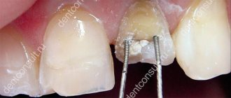
As for the treatment of caries, I agree with this approach:
“In the beginning, in theory, the caries itself should turn white, the dark plaque should be removed, and the cavity filled with dark energies, the visually black area of the tooth, should turn white. First stage.
And when the dark energies are raked out from such cavities-areas, the tooth is cleaned of dark, dirty energies, this caries rot-rust goes away, you can begin to revive and restore it.
This is already the second stage.
You can fill a former diseased tooth with health energies, make balls, inject light etheric energies containing healing information, such ball programs to ensure that your teeth are healthy, white, strong, resistant to heat and cold, overheating and hypothermia. Having healthy, normal teeth, you can drink hot coffee and eat cold ice cream in chunks.
By the way, Frenklin suggested a good technique!
Find in the past, in adolescence, when a person was completely healthy, all his teeth were young and beautiful, a site, place, region, zone where the memories and integral sensations of healthy teeth are localized! It is very easy to find this area in the past.
This section of the VIP is activated, filled, pumped up with good energies and integrated with the present. And then this integral design is constantly supported, like a health program, such as those done on a deuce. And then just fill this structure with different healthy energies!
But at the same time, be sure to ensure that the matrix of a healthy tooth in the program (made by you) is combined with the currently diseased tooth and is constantly filled with good energies.
Or you can introduce an implant, such a matrix, a copy from your own healthy tooth or someone else’s healthy tooth (you can also work with a photograph).
In this case, it is advisable to take into account as much as possible, copy the internal energy structure of a healthy tooth and fill it with different healthy energies, which can also be done from the object.”
RESULT:
As we can see, all techniques have several common points, which in order of priority are arranged as follows:
1. Mental teleportation in time. It is necessary in your imagination or in meditation to transport yourself to the age of 13-15 years, when all the milk teeth are already gone, and the molars are still healthy. Imagine yourself at this time as best as possible, possibly using photographs. Remember as many exciting moments from this period of life as possible...
2. Work with the energy-information field. It is necessary to implant or transfer the “embryo” of a healthy tooth to the place we need. According to Mikhail Stolbov - giving the order to the tooth to grow. Subsequently, there is a constant mental visualization of beautiful, shiny, white teeth.
3. Daily, or better yet hourly, maximum attention to the right place, constant stimulation (both physical and psychological), increased blood flow, massage of the gums with a toothbrush, jaw training. “Hourly (actually every hour for 5 minutes) work with gum cells. Jaw training: clench your teeth for a short time, then release, move them from side to side. Massage the gums with your tongue and fingers.” If there are very few teeth in the mouth, then work should begin, as mentioned above, from the front teeth and then to the edges. If you are working on one or two teeth, then it doesn’t matter... To better visualize a particular tooth, on the right is a modified Foll table showing the number of roots of a tooth.
Bolotov method
Strictly speaking, Bolotov’s method does not concern the regeneration of teeth, but their treatment, but as an alternative it makes sense to consider it in more detail. The regeneration process is based on a combination of vodka tinctures, propolis and calamus. For 0.5 vodka, take one calamus root and crush it, propolis tincture is made at the rate of 20 grams per half liter.
The technique is based on the simultaneous use of both tinctures and involves the following course of action: one tablespoon of each tincture is mixed together and the resulting mixture must be rinsed out of the mouth for three minutes. It is not recommended to swallow this composition; rinsing should be done either at the onset of pain or before bedtime. The duration of treatment is approximately a month, although pain should subside 2-3 after the start of the procedure. Calamus acts as a pain reliever, and propolis is necessary for filling holes in teeth.
Note: This procedure cannot be called regeneration; new teeth cannot be grown with its help, but pain completely disappears and even roots that do not sit well in the gums become stronger.
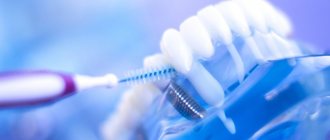
Periodontal regeneration: clinical approaches and opportunities
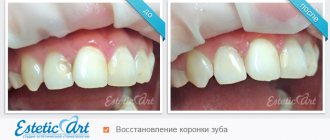
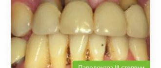
Maximum preservation of one's own teeth is, in fact, a manifesto for all periodontists, and a goal that all dentists have been pursuing for many years. Technological improvements have made this goal more achievable than before, but even despite the possibilities of periodontal regeneration and reconstruction of the periodontal ligament, cementum, gums and alveolar bone, the problem of periodontal lesions has not been completely solved.
In June 2014, 52 international representatives, including clinicians, researchers and scientists, came together at an American Academy of Periodontology (AAP) workshop to collate all available evidence on periodontal regeneration. During the seminar, participants were able not only to discuss the mechanisms of biological reactions in the structure of the periodontium, and how these aspects can be used in practice, but also to identify prospects for further research. The AAP initiated the work of this congress with good reason: at that time in the United States, the prevalence of periodontal pathologies was critically high. Data obtained by the Centers for Disease Control and Prevention (CDC), which were collected for the period 2009-2012, indicate that almost 50% of the entire US population over the age of 30 years have some form of periodontal tissue damage, and 70% Americans aged 65 years and older suffer from destructive forms of periodontitis. Mild to moderate periodontitis has been reported among 38% of the US adult population. Preserving the integrity of the natural dentition while ensuring remission of periodontal disease is an extremely complex clinical task that goes beyond the usual correction of the parameters of aesthetics and function of the structural elements of the oral cavity.
A growing body of evidence suggests a link between periodontal pathology and systemic diseases of the body: for example, an association has been found between moderate-to-severe periodontitis and an increased risk of developing diabetes. Epidemiological evidence also suggests that periodontitis increases the risk of developing cardiovascular disease. Although the available data is still limited, scientists have already noted the possibility of a pathophysiological link between periodontitis and chronic obstructive pulmonary disease, pneumonia, chronic kidney disease, rheumatoid arthritis, cognitive impairment, obesity, metabolic syndrome and cancer. Regenerative methods for correcting periodontal disorders are not aimed at slowing down the progression of the disease, but at restoring existing defects in soft and hard tissues, protecting still intact dentition and preparing affected areas for further rehabilitation using implants. However, the inflammation associated with periodontitis can still be reduced during physiological tissue repair through natural biological mechanisms.
Types of regenerative periodontal intervention techniques
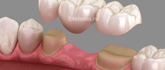
Many new emerging methods of regenerative periodontal interventions, including those that involve the use of delivery systems for certain cells, matrix scaffolds, bone anabolic agents and mesenchymal stem cells, are at an early preclinical stage of testing and require additional scientific research for their further implementation in the clinical setting. dental practice. Hard tissue augmentation procedures allow the restoration of bone tissue, periodontal ligament and root cement, which together provide stabilization of the tooth. Soft tissue augmentation procedures can stimulate the growth of new gums in the area of recession, improving the aesthetic appearance of the mucous membrane. The three most common types of periodontal iatrogenic interventions in clinical practice are bone augmentation, the use of biological agents, and guided tissue regeneration (GTR).
Bone grafting
Bone grafting is one of the longest used techniques for regenerative iatrogenic interventions, which can use the patient’s own bone tissue (autograft), bone tissue from another patient (allograft), bone tissue from different animal species (xenotransplantate), or artificially synthesized bone tissue. tissue (alloplastic graft). Over time, the augment used is replaced by the patient's bone.
Guided tissue regeneration
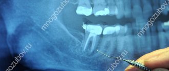
Guided tissue regeneration procedure uses a barrier membrane to protect and stabilize the blood clot during the healing process of the bone graft area. Any interference with the structure of the blood clot during wound healing can cause disruption and the formation of scar tissue instead of new bone and ligament. The membranes used can be resorbable or non-resorbable. The former are ideal for interventions directly on the periodontium, while others work best during the reconstruction of the alveolar ridge and its preparation for future installation of implants. Resorbable membranes eliminate the need for repeated surgical interventions to remove them, which is very convenient for the patient. But at the same time, they must also be characterized by appropriate degradation parameters that will allow maintaining a sufficient amount of space for bone tissue to perform it. In cases where the main barrier function of the membrane may be compromised by anatomical features in the area of intervention, the physician must resort to an alternative method of treatment. So, instead of a membrane, you can use a bone graft, which is characterized by fairly rapid resorption. Membranes that resorb too slowly can impede blood flow to the regenerated structures and are more likely to become infected. As in any clinical situation, the clinician should use NTP only in clinical settings that are optimally suited for this type of intervention. In addition, to ensure the success of scientific and technological progress, the doctor must carry out precision decontamination of the tooth root in the area of the main surgical intervention. It should be noted that proper tooth root disinfection also requires special technical skills and relevant experience.
Biological products
Biological agents have demonstrated significant potential in the process of periodontal wound repair. Such drugs include growth factors, enamel matrix derivatives, and bone anabolic agents that stimulate cells to proliferate, migrate, biosynthesize, or differentiate the matrix. As a rule, most techniques for using biological products are in the early stages of testing, but some of them are already quite widely used in clinical practice. In the future, it is planned to carry out individualized periodontal interventions taking into account the genetic background of the patient, using biomarkers for prognosis, three-dimensional technologies for planning, and cell scaffolds as the main materials for transplantation.
Regenerative procedures for furcation defects and intraosseous defects
As a rule, patients requiring regenerative periodontal interventions are characterized by the presence of tissue defects in the furcation area and intraosseous defects. Classification of defects in the furcation area is carried out by assessing the level of loss of periodontal attachment in areas of “bifurcation” of the tooth roots. Clinicians are often faced with a choice: whether to remove such teeth or to try to restore tissue in the area of the existing defect.
The results obtained by Huynh-Ba and colleagues indicate that the NTR of the furcation defect region provides a high survival rate of treated teeth at the level of 83.3-100% during 5-12 years of monitoring. Intraosseous defects develop with internal periodontal damage to the alveolar ridge. Lang et al recommend assessing the morphology of the intraosseous defect by the number of affected bone walls.
The Goldman and Cohen (1958) classification suggests four classes of defects:
- with damage to three or four bone walls;
- with damage to two bone walls;
- with damage to one bone wall;
- combined defects (for example, with damage to walls 3 and 2, etc.).
The goal of periodontal therapy for any type of defect is the direct regeneration of periodontal tissue, including the apposition of new cement with the penetration of periodontal ligament fibers into it. Reynolds et al found that regenerative treatment of intraosseous periodontal defects increases clinical attachment levels, promotes periodontal pocket reduction, and improves overall periodontal health. The algorithm for the restoration of intraosseous defects, as a rule, involves bone augmentation using different types of grafts, but for the same purpose, methods of directed tissue regeneration and the principles of using biologically active agents can be used.
From theory to practice
Proceedings of the AAP Regeneration Workshop were published in the Journal of Periodontology and included consensus reports and systematic reviews on soft tissue restoration of root surfaces, furcation defects, and periodontal reconstruction. Accompanying practical recommendations were published in the journals Clinical Advances in Periodontics and the Journal of Periodontology, which formed the transition from theory to practice.
Treatments for gingival recession defects represent a key challenge to the potential for periodontal regeneration. Gingival recession is a fairly common condition, and the workshop participants agreed that the results of its correction should be assessed in terms of reducing the depth of the defect, improving the level of clinical attachment and the growth of keratinized soft tissues. In order to restore the coverage of the exposed tooth surface, you can use the techniques of a subepithelial connective tissue flap, a coronally displaced flap, a free gingival graft and its analogues, such as acellular dermal matrix, ketogenic collagen matrix and bioactive agents (recombinant growth factor obtained from platelets or derivatives of the enamel matrix) .
Richardson and colleagues Fr.
For situations that do not require root soft tissue restoration, John et al found that gingival augmentation procedures must be accompanied by patient education on oral hygiene and reduction of potential risk factors: “Understanding that minimal gingival attachment and suboptimal level of oral hygiene are determining clinical conditions - this is the first step towards objective diagnosis. Analysis of patient-specific factors helps to adequately approach the choice of surgical procedure aimed at increasing the parameters of keratinized tissues/attached gums.”
Reynolds et al summarized that the choice of the most effective method of treating intraosseous defects depends on a complete diagnosis and consideration of patient-associated risk factors. However, the authors also summarized that therapeutic approaches involving the use of combined treatment techniques are also quite successful. For example, acellular defect replacement grafts used in a guided tissue regeneration protocol help achieve proper stability of the augmentation site, protect the blood clot, and form new tissue. However, this approach, in fact, does not involve the use of biologically active agents. The choice of treatment method for intraosseous defects should involve the analysis of parameters such as the number of residual intact walls, loss of tissue height, proximity of the defect to other structures, and the possibility of achieving proper vascularization in the future.
Aichelmann-Reidy, when treating pathology in the furcation area, recommend taking into account systemic patient-associated factors, localized anatomical possibilities and characteristics of the furcation area itself. Class II furcation defects can generally be treated predictably using regenerative techniques, while Class I furcation defects can be repaired through a combination of bone augmentation and isolating membranes. Lin et al point out that modern technology is progressively facilitating the development of new periodontal regenerative treatments, and published clinical reports indicate even greater variability in current periodontal intervention techniques. However, one more problem remains - the lack of evidence base, but this aspect depends only on the time and results of the implementation of various approaches.
A systematic consensus report by Cochran et al on priorities for future research in new periodontal technologies can be summarized as follows:
- It is necessary to develop non-invasive methods for assessing the clinical results of regenerative periodontal interventions. This approach should also allow the effectiveness of new treatments to be assessed.
- It is necessary to validate those treatment methods that are used off-label, because some periodontal intervention techniques were originally developed for completely different purposes.
- To personalize treatment, it is necessary to provide an analysis of the patient's genetic and epigenetic profiles.
- To select a specific treatment method, it is necessary to evaluate the influence of pathogenesis, etiology and potential for regeneration in different forms of periodontal lesions.
- For more predictive treatment and restoration of periodontal structure and function, the physician must systematize all potential risk factors associated with iatrogenic intervention.
- The molecular and cellular mechanisms of each treatment modality need to be determined using in vitro and in vivo models.
- It is necessary to determine the paths of development of periodontal lesions to optimize the use of various methods of regenerative therapy.
- Researchers should focus on developing minimally invasive technologies to minimize postoperative pain and discomfort without compromising rehabilitation outcomes.
- There is a need to develop criteria to determine the clinical success of interventions.
- It is necessary to assess the impact of the chosen method of regenerative periodontal therapy on the patient’s quality of life.
Conclusion
Regenerative therapy using soft and hard tissue grafts, biological agents, or guided tissue regeneration has a promising future in the periodontal field. A number of additional regenerative methods, including new cell delivery systems, are highly promising but require further scientific research to determine their effectiveness. The results of consensus workshops on periodontal regeneration indicate a strong evidence base confirming the effectiveness of reconstructive therapy for periodontal defects, allowing to restore not only the structure of soft and hard tissues, but also their full function. The use of regenerative periodontal interventions also helps preserve the patient's own teeth, which is one of the most important goals in periodontics.
Authors: William V. Giannobile, DDS, DMSc Pamela K. McClain, DDS