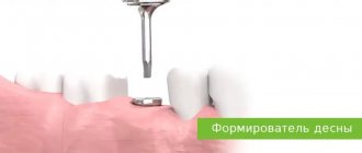
Why cut the gum?
Pain in the gums continues for a long time - this is a natural accompaniment of tooth growth. Contact your doctor if you notice any of the following symptoms:
- Severe pain when chewing food or occurring spontaneously, without reason.
- If you have bad breath, this is a sign of pus under the hood.
- If the gums not only hurt, but also became red and swollen.
- The temperature has risen, severe weakness and headaches are felt.
Dentists will offer to cut the gum, be strong - this is not such a terrible procedure as it seems. After the incision, the rot will come out, the gums will heal, the pain will subside and the tooth will no longer cause concern. In different patients, the gums healed at different times: it usually takes from two days to two weeks.
The drainage system is installed in stages:
- The painful area of the jaw is numbed.
- The gum is cut and a latex drain is inserted into it, through the hole of which the pus is sucked out.
- After suctioning out the pus, an antiseptic drug is injected into the gum incision.
- If a tooth is removed, the gum is not cut, but a drain is inserted into the socket.
How many days should I wear the drainage? Improvement may occur within 24 hours. On average, drainage costs from 3 to 5 days.
What to do if the drainage falls out of the gums
If the drainage randomly falls out of the gums, you should contact a specialist to determine the cause. This is often caused by improper installation of the system, weak fixation, intensive brushing of teeth and frequent rinsing.
In this case, you should pay attention to whether the swelling has subsided or not, whether there is suppuration.
The decision to return the system is made by the dentist; under no circumstances should you install drainage yourself.
Incorrect actions by the patient can lead to the development of complications and relapse of the infectious-inflammatory process. In this case, a rapid overgrowth of soft tissue occurs, blocking the exit to the outside for pus and excess fluid. As a result, the gums hurt, a lump forms, where purulent secretions accumulate abundantly.
If a child pulls out a drain, you should immediately contact a specialist for an examination of the oral cavity and further observation and treatment.
Read also: Basics of remote work as a reader
Rules to follow when having a device in your mouth
Anyone who has had drainage installed after a gum incision or tooth extraction needs to know how to behave during this period. Doctors give the following recommendations:
- you need to follow a certain diet: it is forbidden to eat hot and hard foods and chew on the side of the jaw where the intervention was performed. You need to give preference to soft and liquid dishes, drink more clean water,
- It is undesirable to sleep on the cheek on which the device is installed,
- Brushing your teeth should be done extremely carefully, avoiding the area where the tube is located: for hygiene, you can use a brush with soft bristles and a non-abrasive toothpaste. It is better to avoid using an irrigator for a while,
- Intensive rinsing of the mouth is prohibited: to carry out antiseptic and disinfectant procedures, simply take liquid into your mouth, hold for 2-3 minutes, spit it out,
- It is important to protect yourself from physical activity and sports.
After the procedure
After the procedure, you will be placed in a recovery room. You will need to stay in bed until the sedative medicine wears off.
In the recovery room, the nurse will continue to monitor your condition, heart rate, breathing and blood pressure. They will watch your catheter for possible bleeding.
About your catheter
A black mark will be placed on your catheter at the top of the disc (see Figure 4). The nurse will show it to you. This mark should always be at the same distance from the top of the disc. If the distance changes, the catheter has moved. You should call the interventional radiology department to have a staff member check the position of the catheter.
Figure 4. Black mark at the top of the disc.
The outer end of the catheter is connected to a 3-way stopcock (see Figure 5). It is called a 3-way stopcock because it has 3 connection points and a stopcock that can be turned to control the flow of bile. The drainage bag will be attached to the connection point opposite the catheter. The third connection point has a safety valve cap through which you can inject liquids. This valve is called a needleless connector.
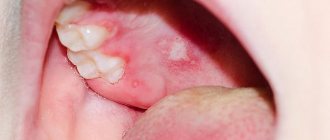
In what cases is drainage installed on the gum?
There are many pathologies in dentistry for which drainage is installed on the gums. These include:
- flux - is a manifestation of periodontitis and is accompanied by severe swelling of tissues
- neoplasms in the roots of teeth - drainage is installed on the gums if abscesses and cysts are diagnosed
- alveolitis - inflammation of the tooth socket
- in order to accelerate the healing of the wound surface after complex tooth extraction
- if necessary, regularly inject medications into the lesion
Gum drainage, photos of which you can see on thematic websites, plays a significant role in dentistry. It ensures the removal of pathological fluids and access of drugs even into the deep periodontal tissues.
There are few contraindications to drainage. The procedure cannot be performed if you are allergic to the anesthetics used during the procedure or have a blood clotting disorder.
How long should drainage remain in the gum?
Patients are most concerned about the question: how long should drainage remain in the gum? This period is determined individually depending on how quickly the swelling of the periodontal tissue subsides. It is the disappearance of this symptom that indicates complete cleaning of the wound and the beginning of its healing.
Only a doctor can determine exactly when to remove the drainage from the gums, taking into account the nature of the pathology. As a rule, the wearing period is 4-5 days. This is enough to clean the wound from pathological fluids. Sometimes the drainage falls out earlier, but complications are not observed - this means that the wound cleared earlier.
If the drainage has been in place for 5 days, and after its removal the wound swells again and causes pain, it is important to immediately consult a doctor. Additional diagnostics and identification of the cause of this condition are required.
Contraindications to gum drainage and possible complications
The procedure is not performed in the following cases:
- bleeding disorders;
- individual intolerance to anesthetic substances;
- unstable mental health.
What to do when drainage is impossible? In this case, alternative methods of disinfection and cleaning of the affected area are used. Even when the procedure is performed as indicated, complications are possible. This may include bleeding, ulcers, pain, or increased body temperature. To identify the cause of the complication and eliminate it, you need to visit a dentist.
When is drainage placed in the gum?
Why is drainage in the gum needed:
- drainage prevents early tissue healing (before the wound is completely cleared of inflammation products);
- drainage on the tooth ensures removal of pus, blood and ichor from the injured area without creating discomfort for the patient;
- often drainage is also used as a channel for direct administration of medication into infected tissue.
If the drainage in the gum is installed correctly, then its design is practically imperceptible and invisible.
The need for such treatment can be caused by almost any disease of the oral cavity if it is accompanied by an infection.
Most often, severe suppuration (and, as a consequence, the need to install gum drainage) is provoked by:
- flux (inflammation of the periosteum);
- pericoronitis;
- alveolitis;
- periodontitis;
- cyst or abscess of a tooth.
Sometimes suppuration occurs against the background of trauma to soft and bone tissues, which is accompanied by complex tooth extraction or mechanical damage to the jaw.
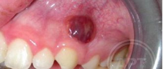
Photo. What does surgical drainage look like to remove pus?
Incision healing time
How long will it take for the gums to heal after drainage is removed from it? On average, tissue regeneration takes 1-2 weeks. The process largely depends on the complexity of the previous operation and the individual characteristics of the patient. For example, in older people, wounds take longer to heal. This fact is explained by the slowdown of metabolic processes with age.
Complete gum regeneration may take more than 6 months. The wound healing process after opening the abscess takes place in several stages:
- the formation of a blood clot that protects the wound from the introduction of pathogenic microorganisms;
- formation of new granulation tissue (within 3-4 days from the moment of intervention);
- maturation of new epithelium (7-10 days from the moment of opening of the purulent focus);
- soft tissue regeneration;
- formation of young bone tissue.
After the operation, the dentist gives the patient a number of instructions that help maintain drainage in the gums and avoid the development of complications after the intervention. In the first 3-4 hours after the procedure, it is prohibited to consume food. During treatment, it is advisable to redistribute the chewing load to the healthy side of the jaw. Doctors also advise giving preference to dietary products.
In the postoperative period, it is necessary to avoid intense physical activity and avoid visiting saunas, swimming pools and baths. It is also important to give up bad habits - smoking and drinking alcohol.
Drainage after cholecystectomy for acute cholecystitis
A large prospective randomized study in 1991 and a meta-analysis of 1920 patients (ACA) summarized 10 similar studies. It was shown that when comparing patients with and without drainage in terms of mortality, reoperation or drainage due to bile accumulation, there were no differences. Wound infection more often accompanied patients with drainage (5). Thus, on the eve of the end of the era of BCI, routine drainage - the sacred cow of biliary surgery - was abandoned in many centers.
What is the trend in emergency LCE? In a recent study of Australian surgeons, drainage was left routinely in 1/3 of cases (6). Another small randomized trial comparing patients with and without drainage for LCE examined the effect of drainage on postoperative pain and nausea, in terms of gas removal, and found no difference (7). If routine drainage is pointless in ACE, why is it indicated in LCE? Therefore, Petrowsky et al. (3) drainage is not recommended for both ACE and LCE. In a prospective study of 100 patients who underwent LCE for acute cholecystitis, all of them underwent cholescintigraphy one day after surgery. Bile leakage was detected in 8, but all of them were asymptomatic (8). Most postoperative collections, be it bile, serous fluid or blood, remain asymptomatic, the fluid is absorbed by the peritoneum and this is well known from ultrasound studies since the time of AChE.
Drainage is much more effective at removing bile than stool or pus. Therefore, it is logical to leave a drain if the surgeon is concerned about possible bile leakage. For example, if a subtotal cholecystectomy is necessary, or when there are difficulties with sealing the cystic duct, or there is a suspicion of additional bile ducts in the area of the gallbladder bed, which manifests itself in the form of bile leakage from the surface of the bed.
Thus, although most patients do not require drainage, if the surgeon is concerned about possible bile leakage or excessive serous fluid, drainage is appropriate. In most cases, almost nothing is separated through such drainage. It is extremely rare that preventive drainage becomes therapeutic in the case of profuse and persistent bile leakage. In cases where the need for an existing drain is questionable, it is extremely important to remove it as soon as possible. “Dry” drainage for 24 hours indicates that it has served its role. Finally, Howard Kelly (1858-1943) said: “Drainage is the recognition of defective surgery.” Clinicians must be careful not to confirm this statement in practice: if it is safer to switch to an open procedure and carefully close the ultrashort cystic duct than to rely on questionable clip closure and safety drainage, then the choice is clear.
Endoscopic method
Using an endoscope, doctors perform nasobiliary drainage of the biliary tract. Indications for endoscopic drainage of the biliary tract are:
- obstructive jaundice caused by malignant and benign neoplasms;
- acute purulent cholangitis;
- external biliary fistulas;
- damage to the walls of the extrahepatic ducts, retroduodenal perforations;
- acute cholecystitis.
There are no contraindications to endoscopic drainage, except in cases where the tube for drainage of the biliary tract cannot be passed through the area of tumor narrowing. The endoscopic kit for drainage of the biliary tract through the nose includes:
- conductor wire;
- drainages of various shapes;
- a connecting tube for collecting bile and flushing the drainage;
- nasal tube, clamp and spatula.
The operation of endoscopic drainage of the biliary tract includes the following steps:
- cholangiography to determine the level and location of drainage;
- introduction of drainage with a metal guide-conductor;
- removal of the guidewire and endoscope;
- control cholangiography;
- assessment of drainage position;
- transferring the drainage from the mouth to the nose and fixing it on the head.
After using the endoscopic method of drainage of the bile ducts, complications do not develop. They may occur due to the progression of the disease.
How to remove the drainage system, why can it fall out?
The stripe is small, sometimes it interferes with eating and creates a lot of inconvenience for its owner. Many people are interested in how long it will take to use the system, and whether it can be delivered ahead of schedule. During the application process, the dentist warns that if the drainage falls out of the gums, it will be safer to seek help from a specialist than to remove it yourself.
If an edge falls out, carefully adjust it and try to put it back in place. If you remove it yourself, you will need to wash your hands, disinfect your mouth, stand in front of a mirror, pull the edge of the strip with your fingers, and pull it completely out. Doing procedures at home is not so much difficult as it is dangerous. It’s better not to take risks and seek professional help. Gently rinsing the treated area will help speed up the tissue healing process. If the tape falls out in the first days of treatment, it must be promptly replaced to avoid possible infection with sad complications.
Self-removal of the structure: possible or not
Some patients experience severe discomfort, so they are interested in how to remove drainage from a tooth on their own. Others do not have time to make an appointment with a doctor on time, so they also want to know the answer to this question. Often, the dentists themselves provide detailed instructions so that the patient can perform the procedure at home. The procedure should be as follows:
- wash thoroughly with soap and disinfect your hands,
- treat the oral cavity with some antiseptic: Miramistin, Chlorhexidine are suitable,
- stand in front of the mirror, open your mouth, carefully grab the tip of the tube protruding from the wound with your thumb and forefinger and pull it towards you without force,
- rinse your mouth with an antiseptic,
- Do not consume food or drink for the next 2 hours.
“On the eve of the New Year holidays, I had a drainage installed after the removal of a dental cyst. The procedure is painless, as it is under anesthesia, but the doctor said that due to the holidays, I will have to pull the tube out of the wound myself in four days. I described in detail how this is done. When the hour struck, I was very worried, I thought it was difficult, but in fact I did everything quickly. However, after ten minutes the pain was so severe that I had to take pills. But within a day I even forgot that I once had this tube. After the holidays, I saw the doctor, he said that I was good, and the tissues were healing normally!”
Irina R, review from 32top.ru
The procedure is simple, but if the device has not fulfilled its main purpose, then it should not be carried out under any circumstances. And, in general, even if your condition has improved significantly, you should not resort to independent measures without consultation with a specialist. It is better to have a professional doctor perform all steps to remove the device. Why? Removing the tube yourself can cause severe pain, prolonged bleeding, damage blood vessels and injure surrounding tissue.