Dental smoother
Description, purpose
This is the main tool in therapeutic dentistry, used for filling teeth.
It is a handle with working parts, which are flat spatulas. They have different angles of inclination to the handle.
The smoother is used to introduce filling material into the treated carious cavity and modeling, restoration (by removing excess filling, smoothing).
Kinds
The choice of tool depends on the group of the tooth, the type, location and size of the carious cavity.
Main varieties:
- approximal teardrop-shaped, sickle-shaped, with an elongated working part;
- leaf-shaped approximal (small, long, wide);
- mini-smoothers approximal drop-shaped;
- combined, with a spherical plugger at one end and a trowel at the other;
- medial curved, thin right and left.
The irons are made of medical oxidized steel. A coating (titanium nitride, tantalum) is also used to prevent the filling from being stained by metal particles and sticking to the instrument.
Views in the photo:
with ball filler
mini medial
teardrop-shaped
sickle-shaped
Manufacturers and cost
Well-known manufacturers: Extradent (Russia), 3M ESPE (USA), Voco (Germany). Cost - from 80 to 1800 rubles.
The LM-Arte set of accessories for artistic restoration contains several types of smoothers. Manufacturer: lm-dental. The set was developed together with Style Italiano by a group of doctors involved in aesthetic dentistry. Made from high quality steel. Each device has its own marking depending on the main task. The set contains 5 tools, the cost of each is from 2500 rubles.
Manual preparation in the practice of a modern dentist
In most cases, the preparation of carious cavities is carried out using the traditional method using a drill. The fear experienced by patients (usually children, pregnant women, elderly and somatically burdened patients), pain during the preparation process arising from the vibration of the bur, its pressure on the tooth, the unpleasant sound of the dental unit: all this affects the patient’s behavior, leading to to the development of dental phobia [1, 3—5]. To reduce the level of psycho-emotional stress in patients, special preparation methods have been developed in accordance with the rules of biological expediency.
In 1994, the Dutch doctor Taso Pilot proposed the ART technique (atraumatik restorative treatment - atraumatic restorative treatment) not as an alternative, but as an addition to the machine method of preparation [1, 3, 4]. Since 1994, the ART technique has been recommended by the World Health Organization for providing dental care to residents of economically backward regions, low-income populations, refugees, and emigrants.
There are general and local indications for the use of the ART technique.
Are common:
- children's, elderly and senile age;
- patients for whom machine preparation is not indicated (with severe diseases of the cardiovascular system, in the post-infarction state, with diseases of the nervous system accompanied by increased excitability);
- patients in a state of prolonged immobilization, disabled people;
- pregnant women, patients with increased individual sensitivity to anesthetics or who have contraindications to their use;
- preparation in field conditions (military conditions, agricultural work, etc.);
- insurmountable fear, categorical refusal of machine dissection at any age [1].
Local (clinical) indications:
- caries of baby teeth;
- extensive areas of carious softening of hard tooth tissues;
- in some cases - hard-to-reach carious cavities (for example, teeth covered with an artificial crown);
- temporary delayed filling; combined use with classical preparation (machine) [1].
Indications depending on location:
- Class I according to Black - the presence of a wide entrance hole into the carious cavity and softened dentin;
- Class II according to Black - localization of the carious cavity below the equator of the tooth;
- Class III according to Black - localization of the carious cavity in the proximal cavities without damage to the vestibular and palatal walls;
- Class IV according to Black - limited use (in combination with machine preparation);
- Class V according to Black - cervical cavities with access to dentin;
- carious root cavities, including teeth under an artificial crown due to gum recession [1].
Contraindications [2]:
- complicated caries;
- exposed dental pulp;
- the obvious presence of a carious cavity, but at the same time its inaccessibility for processing with machine tools;
- indirect signs of a carious cavity, for example, on the approximal surface, but the impossibility of access to the cavity from the occlusal or approximal surface.
Advantages of the ART method [2]:
- minimal preparation of a carious cavity with maximum preservation of healthy tissue and minimal tooth trauma;
- painlessness, absence of psychological trauma for the patient, reduced fear of dental surgery;
- simplified infection control, as hand instruments are easy to clean and sterilize;
- no need for local anesthesia, expensive dental equipment (dental unit), the ability to use the method in areas remote from industrial centers, rural areas, directly in schools, hospitals;
- high economic efficiency; accessibility of restorative dental treatment for all social groups.
Basic principles of the ART method [2]:
- preparation of a carious cavity only with hand instruments;
- tooth restoration with adhesive filling materials (such as glass ionomer cements - GIC).
Despite the fact that a dental office and electrical equipment are not needed to carry out the method, ergonomic rules are observed when performing it. The operator needs to work while sitting, preferably with an assistant. Good lighting of the oral cavity is necessary. In addition to the standard set of tools (dental mirror, probe, tweezers, excavator and spatula), a dental knife for enamel and a carver for removing excess filling material are required [2]. If there is pain, topical anesthesia can be performed [1, 3].
Stages of working using the ART method [2]:
- Isolate the tooth being prepared from saliva using rollers.
- Remove plaque with damp cotton balls.
- Dry the tooth surface with dry cotton balls; if necessary, expand the entrance to the carious cavity with an enamel knife.
- Remove carious dentin with an excavator starting from the enamel-dentin junction.
- Remove overhanging edges of enamel, lacking dentin support, with an enamel knife.
- Clean the cavity with wet and then dry cotton balls.
- Carefully and carefully remove carious dentin near the pulp of the tooth.
- Clean the cavity again with cotton balls soaked in an antiseptic solution.
- Check the occlusal relationships of the tooth being restored by asking the patient to occlude the teeth in bite.
- Dry the cavity with dry cotton balls.
To improve the chemical bond of GIC with tooth structures, the cavity walls must be carefully processed. Dentin is treated either with the conditioner included in the GIC kit, or directly with the liquid for mixing GIC [2]. Then the cavity and fissures are washed several times with cotton balls moistened with distilled water or saline. After this, the cavity is dried with dry cotton balls. GIC is mixed according to the manufacturer's instructions; introduced into the cavity; condenses. Excess material is removed.
After dental restoration using the ART method, observation and evaluation of long-term results are carried out. The patient is asked about the occurrence of discomfort or pain within 4 weeks after dental treatment. Long-term results are assessed after six months, 1 or 2 years. It must be remembered that the restoration of carious teeth by GIC is only part of the ART method, which includes not only treatment, but also the prevention of caries. To avoid new carious lesions, after dental restoration, the patient is given the following recommendations:
- regular removal of plaque;
- rational nutrition, especially regarding the consumption of carbohydrates;
- applications of fluoride-containing solutions, antiseptic drugs (for example, chlorhexidine);
- fissure sealing [2].
An assessment of the long-term results of treatment of carious teeth using the ART method, carried out in Thailand (1991), showed that 71% of restorations were preserved after 3 years. A similar project in Zimbabwe (1993) led to better results: 85% of restorations were preserved after 3 years [2].
Clinical example
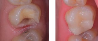
Patient M., 32 years old, complained of the presence of a carious cavity in tooth 3.6 and pain from thermal and chemical irritants when eating. When collecting anamnesis, it was found that the woman was in the 16th week of pregnancy, there was a threat of miscarriage. Anesthesia is contraindicated. It was decided to carry out ART restoration of the tooth. The examination showed the presence of a deep carious cavity in the 3.6 tooth, filled with softened pigmented dentin. The presumptive diagnosis is pulp hyperemia (deep caries). A spherical diamond bur and a turbine tip were used to open the carious cavity. An excavator was used to remove necrotic dentin (Fig. 1).
Rice. 1. View of the carious cavity of tooth 3.6 before filling.
Then antiseptic treatment was carried out. Probing of the bottom confirmed the diagnosis of pulp hyperemia, therefore, before filling the cavity, a therapeutic pad made of a material based on calcium hydroxide was applied to the bottom. The cavity was filled using the classical technique of working with hybrid GIC. The filling was formed using smoothers, fillers and damp, wrung out cotton swabs. After polymerization, the filling was coated with protective varnish (Fig. 2).
Rice. 2. Final appearance of tooth restoration 3.6.
The patient is recommended to undergo a follow-up examination after childbirth. Recommendations on oral hygiene are given.
Thus, the ART method remains relevant today. The method is positively perceived by patients, since dentin preparation is painless or the pain is so insignificant that anesthesia is not required. It is important that the dentist and the patient have a choice: high-tech machine or manual preparation methods, as well as a combination of both.
LITERATURE
- Zapashnik T. A., Gorbachev V. V., Oliferko D. V. Manual preparation in modern dentistry // Modern dentistry. - 2007, No. 3. - P. 55-59.
- Modrinskaya Yu. V., Khramchenko S. N. Methods of minimally invasive treatment of dental caries. ART method. Tunnel restoration / Educational and methodological manual, 2nd edition, expanded. - Minsk: BSMU, 2010. - 31 p.
- Pakhomov G.N., Leontyev V.K. Atraumatic restorative treatment of dental caries. — Incoral, Switzerland.
- Popruzhenko T.V., Yatsuk A.I. // Sovrem. dentistry (Belarus). — 2004, No. 3, 4.
- Chuev V.V., Rachitsky G.I., Al-Alawni S.V., Smetanyak S.M. // Dentist. - 2004, No. 1-3.
Putty knife
Description, purpose
This is a tool that is used for mixing various materials and modeling prosthetic structures.
Therapeutic spatula is used for mixing:
- cement, fillings;
- medicinal dressings.
Mixing the components of the material to the required consistency is done with spatulas, rubbing them on the glass. The laboratory spatula is designed for mixing impression materials, plaster, and casting plaster models. Modeling spatulas, dental and therapeutic metal spatulas are used to model the prosthesis with wax. An electric dental spatula is also used.
Kinds
The spatula can be:
- metal or plastic;
- therapeutic, laboratory, modeling;
- one- and two-sided.
The metal instrument is made of medical steel. Plastic is used when mixing materials that may change color (for example, a chemical composite).
Dental spatula in the photo:
Manufacturers and cost
Manufacturers: HLW Dental Instruments (Germany), IVOCLAR VIVADENT (Liechtenstein). Price – from 120 rubles.
Smoothers for restoration
Smoothers for restoration
Our website presents a wide range of smoothing pads for dentists:
- Tool for modeling fissures light green Silicon Black - designed for modeling the surface of the tooth, forming fissures, tubercles;
- Double-sided plugger, gray Silicon Black, is used to introduce the composite into the formed cavity and distribute it along the walls;
- Double-sided blue trowel Silicon Black - for introducing and distributing the composite into cavities, as well as for forming restoration cusps;
- Brown Silicon Black reverse trowel - for introducing and distributing the composite into cavities, as well as for forming restoration cusps;
- Double-sided trowel with a turn, blue Silicon Black - for introducing and distributing the composite into cavities, as well as for forming restoration cusps;
Also our new ironers made to order, especially for our customers:
- Universal smoother for working in the area of the front teeth, yellow Silicon Black
Bright design, silicone handle with anti-slip effect. It has a wide leaf-shaped shape with a pointed tip for convenient modeling with a composite in the front group of teeth. A thin working part, with two different bends on opposite sides, depending on the surface on which the work is being done. It has a black anti-reflective matte coating for convenience when working with optics. For comfortable work on both vestibular, oral and proximal surfaces.
- Universal smoother for working in the area of chewing teeth, black Silicon Black
The smoother is universal for working in the area of chewing teeth; it has a black coating with an anti-reflective effect for comfortable work with optics. The smoother has a silicone handle with an anti-slip effect. On one side there is a plugger for compacting the composite into the tooth cavity, on the other there is a tool with a sharp tip for modeling fissures on the chewing group of teeth.
- Green filler trowel Silicon Black
Ergonomic design for applying the composite and its subsequent modeling on the tooth surface.
All instruments are made of stainless medical steel AISI 410 and have a comfortable silicone handle, which is very dense and can withstand a huge number of sterilization cycles.
Additionally, you can order your own personalized engraving on any instrument.
We recommend that you adhere to sterilization standards so that your instruments last a long time.
You can order dental instruments at affordable prices separately or in a set on our website dentins.ru.
Stopfer
Description, purpose
This is a tool for compacting filling material in a carious cavity and retrograde filling of the root canal.
Kinds
A dental plugger consists of a metal handle and working parts, which have several structural options:
- spherical;
- cylindrical;
- cone-shaped;
- reverse cone;
- three-cornered.
Made from medical steel. Most often it comes with an ironing board.
