Neuritis
is an inflammation of the peripheral nerve trunk.
The peripheral nervous system refers to the nerve structures outside the brain and spinal cord (which are in turn called the central nervous system). Excitation (signal) is transmitted along the nerves, allowing the central nervous system to receive information about the state of all organs and systems of our body and control muscle contractions. The tissues of various organs are penetrated by nerve endings, which are connected to the brain, and in most cases to the spinal cord using bundles of nerve fibers enclosed in a sheath of connective tissue. Such bundles are called nerves, and large nerves are called nerve trunks.
With neuritis, nervous tissue is involved in the inflammatory process. This leads to the fact that the signal in some part of the nerve passes worse or does not pass at all. Accordingly, the sensitivity of any area may be lost and the mobility of the muscles innervated by the affected nerve may decrease.
Symptoms
With neuritis, the nerve fiber is damaged, and therefore the functions suffer, the main ones being sensitive, motor and trophic, therefore the following symptoms are clinically noted:
- In the phase of irritation of the nerve fiber: pain, burning, feeling of “crawling goosebumps”, feeling “as if many needles are being pricked”.
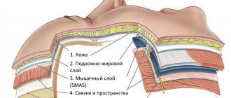
- In the phase of loss of functions: decreased muscle strength, muscle atrophy, decreased reflexes, decreased sensitivity, tissue atrophy including skin, subcutaneous fat, muscles, bone tissue.
The treatment program for this disease at the Bone Clinic may include:
Shock wave therapy
More details
PRP therapy
More details
Acupuncture administration of ozone
More details
Teraquantum therapy
More details
Interference therapy
More details
Causes
The main causes of neuritis:
- infectious (viruses, bacteria, their toxins);
- exposure to toxic factors
- metabolic disorders (diabetes mellitus, deficiency of vitamins B1 and B6);
- allergic reactions;
- injuries;
- oncological diseases;
- hereditary diseases;
- diseases of the spine leading to mechanical compression of the nerve trunk (intervertebral hernia, tunnel syndromes, etc.).
The mechanism of nerve damage when exposed to various unfavorable factors is associated with the launch of a cascade of inflammatory reactions. Swelling of the nerve trunk occurs, damage to the myelin sheath and subsequently to the axial cylinder, which in turn leads to the death of the nerve cell.
Make an appointment
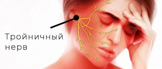
Neuralgia and trigeminal neuritis
Neuritis and neuralgia are different diseases, although (as in the case of the trigeminal and facial nerves) some “specialists” combine them or replace one with the other. The main difference lies in the nature of their occurrence. Neuritis is always associated with an inflammatory process, hence its name: the ending -itis - from Lat. -itis always denotes inflammatory diseases. Neuralgia is irritation of the nerve as a result of concomitant diseases or exposure to external factors. Unlike neuritis, neuralgia does not cause changes in the structure of the nerve.
Most doctors consider neuritis to be a much more dangerous disease, since the inflammatory process affects the inner part of the nerve. At an advanced stage, the disease causes nerve damage and spreads to the auditory nerve, and can also lead to ataxia, a motor disorder. In this case, neuritis and neuralgia can occur together and have some similar symptoms. First of all, these are painful sensations, which, however, vary in nature and intensity.
Clinical picture of some individual nerves
- Trigeminal nerve – pain is sharp, piercing, in series, along one or several branches of the nerve.
- Facial nerve – muscle weakness on one side of the face. It is difficult to close the eye, the corner of the mouth on the affected side is drooping. When liquid food or drink is taken, everything comes out of the mouth.
- Diaphragmatic – feeling of lack of air, shortness of breath, hiccups.
- Median – impaired flexion of the hand, fingers I, II and III and decreased sensitivity on the palmar surface.
- Ulnar – weakness of the flexors of the IV, V and partly III fingers, decreased sensitivity on the palmar surface of the above fingers.
- Radial – impaired extension of the hand and fingers, decreased sensitivity in half of the back of the hand (I and II fingers). It is difficult to move the thumb away.
- External cutaneous nerve of the thigh (Roth-Bernhardt disease) – pain, numbness and burning along the outer surface of the thigh.
- Femoral – impairment of leg extension at the knee joint and hip flexion. Pain and sensory disturbances on the lower 2/3 of the anterior surface of the thigh, the anterior inner surface of the lower leg.
- Sciatic – pain along the back of the thigh and lower leg, weakness of the flexors and extensors of the foot.
- Olfactory – anosmia on one side (lack of sense of smell). When a nerve is irritated, foreign odors may appear.
- Visual – decreased acuity and loss of visual fields. The phenomena of nerve irritation manifest themselves in the form of photopsia (sensation of light, flames, sparks, etc.).
- Oculomotor – drooping of the upper eyelid (ptosis), limited mobility of the eyeball, dilated pupil, diplopia (double vision).
- Block - restriction of the mobility of the eyeball downwards and outwards.
- Auditory – hearing loss, often accompanied by a feeling of noise or ringing in the ear.
- Glossopharyngeal - twitching pain in the tonsils, root of the tongue, pharynx and taste disturbances in the back third of the tongue, impaired salivation and swallowing.
- Wandering – manifested by disturbances in swallowing and speech. On the affected side, the soft palate is lowered, the uvula is deviated to the healthy side. There are also disturbances in the functioning of internal organs - bradycardia, shortness of breath, motility disorders of the esophagus, stomach and intestines (spasms), etc.
- Additional – difficulty turning the head in the healthy direction, the head is tilted towards the affected nerve, the shoulder is lowered.
- Tibial – the foot is extended, but the patient cannot bend it. Cannot stand on toes. Sensitivity is reduced along the back of the lower leg and on the sole.
- Peroneal – the impossibility of standing on the heels and straightening the foot, it hangs down. Sensitivity is reduced on the outer surface of the lower leg and the back of the foot.
- Intercostal nerves – pain in the intercostal space, often radiating into the chest, simulating pain in the heart, chest, lungs, and stomach. Often, pain in the paravertebral muscles is detected at the level of the thoracic spine.
Surgery
Certain surgical procedures can help relieve neuralgia pain when the condition does not respond to treatment.
Examples of surgical procedures that may help treat neuralgia include:
Microvascular decompression : Helps remove an enlarged blood vessel affecting a nerve. The procedure involves placing a soft pad between the blood vessel and the affected nerve.
Stereotactic surgery : This is a non-invasive procedure that directs highly concentrated beams into the root of the damaged nerve. The radiation disrupts the transmission of pain signals to the brain.
Percutaneous balloon compression : This involves inserting a small balloon into the affected nerve. The balloon is inflated, causing controlled, deliberate nerve damage. This procedure prevents the affected nerve from sending pain signals to the brain. However, the effects of the procedure usually wear off after 1-2 years.
Diagnosis of neuritis
The symptoms of neuritis are in many ways similar to the clinical manifestations of various diseases, including non-neurological ones. Therefore, the doctor should conduct a thorough differential diagnosis to make an accurate diagnosis.
Primary diagnosis consists of a thorough collection of patient complaints, identification of possible preceding factors, and direct examination of the patient. Most symptoms of neuritis are specific, so depending on their severity, the doctor can make a preliminary diagnosis.
Stimulation and needle electroneuromyography are used to diagnose peripheral nerve damage. This study makes it possible to answer the questions - which nerve is damaged, in what place it is damaged, what percentage of nerve damage, as well as give a prognosis for its recovery and monitor its recovery.
Diagnostic methods
Electroneurography is used to measure the speed of passage of nerve impulses through the fibers of peripheral nerves from their exit point to the nerve endings in ligaments and muscles. The technique allows you to determine the damaged nerve, determine the location and extent of damage, and identify the severity of the process.
Electromyography – used to study the bioelectrical activity of muscles. This method allows us to answer the question: what is the problem - damage to the nerve or damage to the muscle itself? EMG allows for differential diagnosis of neuropathy with muscle pathology (myasthenia, myotonia, myoplegia, polymyositis).
Ultrasound is a method for diagnosing damage to peripheral nerves. Evaluates changes in nerve diameter, continuity, and deterioration of sound conduction. Ultrasound clearly shows swelling of the nerve trunk and surrounding tissues
MRI – visualizes nerves and soft tissue structure, detects malignancies and provides information about muscle atrophy and nerve damage. With the help of MRI diagnostics, it has become possible to detect nerve damage in areas that are difficult to examine using electrodiagnostics or ultrasound.
Make an appointment
Who will treat you?
Bogdanov Vadim Yurievich
Chief traumatologist-orthopedist..
Read more
Ronami Valery Guseinovich
neurologist, reflexologist, chiropractor, professor, doctor of medical sciences.
More details
Dremin Evgeniy Vitalievich
neurologist, reflexologist, chiropractor.
Read more
Repeated appointments with a neurologist based on examination results in our clinic are free.
Treatment of neuritis
Treatment must be comprehensive. It is necessary to fight both the damaging factor and restore the damaged nerve trunk.
In treatment I use the following groups of drugs:
- Vascular;
- Anti-inflammatory;
- B vitamins;
- Improving the conduction of impulses along the nerve, etc.
Non-drug treatment methods are also used:
- Acupuncture - exposure to biologically active points of the skin leads to signal transmission along the nerve trunk to the spinal cord and brain, resulting in a complex cascade of reactions, which includes improved blood circulation, release of biologically active substances, and hormonal response, which in turn accelerates restoration of nerve fiber.
- Physiotherapy is designed to locally influence reflex zones. The main effects achieved after physiotherapy are: analgesic, anti-inflammatory, antispasmodic, fibrinolytic.
- Massage (improves microcirculation, reduces swelling).
- Therapeutic exercise (by working out muscle groups using a feedback system, the recovery period is accelerated).
To relieve pain and accelerate the recovery of nerve fibers, local injection techniques are used - therapeutic and diagnostic blockades, pharmacopuncture - with various mixtures of drug solutions, if necessary, together with ultrasound navigation. Due to the local administration of drugs (vitamins, anti-inflammatory drugs, anesthetics), it is possible to quickly relieve pain, reduce swelling and the inflammatory reaction, and restore the nerve trunk.
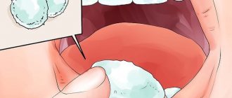
Tooth pain due to neuralgia, causes and treatment
The presence of toothache does not always indicate problems with the teeth and oral cavity. Sometimes tooth pain can occur in people with completely healthy teeth. In this case, there is a possibility that toothache is a symptom of trigeminal neuralgia, which is hidden behind such pain, making it difficult to identify the real cause of the ailment.
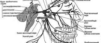
The trigeminal nerve is one of twelve pairs of cranial nerves, which is a continuation of the spinal cord and is responsible for the functioning of many reflexes, such as blinking, sneezing, and eyebrow movements. Also, together with others, this nerve ensures normal breathing. The trigeminal nerve consists of three branches that form it: the first branch is located at eye level, the second above the upper jaw near the nose, and the third in the lower jaw.
There are two types of trigeminal neuralgia - true and secondary. True or primary neuralgia is a self-limiting disease, while secondary neuralgia develops due to the presence of complications of other diseases. The type of inflammation can be determined based on a number of reasons that caused the development of trigeminal neuralgia.
Reasons for the development of true neuralgia:
- displacement of arteries and veins;
- the presence of adhesions or tumors;
- injury to the face.
Causes of secondary neuralgia:
- gingivitis;
- pulpitis;
- periodontitis;
- periostitis;
- after tooth extraction:
- dental caries;
- in the presence of ENT diseases;
- in case of metabolic disorders;
- after hypothermia, etc.
The manifestations of symptoms of trigeminal neuralgia depend on which branch is affected. Toothache, as well as pain in various areas of the head, occurs when the second and third branches become inflamed. However, the nature of the pain differs depending on the inflamed branch, which helps to determine the affected area. With neuralgia of the second branch of the trigeminal nerve, pain manifests itself in the area of the nose, facial muscles and the entire upper row of teeth, while, as with inflammation of the third branch, pain in the neck, chin, cheeks, and lower teeth is characteristic.
The main symptoms of inflammation of the trigeminal nerve:
- paroxysmal toothache, radiating deep into the bones;
- spasm of the facial muscles during an attack;
- discomfort when eating hot or cold food, when brushing your teeth, as well as when touching your face, etc.
If you experience toothache associated with neuralgia, you must consult a doctor. However, if the pain is due to inflammation of the trigeminal nerve, your dentist will most likely not be able to help you. For qualified help, you need to go to a neurologist to avoid the development of complications and not to aggravate the situation.