Exudative erythema multiforme (EEM) - symptoms and treatment
Infectious-allergic MEE, including herpes-associated[13], is characterized by an acute onset of the disease. It can be manifested by fever, general weakness, body aches, headache, sore throat and other symptoms.
Often, a few days before an exacerbation of MEE, a herpes infection is activated in patients - herpes appears on the lips or a relapse of genital herpes occurs[8].
Rashes characteristic of MEE usually begin to appear 1-2 days after the onset of prodromal phenomena. In this case, general symptoms usually subside.
The localization of the rash is very diverse. It can appear on both the skin and mucous membranes.
Skin manifestations
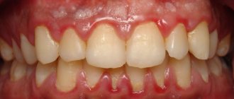
The skin is usually marked by clearly defined rounded red-pink spots and flat, edematous papules, which increase in size from 2-3 mm to 3 cm in diameter. In the infectious-allergic form, the spots are usually somewhat smaller and do not tend to merge. They can cause itching and burning.
The predominant localization of rashes is on the extensor side of the arms and legs, the back of the feet and hands, on the face and in the genital area. Basically, they are located symmetrically on the body, often in groups in the form of arcs or garlands.
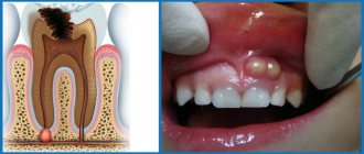
As the papule grows, its center begins to sink and change color to a more bluish color; a red-pink rim remains along the periphery - thus, the elements of the rash acquire the characteristic appearance of a “target” (sometimes they are compared to a “bull’s eye” or “cockade”) [4] .
Afterwards, bubbles form in their center - vesicles and bullous elements. They contain serous or bloody exudate. When they burst, the blisters form yellowish or brown-brown crusts and eroded surfaces.
As a result, elements of varying degrees of development are simultaneously present on the patient’s body - spots, papules and blisters, turning into crusts and erosions. That is why erythema is called multiforme.
At intervals of several days, new groups of rashes may form. This may delay the process. But usually final regression occurs within about two weeks.
The frequency of exacerbations can vary from 1-2 to 5-12 times a year. In rare severe cases, one exacerbation can turn into another, with virtually no clear interval. During this time, previous rashes do not completely resolve.
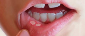
Rashes on mucous membranes
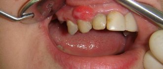
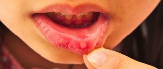
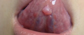
Single elements may appear on the oral mucosa, which do not cause much concern. In more severe cases, the lesions are so extensive and painful. They make it difficult to speak and eat even homogeneous and liquid food[12][13].
The resulting blisters burst quite quickly, so the patient does not have time to notice them - usually erosions are already detected, on which filmy fibrinous deposits of a light or brown hue can sometimes be seen.
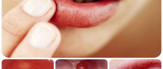
In the area of the red border of the lips, very painful, cracking, bloody crusts may appear, which do not allow the patient to fully open his mouth.
Less commonly, rashes are found on the mucous membranes of the eyes and genitals. In complicated cases, secondary infection may occur, scarring and synechiae (adhesions) may form.
Publications in the media
Exudative erythema multiforme (erythema multiforme) is an acutely developing disease characterized by the appearance of erythematous spots, bullous lesions of the skin, mucous membranes, and a cyclic relapsing course.
Statistical data . The incidence is 0.3–0.5: 100,000 population, severe forms are observed 2–3 times more often in men.
Classification • Infectious-allergic (idiopathic) form is associated with hyperreactivity to allergens and infectious agents • Toxic-allergic (symptomatic) form is associated with hypersensitivity to drugs • Exudative malignant form (see Stevens-Johnson syndrome) • Rheumatic erythema - round or arcuate lesions erythema on the trunk and limbs, sometimes observed during a rheumatic attack.
Clinical manifestations • Local symptoms •• Symmetrical rashes appear acutely on the skin of the extensor surfaces of the forearms, legs, dorsum of the hands and feet, face, genitals, and mucous membranes. Edema, clearly demarcated, flattened papules of pink-red color, round in shape, with a diameter of several millimeters to 2–5 cm, appear, having two zones: internal (grayish-bluish in color, sometimes with a bubble in the center filled with serous or hemorrhagic contents) and external (red in color [cockade-shaped rashes]) •• Diffuse erythema, blisters, erosive areas covered with a yellowish-gray coating appear on the lips, cheeks, and palate • General symptoms •• Burning and itching in the area of the rash, pain and hyperemia of the mucous membranes, especially mouth and genitals •• Fever •• Headache and joint pain • The most severe manifestation is Stevens-Johnson syndrome • In the toxic-allergic form, unlike the idiopathic one, there is no seasonality of relapses of rashes.
Research methods • Conduct laboratory tests to exclude syphilis - serological reactions, studies for Treponema pallidum • Nikolsky, Asbo-Hansen symptoms are negative, there are no acantholytic cells in the fingerprint smears • During histopathological examination, intra- and intercellular edema, hydropic degeneration of basal cells are noted in the epidermis , in the dermis - swelling of the papillary layer, perivascular infiltrates.
Differential diagnosis • Chicken pox • Bullous pemphigoid • Dühring's dermatitis herpetiformis • Herpes zoster • Syphilitic papular rash.
Treatment • For mild cases - antihistamines and desensitizing agents • For blisters and erosions on the skin - ointments with HA and antibiotics (for example, oxytetracycline + hydrocortisone) • For damage to the oral mucosa - warm rinses with 10% sodium bicarbonate solution, local anesthetics ( 2% lidocaine solution), as well as glucocorticosteroids: dexamethasone (elixir, 0.5 mg per 5 ml of water) 4 times a day followed by swallowing • In more severe cases and with common bullous forms - antibiotics (orally or parenterally), GCs (for example, prednisolone 1–2 mg/kg/day followed by dose reduction), proteolytic enzyme inhibitors (aprotinin). For Stevens-Johnson syndrome - see Stevens-Johnson syndrome.
Forecast . The outcome of the disease in uncomplicated cases is favorable. With Stevens-Johnson syndrome, the mortality rate is 10–30%.
ICD-10 • L51 Erythema multiforme
Clinical manifestations
An acute onset is characteristic, like an infectious disease: body temperature rises to 39-40ºC, symptoms of intoxication of the body develop.
Typical complaints: pain, burning, sore mouth, inability to eat, deterioration of general condition, rashes in the mouth and on the skin, etc.
When examining the oral mucosa, extensive erosive surfaces are identified, covered with a fibrinous whitish or grayish-yellow coating. Along the edges of the erosions, fragments of blisters are observed, when pulled, the healthy epithelium does not peel off (negative Nikolsky's sign). There is a primary polymorphism of rashes: papules, erythema, blisters and blisters, after opening of which erosions and aphthae are formed).
In the oral cavity, rashes may vary: hemorrhagic manifestations (bubbles with hemorrhagic exudate, hemorrhages, petechiae and bleeding of the mucous membranes); ulcerative-necrotic (these lesions are caused by allergic alteration of the oral mucosa, the addition of a secondary infection, aggravated by deterioration of hygiene and self-cleaning of the oral cavity due to pain, which leads to significant intoxication and the appearance of a putrid odor); catarrhal (erythema and swelling of the mucous membranes).
The skin is characterized by maculopapular elements of rashes that slightly rise above the surrounding surface. The central part of the element subsequently, after opening the papule, sinks a little and acquires a bluish tint, while the peripheral part retains a pink-red color, forming a “cockade”.
Favorite places for localization of rashes with exudative erythema multiforme: the dorsum of the hands, feet, extensor surfaces of the forearms, legs, elbow and knee joints, palms and soles. A distinctive feature of the toxic-allergic form of exudative erythema multiforme is the absence of seasonal relapses; the history shows a connection with taking medications, after which a relapse occurs.
Stevens-Johnson syndrome is a severe form of MEE. In this case, the mucous membranes of the mouth, nose, eyes, genitourinary organs, and gastrointestinal tract are simultaneously affected. and skin.
Lyell's syndrome or toxic epidermal necrolysis is the most severe form of MEE. In this case, almost all COs are involved in the process, including internal organs; extensive skin surface is damaged with detachment of the epidermis, the formation of hemorrhagic blisters and subsequent erosion.
Nikolsky's sign is positive only in the area of blistering. The course of the disease is continuous, relapsing, accompanied by dehydration, shock, secondary infection and septicemia.
Erythema nodosum
Hepatitis
Arthritis
Colitis
Allergy
18988 08 February
IMPORTANT!
The information in this section cannot be used for self-diagnosis and self-treatment.
In case of pain or other exacerbation of the disease, diagnostic tests should be prescribed only by the attending physician. To make a diagnosis and properly prescribe treatment, you should contact your doctor. Erythema nodosum: causes, symptoms, diagnosis and treatment methods.
Definition
Human skin consists of epidermis and dermis. The epidermis serves to protect the underlying tissues from mechanical, chemical, thermal damage, as well as from the penetration of infectious agents. The dermis is a layer of skin that includes a network of blood vessels, subcutaneous fatty tissue (SAT), connective tissue fibers, hair follicles, sweat and sebaceous glands.
Erythema nodosum is an inflammatory lesion of small vessels of the skin and subcutaneous fatty tissue, which is a type of panniculitis (inflammation of fatty tissue that develops against the background of other diseases or is idiopathic in nature).
Causes of erythema nodosum
Unfortunately, it is not always possible to establish for certain what led to the appearance of erythema nodosum. This poses difficulties in determining treatment tactics for such a condition. Most often, erythema nodosum is found in adults of reproductive age. In children and adolescents, it can develop against the background of a previous streptococcal infection.
The cause of the inflammatory process, which results in erythema nodosum, can be infectious agents, autoimmune diseases, allergic reactions, taking medications (sulfonamides, penicillin antibiotics, oral contraceptives, bromides, etc.). Pregnancy can also provoke the appearance of erythema nodosum.
Under the influence of these factors, lymphocytes are activated, the immune system begins to produce specific antibodies and inflammation develops. However, such a response of the body is not detected in every person, which suggests a special predisposition to such an immune response, and the predisposition can be either hereditary or acquired.
Erythema nodosum usually develops in the presence of two conditions: specific abnormal activity of the immune system and a provoking factor (trigger).
Among infectious diseases, such triggers are often streptococcal infection, tuberculosis, chlamydia, hepatitis B and C, etc.
The incidence of erythema nodosum in autoimmune diseases is high. This is a group of diseases that are based on a violation of the immune system’s recognition of “self” and “foreign” cells. As a result, immune cells begin to show aggression against cells of their own body, just as they would be activated when meeting infectious agents or cancer cells.
Erythema nodosum is common in sarcoidosis. One form of sarcoidosis (Löfgren's syndrome) is a combination of bilateral damage to the lymph nodes located in the roots of the lungs, joints, fever and erythema nodosum.
Other autoimmune diseases associated with the development of erythema nodosum include:
- systemic lupus erythematosus;
- rheumatoid arthritis;
- Behçet's disease (a disease based on vascular inflammation);
- Sjögren's disease (a disease that primarily affects the salivary and lacrimal glands);
- nonspecific ulcerative colitis (severe inflammatory damage to the colon), etc.
Sometimes erythema nodosum develops against the background of various malignant neoplasms, which include leukemia, lymphoma, pancreatic cancer, etc.
Classification of erythema nodosum
Erythema nodosum can be either an independent disease of unknown etiology (
primary or idiopathic erythema nodosum
) or develop as part of other diseases (
secondary erythema nodosum
). This classification is extremely important for determining the patient’s treatment tactics, because the treatment of secondary erythema nodosum is based on the effect on the underlying disease.
Erythema nodosum may have an acute
,
migratory
or
chronic course
.
Symptoms of erythema nodosum
The clinical features of erythema nodosum depend on the etiological factors and the nature of the process.
Primary (idiopathic) erythema nodosum manifests itself acutely - bright red painful confluent nodes form on the legs against the background of swelling of the legs and feet.
In acute cases, the nodes develop rapidly. Characterized by a concomitant increase in temperature to 38-39°C, weakness, headache, arthralgia. The disease is preceded by viral infections or tonsillitis. The nodes usually disappear without a trace after 3-4 weeks without ulceration, relapses are rare.
Migratory erythema nodosum is characterized by an acute course with an asymmetric inflammatory component. The main node is dense, bluish-red in color, delimited from the surrounding tissues. As a result of peripheral growth due to the migration of the inflammatory infiltrate, a ring-shaped plaque is formed with a sunken pale center and a wide, more saturated peripheral zone. Single small nodules may appear, including on the opposite shin. The duration of the disease is up to several months. During the formation of nodes, concomitant general disorders in the form of chills, weakness, arthralgia, and low-grade fever may be observed.
Chronic erythema nodosum usually occurs in middle-aged and elderly women, often against the background of vascular, allergic, inflammatory, infectious or tumor diseases. Exacerbation occurs more often in spring and autumn. Nodes with moderate pain and the size of a walnut are localized on the legs (on the anterolateral surface), swelling of the legs and feet is observed. Relapses can last for months.
Diagnosis of erythema nodosum
The diagnosis of erythema nodosum is made based on the clinical picture. This suggests that the data obtained during the conversation with the patient and examination is sufficient. However, to select treatment, it is necessary to establish causative and concomitant diseases. For this purpose, the patient may be prescribed the following studies:
- A clinical blood test with a leukocyte count, which will allow you to assess the intensity of inflammatory processes in the body, suspect the cause, and identify changes characteristic of leukemia and lymphoma.
Etiology
Etiological factors: with the infectious-allergic form of exudative erythema multiforme, patients exhibit sensitization to bacterial and viral allergens. The source of sensitization is foci of chronic infection (tonsillitis, otitis, sinusitis, cholecystitis). Factors that provoke the onset of the disease and its relapses are hypothermia, fatigue, exacerbation of chronic somatic diseases (tonsillitis, bronchitis, otitis media, etc.).
The cause of the toxicoallergic form is often medications (antibiotics, NSAIDs, synthetic vitamins, etc.), as well as food and household allergens.
The clinical picture is characterized by an acute onset. The disease often begins with prodromal phenomena (fever, malaise, pain in muscles and joints, sore throat).
After the prodromal period, polymorphic rashes on the skin appear in spurts (over 10-15 days or more) - erythema, papules, blisters. Depending on the severity of clinical manifestations, the following forms of the disease are distinguished:
Mild simple (papular) form
The primary morphological elements are hyperemic spots (erythema), papules and vesicles. Papules are round in shape with clear boundaries, ranging in size from 0.3 to 1.5 cm, red-bluish in color, flat, dense on palpation, prone to centrifugal growth with retraction of the central part.
An edematous ridge forms along the periphery of the papules, and the center of the element, gradually sinking, acquires a cyanotic tint (symptom of “target”, or “iris”, or “bull’s eye”. Characteristic are target-shaped lesions less than 3 cm in diameter with clearly defined edges, in structure which are divided into three different zones:
- central disk of dark erythema or purpura that may become necrotic or transform into a dense vesicle
- ring of palpable, pale, swollen area
- outer ring of erythema.
Subjectively, the rash is accompanied by itching. Pathological elements tend to merge to form garlands and arcs. The bubbles are round in shape, small, flat, have a thick covering, are filled with opalescent liquid, and are usually located in the center of the papules.
Secondary morphological elements are erosions, crusts, scales, hyperpigmented spots that do not have clinical features.
The rashes usually appear suddenly, are often located along the periphery, symmetrically on the skin of the dorsum of the feet and hands, palms, extensor surfaces of the forearms and legs, the red border of the lips with the formation of crusts, the mucous membrane of the oral cavity, less often on the neck and torso. Often, only the hands are selectively affected. Damage to the eyes and genitals is observed less frequently. Blisters may form on the mucous membranes, which open to form painful erosions. Resolution of the rash continues within 2-3 weeks, without leaving scars. Pigment spots that appear in place of former papules are yellowish-brown in color.
Vesiculobullous form
The vesiculobullous form occupies an intermediate position between the papular and severe bullous forms. The lesions are represented by erythematous plaques with a bubble in the center and a ring of bubbles along the periphery. Often the mucous membranes are involved in the process.
Severe bullous form
In the severe bullous form (Stevens-Johnson syndrome), the onset is acute, sometimes with prodromal phenomena. Skin, eyes, mucous membranes of the oral cavity, genitals, and perianal area are affected; bronchitis and pneumonia develop. Extensive blisters appear on the oral mucosa with the formation of erosions, which are covered with massive hemorrhagic crusts. The development of catarrhal or purulent conjunctivitis is characteristic, against which blisters may sometimes appear.
Corneal ulceration, uveitis and panophthalmitis with loss of vision often develop. Damage to the mucous membranes of the genitals in men leads to impaired urination with possible involvement of the bladder in the process. Maculopapular rashes or blisters on the skin are noted, less often - pustules, significant in size and number. The period of rash is 2–4 weeks. Skin syndrome is accompanied by fever, pneumonia, nephritis, diarrhea, polyarthritis, etc. Paronychia and otitis media are sometimes associated. Without treatment, the mortality rate for Stevens-Johnson syndrome reaches 5–15%. With all variants of exudative erythema multiforme, there is a tendency to relapse