Tooth enamel is a durable tissue that is resistant to external influences. But even it can be destroyed. For various reasons, cracks and chips appear on its surface. Cracked teeth are damage that crosses the tooth down towards the root. Damage can also extend under the gum, affecting the pulp chamber and tooth root. The cracked unit does not split into two halves, but the child and pulp are more often damaged.
Due to regular stress, a small crack on the tooth begins to gradually increase. The tooth becomes more sensitive to temperature and reacts to sour and sweet foods. Through microcracks, bacteria enter the tooth and even extend beyond its root, which leads to the development of pulpitis, periodontitis and other serious problems. The most common result of an untreated crack is a split tooth. It will simply break into two fragments and the dentist will have to remove it.
Types of dental cracks
Damage can occur on any area of the enamel; the most dangerous is a split of the tooth root. According to the clinical picture and type of damage, the dentist selects a therapeutic regimen. Types of cracks and their degree of danger:
- Vertical cracks in teeth
are usually shallow, small in size, and affect exclusively the enamel. Usually they do not bother a person and rarely require serious treatment, but they must be closely monitored, since against the background of constant loads, the fracture may increase in size. - Diagonal cracks,
regardless of depth
,
quickly become the main cause of tissue destruction and the appearance of chips and chips on the tooth. Moreover, the destruction affects both the enamel layer, dentin and the pulp chamber. - Horizontal cracks in teeth
pose a similar risk to diagonal fractures and require immediate treatment. It doesn’t matter whether the enamel on the front incisor is cracked or the molar is split across, such damage quickly grows, damaging deep dental tissues. They are the cause of large chips, splitting of the tooth to the top of the root.
If a crack has formed on the surface of the tooth, you should immediately go to the doctor. The lower the degree of damage, the less invasive and costly the intervention will be. Today, dentistry offers a lot of techniques that allow you to “patch” a crack before it causes serious damage, including the loss of a dental unit. But if a microcrack can be eliminated with regular grinding, then if there are chips or a deep split in the tooth, the help of an orthopedist will be required.
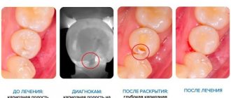
Diagnostic methods
When the first suspicious signs appear, you should visit the dentist. Dentists not only conduct a visual examination, but also use special instruments. To determine the location and extent of tissue damage, an x-ray examination is prescribed. What to do if a crack appears inside a tooth is decided based on the results of diagnostic procedures.
Numerous scratches can be detected without the use of tools. The deep ones can only be seen under the influence of light, while the surfaces are dried with cotton balls. Superficial flaws are detected using a magnifying glass or in a special way - transillumination.
Making an accurate diagnosis can be difficult. This is due to the frequent absence of characteristic symptoms. Specialists take into account all manifestations in their entirety: the patient’s sensations, his medical history, x-rays, exacerbation of symptoms during treatment.
All methods are combined, since this way the dentist will be able to objectively assess the situation and develop a therapeutic regimen. For this we use:
- instrumental examination of the oral cavity;
- examination under a microscope;
- transillumination;
- staining with contrast agents.
In some cases, exploratory surgery is indicated. This is the exfoliation of dentin particles and the removal of granulation tissue for subsequent painting of the damaged layer. In this way, even microcracks are visualized. Sometimes root resection is performed.
Symptoms
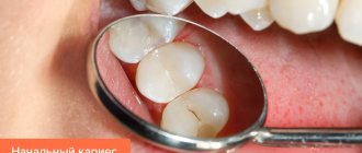
Small, shallow cracks usually do not show themselves in any way. If the damage is voluminous or located in the root area and puts pressure on the neurovascular bundle, the following symptoms appear:
- Increased sensitivity to temperature and chemical irritants;
- pain when chewing, inhaling cool air;
- swelling of the gums around the damaged tooth;
- formation of chips and chips.
As the symptoms become more severe, the danger of the problem becomes clear. If cracks in the front teeth do not extend beyond the border of the crown, this only affects the aesthetics of the smile line. Increased tooth sensitivity or pain indicate deeper tissue damage.
Classification of jaw fractures
Fractures can be classified according to a variety of criteria:
- in relation to the area of injury - indirect and direct;
- according to the number of fragments - double, single, multiple.
Types of jaw fractures by severity of injury
According to the severity of the injury, fractures are divided into closed and open. An open fracture is characterized by:
- damage to mucous membranes and muscles;
- bulging and displacement of parts of the bone;
- violation of the integrity of the skin.
With an open fracture, there is a high risk of infection of the damaged tissue. When closed, in turn, only the bone is injured. In this case, there is no violation of the integrity of soft tissues.
Types of jaw fractures based on displacement of fragments
Displacement injuries are usually the result of severe physical impact. In this case, bone fragments move in relation to other bones, to each other. There are three types of displacement (venital, sagittal, transversal).
Types of jaw fractures by location of injury
In case of single fractures of the articular process of the lower jaw, two fragments of different sizes are formed. The smaller of them moves down (until it comes into contact with the upper units of the dentition).
Important! In case of double fractures, the middle one of the fragments is displaced downward (inside by the mylohyoid muscle attached to it), the larger one - downwards and towards the middle fragment, the smaller one - upwards. In the event of multiple fractures, bone fragments are displaced in different directions (under the influence of the bundles attached to them).
Treatment
The treatment method is selected according to the size and depth of the fracture. In one case, artistic restoration is sufficient, in another, prosthetics with a crown may be required.
- In case of microcracks, surface grinding of the enamel is performed, with remineralization, restoration with light-curing material and coating with dental varnish.
- Cracks in the front teeth worsen the aesthetics of the frontal area. Veneering will help restore the beauty of your smile - fixing thin plates of ceramic or composite onto the enamel. The enamel layer is prepared to a depth identical to the thickness of the plate, and a microprosthesis is attached to the prepared surface with dental glue.
- Deep horizontal and vertical splits are restored using dental crowns. Prosthetics with crowns are also carried out if the damage has affected the pulp. After depulping and filling the dental canals, the tooth is given the desired shape (grinded), and an artificial crown is fixed on it.
A tooth split into two halves with a damaged root must be removed followed by prosthetics. Endodontic treatment of cracks in a tooth is possible if the root remains intact. Deep damage to the pulp with extensive spread of the pathological process to the periodontium also leads to removal of the dental unit.
Provoking factors
A crack is considered one of the most dangerous and painful injuries to the elements of the jaw row. It can form as a result of injury or other predisposing factors.
The main causes of damage include:
- household or sports injuries, which entail not only the formation of a crack, but also damage to the root system;
- biting hard objects (nuts, opening bottle caps, etc.);
- grinding of lateral incisors;
- poor-quality dental treatment, which entails a split of the root into two parts with a further crack in the enamel;
- lack of microelements (potassium and fluorine) in the body;
- congenital or acquired increased thinness of enamel;
- careless play (typical for pediatric patients);
- insufficient oral hygiene;
- alternating very hot or cold foods.
It is worth noting that a crack does not always make itself felt immediately after an injury. Very often it develops gradually, which is why it is considered dangerous.
Untimely treatment threatens the development of serious complications, including complete loss of the problem element.
Is it possible to avoid the appearance of tooth cracks?
The risk of chipping or other damage to teeth cannot be completely eliminated. But certain precautions can reduce this likelihood:
- Regular hygiene care;
- avoiding excessive chewing loads and injuries;
- use of pastes and brushes recommended by the dentist;
- food high in vitamins, minerals, reduction in sugar and salt in the diet;
- quitting smoking and drinking alcohol;
- avoiding temperature fluctuations while eating or drinking;
- taking multivitamin complexes or calcium supplements with vitamin D;
- using teeth for their intended purpose, and not for opening bottles or biting pencils or nuts.
It is impossible to get rid of cracked teeth without the help of a doctor. Traditional therapy is useless and powerless; you should not put off going to the dentist. Delay will only cause an increase in splitting, destruction of dentin and infection of deep tissues, which can lead to the loss of a dental unit.
Prevention of tooth decay
How can you avoid a cracked tooth? No one will give a 100% guarantee here. After all, any pathology can arise individually. However, it is possible to minimize the risks if you follow these recommendations:
- Monitor oral hygiene.
- After consuming coloring foods and drinks, rinse your mouth.
- Avoid harsh mechanical impacts on teeth (do not crack nuts, do not open bottles).
- Use dental floss, as well as toothpastes containing calcium.
- Do not use whitening toothpastes more than 7 days a month.
- Do not eat very hot and very cold foods at the same time.
- Stop smoking.
- Review your diet to include foods containing calcium.
- Visit the dentist's office regularly.
Remember that 90% of the health of your teeth depends on your careful attitude towards them. And timely consultation with a doctor allows you to avoid unpleasant consequences. At our clinic, you can undergo preventive procedures that will keep your smile flawless.
Where to carry out preventive measures
Regular dental examinations (at least once a year) will help diagnose the problem at an early stage. When cracks appear, remineralization therapy aimed at strengthening the enamel will help stop further tissue destruction and prevent complications. The doctor will treat the teeth with a composition with a high concentration of fluoride, phosphorus and calcium. Next, the crack is sealed with varnish to strengthen and protect the enamel layer. You can carry out preventive remineralization of enamel in Moscow at the Ilatan clinic.
First aid
If unpleasant symptoms appear, it is best to immediately seek help from a dentist; timely treatment will reduce the risk of developing complications in the future.
If this is not possible, then it is important to provide first aid correctly. These activities include:
- rinse the mouth with antiseptic solutions (Chlorhexedine, Furacilin, etc.);
- for severe pain, you can take local analgesics (Nurofen, Ibuprofen, etc.);
- limit any contact of the cracked element with food or tongue, otherwise this may lead to chipping of the enamel;
- limit temperature changes.
It is worth noting that such measures will not help get rid of the problem, but will only relieve unpleasant symptoms. Self-medication can lead to the development of serious complications.
Recovery methods
The choice of treatment method depends on the type and depth of the damage. Only a doctor can determine the correct treatment regimen after a thorough examination of the patient.
It is important to understand that a crack does not always lead to a split tooth and requires serious treatment.
Enamel restoration
This includes procedures for remineralization and fluoridation of enamel. In the first case, I saturate the surface with fluorine, calcium and other necessary minerals and trace elements. This helps strengthen the enamel and prevent further cracking.
Fluoridation is carried out in several stages; this method is considered more durable than the previous one.
Sealing
This procedure is performed primarily on molars. Applying filling material helps solve the problem of small cracks, chips and discoloration of the enamel.
Before installing a filling, the root canal must be cleaned, etc. As a rule, the doctor eliminates shallow damage in one session.
Covering with veneers
The procedure involves placing a veneer, made primarily of ceramic, on the outer surface of the tooth.
They do not require periodic correction and have a good aesthetic appearance. The procedure is carried out in several stages; veneers are made to the patient according to individual sizes.
The disadvantage of this method is its high price. Among the advantages are long service life and beautiful appearance (people around will not distinguish a veneer from an anatomical organ).
Building up
To restore the fragment, a special composite resin is applied. The color can be selected individually, thus ensuring an attractive appearance.
The advantage of this method is its low cost and the absence of the need for frequent visits to the dentist. The disadvantage is the fragility of such material.
The duration of the entire procedure does not take more than 30 minutes, during which the patient does not feel any discomfort.
No special preparation is required before the extension, but after restoration it is recommended to avoid the use of any coloring products. This will significantly extend the service life of the material.
Diet
If the mandibular bones are fractured, immediately after the injury the patient is unable to chew. Therefore, he receives exclusively liquid food. Patients are fed through a straw or nasogastric tube.
There are two types of diet recommended for a fracture of the lower jaw:
- first table. It is prescribed for problems with swallowing and chewing food. The recommended calorie intake is 3000 kcal per day. The consistency of the food is similar to heavy cream. The first table provides feeding through a tube;
- second table. It is used if the patient is able to open his mouth. The consistency of the food is similar to thick sour cream.
After the victim is discharged from the medical facility, the following products are recommended:
- vegetable and fruit juices;
- fermented milk products;
- meat broth;
- fruit drinks and compotes.
When preparing food after the patient is discharged from the hospital, a blender is used. This device helps grind food to a liquid consistency.