Date of publication: 06/22/2012
Jaw bone atrophy is a progressive loss of bone tissue with a visible decrease in the height and width of the alveolar process of the jaw. Most often, bone atrophy develops after tooth loss, and this can also be facilitated by long-term inflammatory diseases of the oral cavity, anatomical features, age-related changes, congenital anomalies, various injuries of the maxillofacial area, tumor processes in the jaws, and general diseases.
Also, the jaw, deprived of load, decreases in size and hence the premature appearance of wrinkles and changes in the shape of the face. If one tooth is removed, the other teeth take on the load. As you know, nature abhors a vacuum. Neighboring teeth begin to shift towards the resulting defect. This leads to changes throughout the entire dentition. As a result, chewing function is impaired, resulting in bone tissue atrophy.
Two-stage implantation with delayed loading ROOTT - RUB 32,000.
One-stage implantation with immediate loading in 3 days - 44,000 rubles.
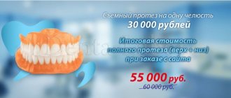
The price for complete restoration of teeth on 1 jaw in 3 days is 265,000 rubles.
Expert opinion
Emir Romanovich Omerelli
Maxillofacial surgeon, implantologist
Experience: more than 13 years
It has been proven that maximum atrophy of bone tissue occurs in the first year after tooth loss, and begins already three months after tooth loss, so you cannot delay dental prosthetics. The fact is that the lack of just one tooth in the jaw often causes quite serious problems. Each tooth performs its own function. The absence of even one tooth leads to disruption of chewing function, and can cause diseases or exacerbations of diseases of the digestive system, periodontitis, traumatic occlusion, displacement of the remaining teeth into free space, aesthetic defects, changes in speech, deterioration of the condition of the teeth, making subsequent prosthetics difficult.
Atrophy of the alveolar process of the upper jaw and parts is a natural process that occurs as a result of tooth loss.
Most often, bone loss occurs within a few weeks after tooth extraction. The blood clot that forms in the hole after tooth extraction often fills it completely. It may be lost as a result of pushing out by the tongue, as well as infection with subsequent development of suppuration.
Types of alveolar bone defects and treatment options
There are three topographic forms of alveolar process atrophy:
- single (loss of bone tissue within one tooth);
- segmental (within several missing teeth);
- complete (of the alveolar process of the jaw as a whole).
Seibert (1983) divided alveolar bone defects into three classes:
- Class I. Bone deficiency in the buccolingual plane with normal thickness of the bone margin in the apical-coronal plane.
- Class II. Deficiency of bone tissue in the apical-coronal plane with normal thickness of the bone edge in the bucco-lingual plane.
- Class III. A combination of buccolingual and apical-coronal tissue deficiency, leading to loss of normal thickness and width of the bone margin.
Evers et al (2010) proposed a new classification of methods for increasing bone volume, which depends on the degree of its vascularization:
- Class I - free bone grafts with microanastomosis;
- Class II - distraction osteogenesis method;
- Class III - use of a pedicle flap;
- Class IV - morphogenetic induction bone grafts;
- Class V - non-vascularized bone grafts.
Nicolas Caplanis in 2009 proposed a new classification of bone defects, which allows for a qualitative clinical assessment of the defect immediately after tooth root removal and to determine recommendations for further orthopedic rehabilitation using dental implantation.
The Caplanis classification distinguishes 4 types of defects:
- type 1 is characterized as a clean socket of an extracted single-rooted tooth with unaffected socket walls;
- type 2 refers to minor destruction of the alveolar ridge and loss of septal bone tissue of no more than 2 mm;
- type 3 is characterized by loss of soft tissue and bone from 3 to 5 mm, accompanied by the destruction of one or two bony walls of the socket;
- type 4 is accompanied by soft tissue injury with a loss of more than 5 mm.
The success or failure of orthopedic treatment largely depends on the volume of the alveolar process. A lack of width makes it difficult to carry out dental implantation or makes it impossible to carry it out without additional measures.
Adaptation of a patient to orthopedic structures with significant atrophy of the jaws due to their unsatisfactory fixation is associated with serious problems, and sometimes may not occur at all.
Until recently, such patients were repeatedly subjected to orthopedic treatment, and sometimes this entire cycle was unsuccessful. But currently, in such cases, reconstruction of the atrophied alveolar process is recommended.
Indications for surgical interventions to increase bone volume:
- preparing the oral cavity for orthopedic treatment;
- adaptation of the atrophied alveolar ridge to the orthopedic structure;
- planning subsequent dental implantation;
- replacement of bone tissue defects that formed after the removal of benign tumors or impacted teeth;
- raising the floor of the maxillary sinus;
- injuries of the facial bones in patients with osteomyelitis of the lower jaw;
- fractures of the bones of the middle and upper zone of the face;
- post-traumatic deformities of the bones of the facial skeleton.
Methods for increasing bone volume
Horizontal and/or vertical augmentation of the alveolar process can be performed immediately after tooth extraction or after a certain period of time.
For each surgical treatment method, different techniques and materials are used, separately and in combination. The ultimate goal of treatment is to restore the functional and aesthetic dentition, taking into account the characteristics of the clinical case.
The operation of raising the maxillary sinus (sinus lift elevation, sinus lift) makes orthopedic treatment possible using intraosseous implants in the lateral area of the upper jaw.
The sinus lift procedure was first performed in 1975 by Dr. Hilt Tatum, and the first comprehensive scientific description of the method was carried out by Boyne and James in 1980.
Yamurkova N.F. et al. (2015) note in their studies that volumetric reconstruction using the developed methods of surgical treatment of severe atrophy of the alveolar process of the upper jaw ensures the creation of sufficient parameters for the installation of dental implants and orthopedic restoration of chewing function.
Among these methods, the authors highlight plastic surgery using an L-shaped autograft, reconstruction of defects using local bone tissue (sandwich plastic method, sliding bone fragment method, intercortical osteotomy method and splitting bone tissue in the defect area).
These authors have proven the effectiveness of using in clinical practice three developed methods for plastic closure of the Schneiderian membrane, which are used during open sinus lift surgery.
The use of these techniques makes it possible to isolate defects in the mucous membrane of the maxillary sinus and continue vertical augmentation.
Razmyslov A.V. in his study (2011) indicated that the use of osteoplastic materials in dental practice enhances osteogenesis and allows the creation of a bone matrix of new bone tissue of optimal density in a period of 6 to 12 months.
When the size of the alveolar process was increased with an autogenous bone graft, a tendency to recovery was noted in the period from 3 to 6 months.
An increase in the alveolar process of the upper jaw (sinus lift) with the complex use of osteoplastic materials, resorbable membranes and autogenous bone filings is characterized by an early and significant increase in the density of the formed bone tissue.
In other works, Kozlova L. (2015) suggests, for atrophy of the alveolar processes of the jaws with a decrease in bone density, complex anti-osteoporotic therapy with bisphosphonates in combination with calcium and vitamin D3 preparations, which helps to normalize the architecture of trabecular bone.
To predict the effectiveness of orthopedic treatment of patients with partial and complete edentia, a clinical diagnostic algorithm is used, which includes:
- study of the microarchitecture of bone tissue according to cone-beam computed tomography data;
- detection of crystallization of the patient’s saliva to assess possible systemic disorders of bone remodeling;
- densitometric determination of bone mineral density.
Meltzer (1979) first published a clinical report on the use of soft tissue flaps exclusively for aesthetic correction of vertical marginal defects. The method is performed by forming a free gingival flap, with the help of which an increase in the thickness of the soft tissue of the alveolar process is achieved.
Garber and Rosenberg (1981) developed a technique for correcting horizontal bone deficiency by using a connective tissue graft and placing it under the epithelial surface, which in turn stabilizes and increases the volume of the alveolar ridge.
In 1985, Allen et al., analyzing the results of early studies, developed an improved surgical technique for local marginal defects. It consists of creating a mucoperiosteal flap using two vertical incisions, which are connected by a horizontal one.
Next, the flap is separated from the bone tissue, and the resulting cavity is filled with one of the hydroxyapatite or graphite-based allomaterials. After this, the cavity is sutured.
Shcherchkov S.V. (2013) to increase the effectiveness of implant treatment of patients with alveolar process atrophy, proposed a modification of the method of autogenous bone grafting similar to the veneer technique - through osteoperforation of the autograft and the recipient site.
This modification with delayed installation of dental implants allows for orthopedic treatment to be carried out within a period of up to 8 months, and the technique is indicated for bone widths of at least 3 mm and cortical thickness of no more than 1.5 mm.
With the immediate installation of dental implants, orthopedic treatment can be carried out after 5 months, the important condition being that the bone width is more than 3.5 mm and the thickness of the cortical layer is no more than 1 mm.
G.A. Ilizarov developed the method of distraction osteogenesis in the late 1960s. The first attempts to use this principle for vertical bone distraction in maxillofacial surgery were made about 15 years ago.
Distraction osteogenesis does not require obtaining a bone graft, suturing the donor site, or performing additional surgery to correct the mucogingival attachment, therefore this method is associated with fewer postoperative complications compared to other regenerative interventions.
However, when using this method in long-term studies, bone resorption was observed after vertical callus distraction. Another disadvantage is the possibility of exclusively vertical regeneration.
When simultaneously increasing the height and width of the bone crest, additional bone block transplantation or directed bone regeneration is required.
The sandwich osteotomy method has become an alternative to distraction osteogenesis.
With this technique, the mobilized bone fragment is fixed in the desired position with plates for osteosynthesis, and the gaps are filled with autogenous bone chips. This method does not require the installation of extrawell distractors.
However, the relatively dense mucous membrane of the palate limits the capabilities of this technique. This method allows you to increase the height of the alveolar process by a maximum of 5 mm, while minimally increasing the width of the bone.
Gerasimenko A.V. et al. (2013) proposed a technique for augmentation of the alveolar process by subperiosteal injection of osteoplastic materials.
In this technique, blood is drawn from a patient's vein, after which the blood plasma is enriched with platelets, thus obtaining platelet-rich plasma.
The latter, using an injection needle, is injected subperiosteally from the vestibular side in the projection of the tooth, infiltrating the area of deformation of the alveolar process.
To enhance the result, after 10-15 minutes, a suspension of powdered bone material in an isotonic sodium chloride solution is injected subperiosteally into the tissue of the same area.
The literature describes a method of augmentation of the alveolar process in difficult anatomical conditions in the area of the chewing teeth of the upper jaw, proposed by D.S. Avetikov. et al (2014).
It involves taking a bone autoblock of the desired shape and volume from the anterior surface of the body of the upper jaw using a piezo scalpel. Then the bone fragment is fixed with screws to the vestibular surface of the alveolar process of the upper jaw, and the interosseous spaces are filled with autologous and xenogeneic bone chips and stabilized with a collagen membrane.
Researcher Malanchuk V.A. (2002) proposed an alternative treatment method.
After tooth extraction, the mucoperiosteal flap of the alveolar process is detached from the vestibular side with mobilization of the vestibular mucoperiosteal flap. Using a dental bur, the cortical layer of the side walls and top of the tooth socket is additionally removed, revealing the bone marrow spaces, leaving shavings of the cortical layer in the socket and suturing it.
Open and closed sinus lift
The closed sinus lift procedure is carried out by separating the periosteum, after which a pilot canal is formed in the area of the missing tooth without perforating the bottom of the cavity with a cutter. Next, carefully continue preparation using a cutter with depth marks until contact with the bottom of the cavity is felt.
After this, a bed for the dental implant is formed. The last stage consists of perforating the floor of the paranasal sinuses, leaving the mucous membrane of the maxillary sinus intact. Finally, mobilization and elevation of the shell are performed and a resorbable membrane and bone material are introduced through the formed bone bed, the length of the corresponding implant is measured and it is installed.
The open sinus lift procedure is recommended when the residual bone height is up to 3-4 mm. The incision configuration depends on the prosthetic planning.
If you plan to make a fixed bridge structure, the incision must be shifted from the center of the alveolar process by 2 mm to the palatal side. If a removable structure is planned in the future, then the incision is made in the center of the alveolar process.
Next, the mucoperiosteal flap is separated. After exposing the lateral bone wall, corner points are applied, connecting them with a diamond bur to form a bone window. Through the window, the mucous membrane of the maxillary sinus is mobilized and lifted and bone material is introduced, after which the wound is sutured.
The double window technique is an open sinus lift technique.
The indication for the use of this technique is an enlargement of the maxillary sinus, which usually occurs with prolonged absence of teeth. The bone partition created with this method between the two windows corresponds in its location to the zygomaticalveolar ridge.
With the double window technique, the maxillary sinus mucosa can be mobilized from both the medial and distal windows, after which bone material is injected.
Raising the floor of the nasal cavity is used before installing dental implants, in case of insufficiency of bone tissue in the frontal area of the upper jaw, or in order to prevent its perforation.
The mucous membrane of the bottom of the nasal cavity is thicker, in contrast to the mucous membrane of the maxillary sinus, and has a stronger fixation to the bone.
This procedure is carried out through a formed bone canal for installation of a dental implant. The mucous membrane of the bottom of the nasal cavity can be separated, protecting it from perforation during the formation of the implant bed.
Kasiyanchuk M.V. (2009) proposed a method of performing a combined sinus lift, involving trepanation, preparation, lift and peeling.
In this case, trepanation is carried out slit-like along the alveolar ridge. The preparation is carried out separately along the vestibular and palatal walls. The lift is performed in an open way, detachment of the Schneiderian membrane from the bone walls is done in a closed way, after which it is performed with osteotropic material and a membrane.
Sennikov A.N. and co-authors (2009), when performing a sinus lift, propose preparing the anterolateral wall of the maxillary sinus with mutually intersecting lines that connect along the perimeter of the window to form a perforation hole.
The depth of the cuts is determined visually when the mucous membrane of the maxillary sinus appears adjacent to the bone tissue, and the peeling of the mucoperiosteal flap is carried out only along the periphery of this opening.
Research Kekukh E.O. (2013) are focused on the use of endoscopic sinus lift, which can reduce surgical trauma to soft and bone tissues, and reduce the recovery time for patients after surgery
Endoscopic sinus lift eliminates many of the disadvantages inherent in the traditional procedure on the maxillary sinus, prevents scarring of the mucous membrane, reduces blood loss and preserves local blood microcirculation.
This method involves restoring the integrity of the oral mucosa with the immediate elimination of the defect in the bone tissue of the alveolar process and the creation of the necessary volume of bone tissue for the subsequent rehabilitation of patients using delayed dental implantation.
Modern clinical, endoscopic and radiological research methods, as well as analyzes of long-term results of surgical treatment of patients using this method prove the effectiveness of endoscopic sinus lift.
Conclusions on the treatment of alveolar ridge atrophy
The above literature review allows us to draw the following conclusions:
- The data obtained as a result of a systematic retrospective analysis do not provide an adequate evidence base for identifying clear advantages of one method of surgical treatment of atrophy of the alveolar process of the maxilla over alternative methods.
- Each of the above-mentioned techniques should be selected according to the anatomical characteristics of the patient. The scope of surgical interventions depends on the severity of jaw bone atrophy. It is especially important to obtain research results in combination with concomitant diseases.
- To obtain reliable results, prospective clinical studies are needed, especially among postmenopausal patients, who account for a significant portion of the known anatomical and functional disorders.
Taking into account the results of studies in the future may make it possible to adjust the process of bone restoration during surgical treatment of alveolar atrophy and significantly increase its predictability.
Manifestations of bone atrophy
The patient himself, as a rule, does not notice the initial manifestations of jaw atrophy. He encounters this pathology only when the question of dental prosthetics arises.
Classification of jaw atrophy
The most widely used classifications are Schroeder for the upper edentulous jaw and Keller for the lower edentulous jaw. Schroeder distinguishes three types of upper edentulous jaw:
- The first type is characterized by a high alveolar process, which is evenly covered with a dense mucous membrane, well-defined puffs, a deep palate, and the absence or weakly defined palatine ridge (torus).
- The second type is distinguished by an average degree of atrophy of the alveolar process, mildly expressed tubercles, average depth of the palate, and a pronounced torus.
- The third type is the complete absence of the alveolar process, sharply reduced dimensions of the body of the upper jaw, poorly developed alveolar tubercles, a flat palate, and a wide torus. In terms of prosthetics, the first type of toothless upper jaws is most favorable.
Keller distinguishes four types of edentulous mandibles:
- The first type is a jaw with a clearly defined alveolar part, the transitional fold is located far from the alveolar ridge.
- The second type is a uniform, sharp atrophy of the alveolar part, the mobile mucous membrane is located almost at the level of the alveolar ridge.
- The third type - the alveolar part is well defined in the area of the anterior teeth and sharply atrophied in the chewing area.
- The fourth type - the alveolar part is sharply atrophied in the area of the anterior teeth and is well expressed in the chewing area.
With regard to dental prosthetics, the most favorable are the first and third types of toothless mandibles.
Modern techniques of bone grafting and plastic surgery create conditions for prosthetics and dental implantation in cases of existing bone tissue atrophy
Classification of the mucous membrane according to Supple
One of the popular and universal ones in cases of radical absence of teeth is the well-known classification according to Supple, which is focused on the condition of the mucous surface of the prosthetic bed. Within the classification, there are the following categories of compliance:
- The ridges of the alveoli are lined with the pliable mucous membrane of the prosthetic bed.
The palate is equally covered with mucous membrane. Physiological folds of the mucosa are distant from the apex of the alveolar ridge. This category of mucosal compliance is suitable for a prosthesis made of any material.
- The atrophied mucous surface lines all the ridges of the alveoli and the palate.
The slightly thinned shell has some inconveniences for installing structures with a metal base.
- The mandibular structures and hard palate are lined with an uneven mucous membrane.
Patients quite regularly require additional treatment.
- The strands of the mucosal surface are mobile and move with minimal pressure.
In such a situation, using a prosthesis will be uncomfortable. Prosthetics for such pathology are possible only as a result of excision of the alveolar edge.
The classification naturally confirms the medical assumption that when studying the condition of the mucous membrane in each specific case, it is important to take into account the patient’s physique and general health.
Questions and answers
Is it possible to install a removable denture on implants if 5 teeth are missing in a row, and the outer one on one side is also missing.
How strong will it hold if the bone atrophies? Are they mobile when chewing? Life time? Will the diction change? Is it possible to install a removable denture on implants if 5 teeth are missing in a row, and the outer one on one side is also missing. How strong will it hold if the bone atrophies? Are they mobile when chewing? Life time? Will the diction change?
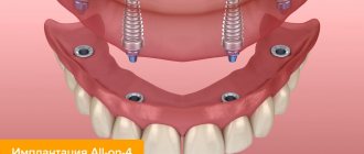
Hello! Theoretically, such prosthetics are possible, but the cost of such a design will not be much less than a fixed bridge on implants, while the service life is significantly shorter. The average service life of removable dentures is no more than 5 years; accordingly, every 3-5 years such a denture requires replacement, and even more often it may require adjustment. Removable dentures attached to implants are motionless during chewing; they are held quite securely. However, if you have bone tissue atrophy, it is necessary to conduct a diagnosis to understand whether installation of implants is possible in your case, and if so, what kind of implants can be used. Diction disorders can be observed, as a rule, in patients who require restoration of the front teeth or the entire dentition. An orthopedic doctor will tell you after an initial examination whether such an effect is possible in your case. The best option in terms of price-quality ratio when restoring several teeth in a row is basal dental implantation, in which a fixed prosthesis is installed already 3-4 days after implantation. In addition, this method is suitable for patients with bone atrophy. You can obtain more detailed information about the possibilities of implantation and prosthetics in your case from our doctors following a free consultation. You can make an appointment by calling + 7 (495) 789-42-02. Sincerely, Patient Support Center SIMPLADENT+ 7 (495) 789-42-02+8 800 333-53-41
3 years ago I had implants placed on my lower jaw and the remaining teeth were removed. And in March, all the implants were removed. The result is changes in bone tissue and flat gums. Is it possible to install a flexible prosthesis there? On top of my head there is a removable denture
3 years ago I had implants placed on my lower jaw and the remaining teeth were removed. And in March, all the implants were removed. The result is changes in bone tissue and flat gums. Is it possible to install a flexible prosthesis there? On top of my head there is a removable denture
Hello! Prosthetics with soft dentures in such cases is possible, but a preliminary examination of the oral cavity is necessary. Come visit us for a free consultation. Based on the results of the examination, our orthopedists will offer you the best options for dental prosthetics in your case. You can make an appointment by calling + 7 (495) 789-42-02. Sincerely, Patient Support Center SIMPLADENT++8 800 333-53-41
Other questions
Other jobs
Degree of expression
Pathology is expressed in three degrees - mild, moderate and severe. The lung is characterized by thickening of the mucous membrane and pronounced bumps. At this stage, prosthetics are possible, since there is enough bone tissue to install the structure.
The middle stage is expressed in the form of thinning tissue and a decrease in the size of the hole. Progression of atrophy is observed; special preparation is required for prosthetics.
The acute stage is characterized by complete atrophy, that is, the alveolar process is absent. There are no tubercles, there is a narrowing of the jaw bed.