From this article you will learn:
- what is aesthetic dental restoration,
- how it is carried out, patient reviews,
- price for restoration of front teeth in Moscow.
Artistic dental restoration is an aesthetic method of restoring damaged teeth, which gives them a natural appearance by simulating dental tissues with various materials. This method differs from conventional tooth filling in that in this case it is not just about restoring the shape and function of a damaged tooth, but when the doctor tries to achieve high aesthetics (so that the restored tooth cannot be distinguished from a healthy one). In dentistry, the terms aesthetic or cosmetic can also be used to refer to this type of restoration.
The artistic restoration method is more expensive compared to conventional traditional dental fillings, which has its justification. To achieve good aesthetics, it is important to restore not only the shape of the tooth, but also its color and transparency. Restoring the latter is the most difficult, because... Human teeth have different shades and transparency all the way from the cutting edge to the neck of the tooth. Therefore, making a tooth natural in color and transparency, as well as indistinguishable from neighboring teeth, requires not only skill and more expensive filling materials, but also a lot of time.
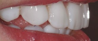
Dental restoration: photo (restoration of the 14th tooth, i.e. the first premolar of the upper jaw)
It should be clarified that the method of artistic restoration is used not only to restore teeth destroyed by caries, but also for congenital or acquired changes in the color of the enamel, age-related abrasion of teeth, chipping of the incisal edge, as well as for changing the unsightly shape of a tooth or moderate correction of its inclination. An alternative to the aesthetic restoration of teeth with filling materials can be other methods of cosmetic dentistry - ceramic and metal-ceramic crowns, as well as veneers.
Methods of aesthetic dental restoration –
Probably, as you already understood, this article will talk about “direct restorations,” i.e. when tooth restoration is carried out using filling materials. However, there are also so-called “indirect restorations” (see below).
- Direct dental restoration – also known as dental restoration using light-curing composite material (i.e., photopolymers), and is done by dental therapists. It is this type of restoration that we will examine in detail in our article. This method also includes restoration of teeth with composite veneers, more about which you can read in the article at this link.
- Indirect dental restoration - in the manufacture of this type of restoration, an orthopedic dentist (prosthetist) takes part, as well as a dental technician sitting in a dental laboratory.
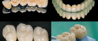
This includes prosthetics with ceramic veneers, metal-ceramic ceramic crowns, restorative ceramic inlays. In this case, the tooth is first prepared and then an impression is taken of it. Based on this impression, an inlay, veneer or crown is made in a dental laboratory, and then fixed to the teeth using cements. For indirect restoration methods, mainly only ceramics are used - this can be Emax, zirconium dioxide, feldspathic ceramics. If we are talking about aesthetic restoration with metal-ceramic crowns, then for their metal frame they use not an ordinary cobalt-chromium alloy, but alloys of precious metals (for example, gold-palladium). Alloys containing gold give metal-ceramic crowns a more natural shade. Read more about indirect restoration methods in the articles:
→ Restoration with ceramic veneers → Restoration with metal-ceramics → Restoration with ceramic crowns
Example 3
| Before orthodontic treatment | After orthodontic treatment |
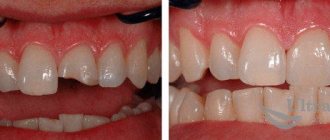
Here, orthodontic treatment with braces has significantly improved the appearance of the teeth, but when everything is “not very good,” then little things do not attract attention. When the teeth were aligned, the uneven edges of the teeth became very noticeable (1), and the asymmetrical corner of the second incisor also stood out (2). Since this problem is easily solved, and the necessary specialist is in the same clinic, then why leave any imperfections? A comprehensive approach to dental aesthetics is necessary for the best results.
This is what happened as a result of the work of an aesthetic dentist after orthodontic treatment:
In this case, laser teeth whitening and restoration of the front teeth with light-curing material Estelite (Japan) were performed.
Restoration of anterior teeth using composite filling materials –
In this chapter we will talk about how artistic restoration of the front teeth is carried out using specific examples - 1) in the first video you will see the replacement of an old, darkened restoration of a front tooth with a new one, 2) in the second video the restoration of an extensive chipped front tooth is shown. These videos perfectly demonstrate how artistic aesthetic dental restoration differs from conventional tooth filling.
For example, a special cast is used here to accurately restore the palatal surface of the tooth, as well as a layered restoration technique, which allows, by combining filling materials of different shades, to obtain an aesthetic result that is close to ideal (so that the restored part of the tooth does not differ in color and transparency from the natural tooth tissues). In fact, not all dentists have such skills, and there is no doubt that a certain artistic talent is needed.
Restoration of anterior teeth: video
The main stages of dental restoration:
- Preparation for restoration - at this stage it is necessary to hygienically clean the teeth from plaque and tartar, determine the color of the tooth being restored using a special scale and, in accordance with this, select the colors of the composite filling materials that will be used in the restoration process.
- Local anesthesia (if necessary)
- Drilling out tissues affected by caries - if an old unsatisfactory-looking restoration is being replaced, then the old filling is drilled out.
- Isolating a tooth from saliva - the use of cotton balls is almost universally a thing of history.
Their use does not allow reliably isolating the tooth from saliva, as well as from the patient’s wet breath, which also significantly worsens the quality and reliability of fillings and restorations made of composite filling materials. Now a rubber dam is used for this purpose. It is a latex scarf with holes for the teeth, which is pulled over the teeth. Composite dental restoration performed without reliable isolation from saliva and wet breath may not last long. Firstly, excessive moisture will cause a violation of the marginal seal of the filling to the tooth tissue, which can lead to the appearance of a dark strip or caries at the filling/tooth interface. Secondly, your restoration may simply fall out at one point, because... moisture impairs the adhesion of filling material to tooth tissue. - Fixing the pin in the canal - this stage is carried out if the crown of the tooth is destroyed by more than 1/2 and the tooth is pulpless. In this case, it is necessary to strengthen the filling by installing a pin, otherwise the restoration may fall out under load (24stoma.ru).
- Restoring the shape of a tooth with filling material – to make the tooth look natural, the layered restoration technique is used. In this case, layers of filling material of different shades and different transparency are applied to each other, which together then give the tooth a natural appearance. And this is real art.
- Finishing of the tooth – finishing should be understood as the final modeling of the tooth shape using burs, as well as grinding and polishing of the filling.
Price
Prices for aesthetic
dental restoration are based on several indicators:
- Status of the clinic and qualifications of specialists.
- Scope of dental crown restoration (full, partial, chipped restoration).
- Anesthesia depending on the drug.
- Rubber dam applications.
- Pin fixation, depending on the material and manufacturer.
- Consumables, including a sterile kit (gloves, mask, shoe covers, bib).
- Procedures for preparing a tooth for installation of a veneer (shaping the tooth, taking an impression, making a veneer).
| Service | Price, rub.) |
| Restoration of a chipped incisal edge of a tooth | from 1600 |
| Restoration of ½ tooth | from 3500 |
| Complete restoration of the crown with installation of a fiberglass pin | from 5000 |
| Complete restoration of the crown part of the tooth on a metal pin | from 4000 |
| Anesthesia | from 200 |
| Application of rubber dam | from 350 |
| Sterile kit | from 100 |
| Composite veneer | from 3000 |
| Ceramic veneer | from 12000 |
Sources:
- https://dentoland.com/protezirovanie/restavraciya-perednix-zubov.html
- https://Zuub.ru/plombirovanie-i-restavraciya-zuba-glavnye-otlichiya.html
- https://protezi-zubov.ru/restavracija-zubov/jesteticheskaja-restavracija-zubov.html
- https://protezi-zubov.ru/restavracija-zubov/
- https://DentConsult.ru/front-banner/metody-restavracii-perednih-zubov.html
- https://expertdent.net/lechenie-zubov/restavratsiya-zubov/restavratsiya-perednih-zubov-plombirovochnyim-materialom.html
- https://www.StartSmile.ru/esteticheskaya-stomatologiya/restavraciya-zubov/
- https://anZub.ru/lechenie-zubov/plombirovanie-perednikh-zubov/
- https://stoma.guru/lechenie/esteticheskaya-restavraciya-perednih-zubov-vidy-i-preimuschestva-cena.html
- https://infozub.ru/protezirovanie/restavraciya-zubov.htm
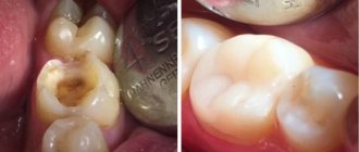
Restoration of chewing teeth –
In videos 3 and 4 you can see how the restoration of chewing teeth is carried out with composite filling material. In video 3, the restoration can hardly be called “artistic”, because... The chewing surface of the tooth with all its fissures was restored only “satisfactorily”. However, this video does a good job of showing the restoration of the side walls of the tooth and the contact points between the teeth. Video 4 shows the final stage of a truly artistic restoration.
How to restore a beautiful smile?
The front teeth include six upper and six lower teeth. Our dentists use all possible methods for their restoration:
- Ceramic or composite inlays,
- Veneers are permanent “onlays” that are attached directly to an imperfect tooth.
- Restoration by filling is called aesthetic or artistic. Photopolymer fillings are used.
Only your attending physician should choose between the presented methods, since each procedure has its own characteristics and contraindications. In addition to the chosen technique, special attention should be paid to the material. The maximum resemblance to real enamel will depend on this.
With the help of artistic restoration, you can restore the front teeth and canines so that no one can distinguish the restored tooth elements from their own, except the dentist.
Dental restoration: prices in clinics
For dental restoration, the price in different dental clinics will vary greatly, but truly high-quality aesthetic work in this case definitely cannot be cheap. To obtain a close to ideal aesthetic result, you need an experienced dentist with certain skills, training for which costs a lot of money, more expensive filling materials are also required, as well as a significant investment of time. All this increases the cost of the service.
When choosing a doctor, it is not enough to rely on reviews - ask to see before and after photos (of real patients treated in the clinic). It’s usually not worth relying on photographs on clinic websites, because... Most dental clinic websites do not post their own work, but pictures copied from the Internet. Below we will calculate the cost of dental restoration in a mid-price clinic (prices in premium segment clinics can be 2 or more times more expensive).
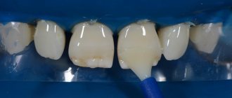
1) Complete restoration of the tooth crown –
In photos 7-9 you can see a real case of artistic restoration of the front teeth, performed with the crown part completely missing. When restoring tooth crowns, fiberglass pins and a composite light-curing filling material were used. For such dental restoration the cost will be no less than 6,000 rubles per tooth.
Restoration of anterior teeth: before and after photos
The cost of a complete tooth restoration consists of the following items.
- Anesthesia – from 400 rub.
- Applying a rubber dam to isolate a tooth from saliva – from 700 rubles.
- Sterile kit – from 150 rub. this includes: a mask and gloves for the doctor, shoe covers and a bib for the patient, the cost of sterilizing the instrument.
- Restoring a crown from a light composite costs about 3,500-5,000 rubles.
- Fixation of a WDW fiberglass pin in the channel – RUB 1,500.
this is a very important stage (Fig. 8)! The pin is necessary to prevent premature breakage of the restored tooth crown due to the fact that it will experience significant mechanical stress when chewing and biting off food. For the restoration of front teeth, it is very important that the pin is fiberglass and not metal - otherwise the metal will show through the filling material and the crown of the tooth will be bluish in color. Of course, you can install cheaper pins, for example: a) a metal pin (cost 50 rubles), but in this case, the restored tooth will not have the same transparency and natural shade “like natural teeth.” Although for teeth that are not included in the smile line, this is not so scary. b) Russian-made fiberglass pin (cost 50 rubles), but such pins also do not have proper light conductivity, and in addition have insufficient adhesion (stickiness) to filling materials. This leads to their poor fixation in the root canals and the risk of the “extended” tooth falling out along with such a pin. Total: the cost of tooth restoration is from 5,000 to 7,000 rubles.
2) Restoration of half a tooth –
If a full crown restoration (using a fiberglass pin) costs 5-7 thousand rubles, then restoring half a tooth crown without a fiberglass pin, but taking into account all other costs, will cost approximately 1500-2000 rubles less. Total: from 3500 to 4000 rubles.
3) Restoring a chipped cutting edge of a front tooth –
Restoring the angle of a tooth crown using a light-polymer filling material will cost from 1,500 to 3,000 rubles (depending on the amount of work/size of the chip).
Incisal edge correction for optimal function and esthetics
Problems with which a patient seeks help from a dentist can sometimes turn out to be a very difficult clinical task for the doctor to solve. For example, sometimes a patient may think that longer teeth will help them achieve a more youthful appearance, but the dentist actually needs to spend a huge amount of time planning such interventions. Such situations determine all the skills of a dentist. After all, by carrying out a simple and thoughtless correction of the incisal edge of the anterior teeth of the upper jaw, the doctor can provoke the development of a number of unpredictable aesthetic, functional and phonetic consequences that will have to be faced in the future. Correcting an inappropriate position of the cutting edge is not different, perhaps, at first glance, less, but also not reasoned - this is, simply put, “the path to nowhere.” Determining the proper incisal position is primarily the responsibility of the dentist, who must also communicate closely with his technician to achieve a truly clinically successful result. During this process, both the technician and the doctor are greatly assisted by temporary restorations.
Planning for Functional Success
Sometimes a complex clinical problem can be solved simply by breaking it down into a series of successive and less complex steps. The predicted treatment outcome is the outcome of correctly conducted diagnostic and planning processes, in which the doctor and the patient himself participate. Before starting dental intervention, in addition to the anamnesis, the doctor should also take clinical photographs of the initial situation of the dental status, obtain diagnostic impressions, radiography data, periodontal examination, facial arch analysis, bite registers in the centric relation and maximum fissure-tubercle contact. It is mandatory to study the area of the temporomandibular joint, which largely determines the entire function of the maxillodental apparatus. It is extremely important that the dentist understands the importance of the centric relation in the process of orthopedic treatment, because if the role of the CS is not fully understood, the treatment itself is doomed to functional failure. Clinical cases involving correction in the area of the anterior teeth are characterized by a certain level of occlusal instability. That is why, before starting any prosthetic interventions, it is necessary to examine the joint area for the presence of hidden pathologies that limit the possibility of achieving CV.
Photographs are another important tool in treatment planning. When reconstructing an incisal area, it is critical to obtain the following six types of photographs:
- Full face photo (examples: photo 1, photo 14, photo 15 and photo 31)
- Photo of a smile in a 1:3 ratio (examples: photo 3 and photo 20)
- Photo of a smile from top to bottom in a 1:3 ratio (example: photo 16)
- Photo at rest in a 1:3 ratio (examples: photo 6 and photo 18)
- Photo of a smile from the side at an angle of 90° in a ratio of 1:3 (photo: Fig. 17)
- Photo of a smile when pronouncing the sound “I” in a ratio of 1:3 (example: photo 19)
Factors affecting the position of the incisal edge
The envelope curve, which is formed during the function of the anterior teeth, largely determines the position of the cutting edge, which, in turn, is an extremely important component of the anterior route of insertion. The combination of lingual contours, position and inclination of the maxillary anterior teeth influences the harmonious relationship of the anterior guidance path and the envelope function curve. Incorrect positioning of the incisal edges may provide an adequate anterior insertion path but disrupt the normal curve of incisal function.
The neutral zone is a complex of soft tissues and muscles surrounding the oral cavity that counteract the pressure of the tongue. Unreasonable prosthetic manipulations for the purpose of correcting the incisal edge, violating the neutral zone, provoke the development of a feeling of discomfort in the patient and the development of various types of compensatory reactions. During treatment, the physician must always consider the path of lip closure and the relationship between the teeth and the masticatory muscles that determine the movement of the lower jaw. An adequate relationship between the lower lip and incisal edges ensures the patient's normal phonetic function, while an imbalance between them already alters the pronunciation of certain sounds.
Determining the length of the incisors
Vertical position of the cutting edge
Often the upper incisors are the very first starting point from which the rehabilitation of the smile profile begins. In this case, the doctor should take into account all the parameters of the smile at the planning stages, because ignoring the existing features of the position of the incisors can provoke the development of further asymmetry, imbalance and function of the front teeth. The final length of the front teeth depends on the influence of many factors, including the position of the lower lip at rest and smiling, the volumetric characteristics of the upper lip, the condition of the soft tissue of the gums surrounding the teeth, the nature of the envelope curve of the incisors during function, and, of course, the proportions of the patient’s face. It is also necessary not to forget about the need to maintain a certain percentage ratio between the length and width of the teeth, which are interdependent. The acceptable ratio of width to length is about 80%, being a kind of golden ratio for teeth. Moreover, this indicator for the lateral incisors is 1, for the canines – 0.6, and for the central incisors – 1.6. These data are indicative only and should only be taken into account when analyzing the smile profile directly from the front. When changing the angle of the smile analysis, these numbers may change: thus, upon lateral examination of the canine, it becomes obvious that the ratio of crown width to length exactly exceeds 0.6.
It is quite difficult to determine the correct crown length in patients with signs of pathological wear. In such cases, the selection of length is carried out at the stage of provisional structures, to which the patient has to get used to. Studies devoted to determining the absolute length of teeth helped to establish that the average vertical value of the central incisor is 11.69 mm, while with pathological abrasion this average decreases to 10.67 mm. In photo 6 you can see the patient's smile at rest. The same position is achieved after the patient purses his lips with relaxed facial muscles, and then opens them slightly to be able to breathe through his mouth. According to Vig and Brundo, men in this position show an average of 1.91 mm of central incisors, and women - 3.4 mm. The level of tooth exposure at rest may decrease with age simply due to the effect of gravity on soft tissue. Photo 19 illustrates the position of the teeth in the so-called “E” position. This is the resting position created by the lips and muscles when the patient says "E". It is similar to the smile position, but the soft tissues are in a more functional position. Under such conditions, the cutting edge of the central incisor occupies 50-60% of the position between the upper and lower lips. The author of this article believes that the length of the central incisor of 10.5 mm can be considered acceptable for taking it into account when planning further dental treatment. In this case, the position of the cutting edge at rest and when pronouncing “E” allows you to plan the most suitable spatial position, which will be as harmonious as possible for the patient. However, when adding length to the cutter without justification to achieve 10.5 mm, problems begin in the envelope curve of the function and the neutral zone. In addition, phonetic changes may also be noted. The lower lip should interact with the cutting edge of the upper incisors in a relaxed and natural way, which is especially evident when pronouncing the sounds “V” and “F”. In this case, the contact of the lip with the cutting edge should be formed on the inner side of the red border of the lips and in no case on the skin. The last mistake is quite often observed with too long restorations of the anterior teeth of the upper jaw.
Determining the horizontal position of the cutting edge
Doctors often pay attention to the vertical position of the incisal edge, forgetting about the importance of the horizontal position of the coronal edge of the incisors. This position should also be consistent with the envelope curve of the incisors during function and the overall profile of the neutral zone. A too anterior position of the incisal edge can cause changes in the relationship with the lower lip, limiting the normal closure of the lips with each other. An overly labial position of the incisal edge pushes the labrum anteriorly and causes it to “work” around the displaced crown position in a most unnatural manner. Typically, patients in such cases complain of muscle fatigue and discomfort during function. Displacement of the cutting edge orally provokes anteroposterior restriction in the envelope curve of the function. As a result, when the jaw closes, a zone of interference will develop, concentrated precisely in the area of the cutting edge. Additionally, phonetic changes in the pronunciation of the sounds “V” and “F” are also developing. In addition, the horizontal position of the cutting edge is extremely important for the formation of the “C” sound, since when pronouncing it, the air literally compresses and slips between the upper and lower incisors. According to the author's observations, errors of too vestibular positioning of the frontal teeth of the upper jaw are most often observed. Typically, such errors are caused by inadequate design of tooth preparation by underpreparing and reducing tissue in one plane, instead of three. When the middle and incisal thirds of the crown are underprepared, the dental technician has to model the design with excess volume of material, based on the existing conditions. Therefore, to control the reduction of hard tissues, it is necessary to use diagnostic wax keys, which allow preparation in the natural three planes of the tooth, ensuring the proper design of the stump (photo 26-27).
Teamwork with a ceramic technician and choice of restoration
A successful treatment outcome is the outcome of a team approach. Moreover, the responsibility for organizing such an approach lies with the dentist. The physician should provide the technician with all necessary information, including clinical photographs before and after preparation, as well as photographs of the fixed temporary structures. The key element of team interaction remains an adequately obtained impression. After trying on provisional crowns, the silicone key allows you to duplicate or adjust in a controlled manner the required position of the cutting edge. When working with provisional crowns, it is also important to connect the facebow. It allows for a three-dimensional transfer of the maxilla (including the position of the cutting edges) and the joint into the articulator. In addition, thanks to the face bow, it is possible to transfer the position of the condylar axis when closing the jaws in the structure of the overall treatment plan. This aspect is extremely important when recording the central relationship of the jaws. Without all this data, the technician simply has to guess which jaw position is optimal physiologically for a given patient. Another important aspect of interaction is the choice of orthopedic design. The dental technician is more deeply familiar with the advantages and disadvantages of each of the materials used, and the predictability of their use during aesthetic rehabilitation.
The author of this article takes into account the following six criteria when choosing a material for future aesthetic designs:
- aesthetic risk
How many teeth are visible when smiling? Is the cervical part of the teeth exposed? Is it necessary to cover the area of the black triangles? Is it possible to provide the most natural restoration of a patient’s smile, taking into account his wishes and existing risks?
- occlusal risk
What are the risks of restoration failure under normal function of the patient’s dentofacial apparatus and under conditions of dysfunction? Has the clinician monitored the joint position? Does the patient have signs of bruxism? These factors determine the choice of restoration based on its strength, because beautiful crowns, but with a high risk of fracture, are essentially not functionally significant.
- enamel quality
To ensure adequate adhesion of restorations, especially veneers, the structural health of the enamel is necessary. The deficiency of enamel justifies the choice of other fixation methods and a revision of the approach to treatment.
— quality of residual dentin
The quality of residual dentin is especially important in the area of distal teeth. Affected or sclerotic dentin provokes a decrease in adhesion strength. Therefore, it may be necessary to use alternative methods of fixation of prosthetic structures.
— the ability to ensure adequate isolation of the working field
Insufficient isolation of the working field compromises the quality of adhesion of the restoration. Therefore, the doctor must decide whether he can maintain the cleanliness of the working field for a long enough time and with high quality?
- desire to preserve the structure of the teeth
The use of different materials determines how much preparation must be done to ensure adequate strength and esthetics of the restorations. Preservation of the tooth's own structures should be a priority for targeted reduction of enamel and dentin. The doctor also needs to decide on a material that can satisfy both the strength and aesthetic criteria of the restoration.
Detecting errors in cutting edge position
At the planning stages, it is important to ensure not only the process of analyzing all parameters of the clinical situation, but also the detection of possible errors during diagnostic manipulations.
Errors of too vestibular position of the cutting edge
When the incisal edge is positioned too vestibular, the patient may complain of too long incisors, constant dryness and discomfort in the upper lip. In this case, sensations of overload and changes in the relationship with the upper lip may form in the lower lip. Patients become uncomfortable pronouncing the sounds “F” and “V”. In addition, problems with the “C” sound may occur, since the lower jaw will try to find the correct position with the upper incisors when pronouncing it. Thus, we can state the error of too vestibular position of the cutting edge.
Errors of too lingual position of the cutting edge
These types of errors are most often associated with unsuccessful restorations. If the positioning is too lingual, the first contact during the closure of the jaws is formed precisely in the area of the cutting edges of the incisors. This increases the risk of the restorations becoming chipped or debonded. The first such problems, which can be noted even at the stage of using provisional structures, should raise doubts among the dentist, and the need to review the formed position of the cutting edges and change them to a new, more optimal one. In addition, there is a lingual displacement of the lower lip and disturbances in the pronunciation of the sound “C”. To monitor the adequacy of positioning of the incisal edge, temporary structures can be successfully used. Patients often want to minimize the time they use temporary restorations and are in a rush to receive definitive crowns. But there is no need to rush either. The physician should explain to the patient all the risks and benefits of controlling the position of the incisal edge using provisional crowns, thus increasing the prognosis for obtaining a final successful treatment result.
Clinical case 1
This patient requested to improve the appearance of his smile (Figure 1). He was embarrassed about his diastemas, especially those that were visualized on the right side (photos 2-3). From an aesthetic point of view, his anterior teeth were characterized by an imbalance of width and length (photos 2-3), and the patient had previously undergone orthodontic treatment, as a result of which the best position of the incisors had already been achieved (photos 4-5).
Photo 1. Photo of the patient's entire face.
Photo 2 - 4. Photo of a smile in a ratio of 1:3.
Photo 5. Visualization of the diastema.
Evaluation of the photograph at rest allowed us to determine that the teeth were too long with vertical displacement of the incisal edges. The length of the central incisors exceeded 14 mm, which made them dominant in the smile profile. The horizontal position of the cutting edge of the teeth was within normal limits. The rehabilitation protocol using veneers was chosen as a treatment method. Taking into account the need to close diastemas, the preparation design involved reduction of the hard tissues of the anterior teeth not only from the vestibular, but also from the proximal sides. In this way, it was also possible to control the required length-to-width ratio (photo 7-8).
Photo 6. View of the patient's teeth at rest: signs of excessive visualization of the incisors.
Photos 7 - 8. View of prepared teeth: minimizing tissue reduction on the vestibular side.
The preparation depth on the vestibular side was approximately 0.3 mm in order to preserve the enamel layer as much as possible. On the cutting edge side, the preparation level was formed so that the length of the incisors after restoration was about 11.5 mm. The preparation margins between teeth 6 and 7 were positioned below the gingival level (Figure 9) to achieve the most natural-looking restoration possible. The treatment algorithm was planned in such a way as to avoid the effect of black triangles between the teeth. IPS Empress press-ceramic veneers (Ivoclar Vivadent) were used as restorations. This material is characterized by higher strength compared to glass ceramics and fairly high aesthetic values. As a result of the treatment, it was possible to correct the aesthetic profile of the smile with the closure of diastemas and normalization of the symmetry of the teeth (photos 10-14).
Photo 9. The finishing line of the preparation was positioned slightly below the level of the gums.
Photos 10 - 12. View of the patient’s smile after fixing the restorations. Correction of the ratio of tooth sizes, their axial inclination and closure of diastemas was carried out.
Photo 13. Adapted preparation design allowed for natural-looking restorations.
Photo 14. View of the patient’s face after dental treatment.
Clinical case 2
In some clinical cases, to correct a smile in the frontal area, it is necessary to carry out preparatory complex treatment in order to minimize possible aesthetic and functional risks in the future. In this clinical case, the patient was referred to our clinic to improve her smile, but at the same time her complaints also included the constant loss of crowns in the area of the 8th and 9th teeth (photo 15).
Photo 15. View of the patient before treatment: signs of a gummy smile.
In her youth, the patient underwent orthodontic treatment, which included the removal of maxillary premolars. Teeth 8 and 9 were endodontically treated after a sports injury. Also, after this injury, partial ankylosis of the upper central incisors was noted, which complicated the process of their movement during orthodontic treatment. The horizontal position of the cutting edges was too vestibular. This is clearly visualized in photographs taken from top to bottom (photo 16) and in profile photographs (photo 17).
Photo 16. Top-down view of a smile: the position of the cutting edges of the 8th and 9th teeth is too vestibular.
Photo 17. Lateral view of the position of the patient’s teeth: the position of the incisors is too vestibular.
During the conversation, the patient noted some discomfort, since the cutting edge of the upper teeth touched the skin of the lower lip. In the vertical plane, the position of the cutting edge was also too long. This symptom was noted at rest (photo 18) and at the so-called “E” position in the photograph (photo 19). The photo also revealed that the front teeth looked too short and square. The patient also had a gummy type of smile (photo 20).
Photo 18. View of teeth at rest: the ideal level of visualization should reach about 3.5 mm.
Photo 19. The appearance of a smile when pronouncing the sound “i”.
Photo 20. Photo of the patient’s smile before treatment in a 1:3 ratio: dominance of the incisors in the profile, incorrect position of the occlusal plane, displacement of the position of the gingival zeniths.
The solution to many aesthetic problems is to add a length parameter to problem teeth. However, in this clinical case, the length of the incisors was already too long. After recording the centric relation using the facebow and transferring these parameters into the semi-adjustable articulator, wax-up modeling of the future restorations was carried out (photos 21-22).
Photos 21 - 22. Formation of an optimal visual profile on a wax reproduction.
This approach allowed the periodontist to accurately assess how much soft tissue needed to be moved apically to achieve the desired smile harmony (Figures 23-25).
Photos 23 - 25. View of teeth after the crown lengthening procedure.
In addition, during the manufacture of provisional structures, the needs of reducing the size of the teeth themselves and their mesialization were taken into account. Thus, provisional structures have also become an important diagnostic tool. Particular attention was paid to the three-plane preparation of teeth from the vestibular side (photo 26). A new facial profile and adequate position of the incisal edge were created. To control the preparation using the wax models, silicone keys were obtained (photo 27).
Photo 26. Preparation of teeth in three planes in order to lingualize the position of the cutting edges.
Photo 27. Control of the volume of preparation using a silicone key obtained from a wax reproduction.
In this way, the doctor controlled how much tissue needed to be reduced in order to achieve adequate thickness for future restorations. All corrections were made on provisional restorations and an impression was obtained from them, from which the technician could confidently make the final crowns. Among other things, the problem was that the shade of endodontically treated teeth 8 and 9 was too dark (photo 28). The preparation depth reached 0.5 mm, which made it possible to adjust the shade of the teeth and at the same time provide the opportunity for the most conservative treatment. On teeth 6, 7, 10 and 11, lithium disilicate veneers were made from IPS e.max (Ivoclar Vivadent). To cover the gray shade of the crowns, maximally opaque ceramic blocks were chosen. To correct the shade of the stump in the area of 8 and 9 teeth, special Captek crowns (Captek) were fixed on top of them.
Photo 28. View after tooth preparation.
The aesthetic and functional success of the treatment was achieved through a thorough preliminary analysis of aesthetic needs and functionality, taking into account the specific vertical and horizontal positions of the incisal edge. At the same time, the volume of intervention was minimized, which allowed for maximum savings in the patient’s financial costs (photos 29-31).
Photo 29. View after fixing the restorations.
Photo 30. Captek crowns were used to mask discolorations. E-max veneers were fixed in the area of the lateral incisors.
Photo 31. View of the patient after dental treatment and correction of the position of the teeth in three planes.
conclusions
Clinical cases requiring correction of the incisal edge are quite complex from the point of view of an integrated approach to the treatment algorithm. Understanding the features of cutting edge positioning allows you to achieve the desired result even in particularly difficult clinical conditions. The predicted result of dental treatment is based on the use of modern materials, implementation of the principles of digital smile design and taking into account all the necessary aesthetic and functional criteria.
Author: Leonard A. Hess, DDS
Dental restoration: reviews
Very often, patients are dissatisfied with the way the restoration of the front tooth was performed (in terms of its shape, color and transparency).
Those. First of all, complaints are made specifically about insufficient aesthetics, and often the restored tooth simply looks like a real plastic crown, i.e. artificial and unnatural, standing out against the background of natural dental tissues. Examples of unsuccessful dental restorations: photos
Restoring aesthetics during restoration has 3 key points:
- Tooth shape - in fact, the work of restoring the anatomical shape of a tooth is very similar to the work of a sculptor who sculpts his model, but not from clay, but from filling material. Try to mold a tooth from simple plasticine with all its surfaces, cusps and fissures. I think that you will immediately understand that this requires a certain talent and artistic flair.
- Tooth color – it is necessary to achieve full matching of the shade of the restored tooth with the shades of neighboring teeth.
Filling materials have numerous shades (colors). For example, one factory package of Filtek filling material may contain 20 syringes of the material, each of which will have its own shade. The possibility of layer-by-layer application of a material with one shade onto a material with another shade (i.e., their combination) allows you to ultimately obtain all kinds of color combinations. Unfortunately, most dentists simply do not know how to use all the possibilities of modern filling materials and “play” with colors. The problem is further aggravated by the fact that the color of the tooth in the direction from the cutting edge to the neck of the tooth is always different, it changes, and this also needs to be taken into account. Otherwise, the restored tooth will be completely different from the patient’s neighboring “native” teeth. - Transparency of the tooth - in addition to the fact that the tooth changes its color in the direction from the cutting edge to the neck of the tooth, the transparency of the tooth also changes in exactly the same direction. Therefore, when restoring the cutting edge and cusps, those filling materials are used that, after exposure and curing, will have higher transparency.
Features of the restoration of the frontal group
To restore the smile area, the material is selected taking into account the natural shade of the enamel. In addition, such characteristics as shade and translucency in different parts of the tooth must be taken into account. Thus, the restoration procedure must take into account the following visual features:
- The dentin in the area of the cutting edge is thinner, and the enamel layer is dense. This explains the increased translucency in this part,
- closer to the gum, the enamel becomes thicker, and the dentin becomes thinner,
- in the cervical area, the enamel is thinner, and the volume of dentin is larger - here the tooth appears darker and less transparent.
The material is selected taking into account the natural shade of the enamel.
This heterogeneity of the surface of the incisors significantly complicates the task of the specialist. It is important to achieve a perfect resemblance to natural enamel, otherwise any minor flaws will immediately be evident. And this is one of the reasons why restoration of incisors usually costs more than restoration of premolars and molars.
Disadvantages of restorations –
- Restorations darken and lose their shine over time - fillings and restorations made from light-polymer filling materials tend to gradually darken. In addition, they lose the shine characteristic of healthy tooth enamel. If the loss of aesthetics on chewing teeth is not so terrible, then on the front teeth it is very unpleasant. Not very good restorations will have to be replaced every few years, but high-quality restorations should require periodic care (you need to visit the dentist once a year to polish them) - then their aesthetics will last longer.
- Risk of breakage of restorations - if your tooth is destroyed by more than 1/2, and especially if it is also depulped (nerve removed), and you still want to chew meat, then there is a risk of breakage of the restoration.
Depulped teeth are much more fragile than living ones. Particularly often, restored teeth break when the tooth is restored completely from the root. If the chewing load suddenly exceeds the strength limit of the restored structure, the restored part of the tooth breaks off. Sometimes this even causes a fracture of the tooth root (with the need for its subsequent complete removal). What is the best way to restore a tooth if it is destroyed by more than 1/2 - this is where indirect aesthetic restorations will help us, because ceramic crowns, veneers, etc.
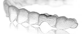
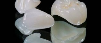
Ceramic veneer
Photo: before and after restoration with veneers
Veneering is a new procedure, but it has already gained popularity among visitors. This is a material that has virtually no flaws, does not fade or change color over time.
If you need to achieve
- Restore the front tooth by more than one third
- Ideal aesthetic appearance
- It is necessary for the restoration to serve for a long time and at the same time not lose its aesthetics
- If you need to remove cutting edges
As a result of using ceramic veneers, teeth become light, and their texture is very natural and does not differ from the main row. Plus, several years after using this type of restoration, after 3 years, you can see the same teeth as they were after leaving the dentist’s office. But this cannot be said about composite materials, since their appearance changes.
This is the main difference between composites and veneering - the latter last longer and are more practical.
But each individual case must have an individual approach, and therefore the dentist knows better what kind of restoration to offer the patient. The doctor must have knowledge of any technique and offer the patient all suitable options with an indication of the price. By the way, if we talk about price, this is an advantage of composites. Restoration of composites costs an order of magnitude less than veneering, and therefore it is more accessible.
Alternatives to composite restorations –
An alternative to composite filling materials is ceramics, i.e. veneers, crowns, restorative inlays made from ceramics. The price corresponds to the quality, and therefore the cost of ceramic restorations will be significantly higher than composite dental fillings.
- Veneers – if your front tooth is destroyed mainly only on the front side, and the lingual part of the tooth is not very damaged, then in this case the best alternative to restoration would be a ceramic veneer. Precisely from ceramics, because ceramics do not darken at all and do not lose their shine over time. The only drawback of this type of prosthetics is its cost.
- Crowns (metal-ceramics or ceramics) - if your tooth is destroyed by more than 1/2 and especially if it is also depulped, we recommend abandoning restoration in favor of an artificial crown. By the way, restoring a tooth “from the root” costs the same as it costs to restore a tooth with a metal-ceramic crown.
- Inlays - if you need to restore a lateral chewing tooth, then an alternative to restoring a tooth from a composite filling material can be a restorative inlay. In terms of reliability, service life and aesthetics, ceramic inlays are at least 3-4 times superior to restorations. We hope that our article was useful to you!
Sources:
1. Personal experience as a dentist, 2. National Library of Medicine (USA), 3. Harvard Medical School (USA), 4. “Adhesive ceramic restorations” (Manier P.), 5. “Aesthetic dentistry and ceramic restorations ( Tuati B.).
Example 2
In this work, after orthodontic treatment, a chipped corner of the second upper incisor on the right (2) and an asymmetrically short incisor on the left (1) became more noticeable; the teeth had an uneven color, the upper front tooth was darker (3). It was not possible to change the color of the front tooth only by bleaching, since the dark tooth was previously depulped and darkened from the inside. After some teeth whitening to brighten them overall and restoration of the four upper teeth with veneers, the smile became perfect. By installing veneers on the teeth, we solved several problems at once: evening out the color of the front teeth, eliminating chips and uneven edges, and correcting the length of the second incisor.
Preparation for restoration: stages
If the patient has indications for such treatment, then:
- Examination and consultation with an orthopedic doctor who will help you choose the necessary recovery option.
- Carrying out additional diagnostics - computed tomography, which will allow us to examine all structures in detail and identify contraindications to this or that type of restoration.
- Professional oral hygiene performed by a dental hygienist. It consists of removing plaque, stones from the surface of the enamel, eliminating bleeding gums, etc.
- Treatment: sanitation of the oral cavity is necessary in order to eliminate foci of infection and caries.
- Whitening can be carried out before the restoration, since if it is carried out after, the color of the bleached teeth will not match the previously carried out restoration.
- The Vita scale is used: the orthopedic surgeon determines the color of the teeth.
Indications
Most often, patients resort to artistic restoration of teeth in order to improve, first of all, their aesthetic appearance. But at the same time, you need to understand that the procedure pursues another main goal, namely to restore the tooth completely, returning functionality.
Let's consider the indications for artistic restoration:
- large gaps between teeth;
- the presence of irregularities in the dentition;
- presence of chips on the front teeth;
- severe wear of the teeth.
When is dental restoration required?
Indications:
- Presence of caries.
- Aesthetic defects of the dentition: chips, cracks, darkening.
- Old fillings that are noticeable and stand out when you smile.
- Presence of large interdental spaces.
- Non-carious lesions: fluorosis, enamel hypoplasia, enamel necrosis and others.
- Minor crooked teeth that can be corrected without installing braces.
What problems may appear after recovery?
Over time, even modern light-curing materials darken, lose their shine and begin to stand out against the background of living enamel. For lateral molars, this may not be as critical as for the teeth of the frontal group, where such defects can clearly ruin a beautiful smile. If a larger-scale restoration was carried out, for example, a large part of the pulpless crown was restored, then the risk inevitably arises that the artificial material will break off. It must be taken into account that teeth with removed nerves are especially fragile. If the root is fractured at the same time, complete removal will be required.
Artificial material may break off
Method of layer-by-layer application of composite
In recent years, when restoring class IV defects, the technique of layer-by-layer application of a composite has been used, for which there are a large number of terms: “anatomical superstructure”, “three-layer concept” or “natural layer concept” [2]. The essence of this technique is to imitate natural enamel and dentin [3]. However, not all composite materials available on the dental market can perfectly imitate tooth tissue.
The main problem with traditional composite materials is the dependence of the light transmission of transparent composite materials on the layer thickness. Therefore, the doctor has to very carefully calculate the thickness of the applied dentin and enamel shades of the material to obtain a satisfactory aesthetic result.
However, clinically it is very difficult to determine and limit the thickness of the layer with an accuracy of tenths of a millimeter, therefore, as a rule, a thicker layer of mass is applied, which is then ground off. The slightest fluctuations in layer thickness can significantly affect the final appearance of the restoration [4].
The main problem of traditional composite materials is the dependence of the light transmittance of transparent composite masses on the layer thickness