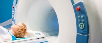
Today, only the laziest have not listened to magnetic resonance imaging. This diagnostic technique is considered the most informative and accurate for identifying pathologies and drawing up a correct medical report. Doctors of different specialties can refer you for examination if the patient has characteristic symptoms. What if you need to undergo an MRI, but you have metal implants in your body or crowns on your teeth?
Even today, there is a widespread belief that patients with such devices in the body should not undergo tomography. However, in reality this is not entirely true. A similar situation existed decades ago, when at that time patients were fitted with prostheses made of steel, cobalt or nickel. At that time, conducting electromagnetic research could harm the patient's health.
In 2022, this situation has changed dramatically. Today, individuals with endoprostheses, screws, fixation plates, pins, breast and dental implants are allowed to undergo MRI. It all depends on the composition of these items. In the article, the team will look into the intricacies of diagnostics for patients with implants. Go!
Operating principle of tomography equipment
First of all, you need to understand how exactly an MRI device works. The equipment is designed for targeted screening of soft tissues, bones and blood vessels. Advanced devices in diagnostic centers today have enormous technical capabilities. Thus, the power of the equipment is usually 1.5–3 Tesla, thanks to which doctors can examine anomalies whose dimensions are less than 0.2 mm.
MRI technology is based on the generation of radio waves and the ability to magnetize hydrogen elements present in different tissues of the body. The nuclei are able to respond to the influence of a magnetic field, that is, enter into resonance, and a computer program records all the data and converts it into detailed images. The research results are generated from many virtual sections of the selected department, for example, the brain, the vascular system, the abdominal cavity, and so on. Based on them, the radiologist draws up a medical report and a preliminary diagnosis.
Preparation
Many types of MRI examinations do not require special preparation, but there are exceptions.
Before an MRI of the abdominal cavity, it is advisable to follow the following diet:
- for two days, eliminate gas-forming foods, fresh fruits and vegetables;
- on the day of the procedure, avoid tea and coffee;
- It is advisable not to eat for at least 6 hours before the examination, and not to drink for 4 hours.
The doctor will recommend special medications for constipation and flatulence. To exclude artifacts from intestinal motility, two hours before the procedure you need to drink 2 tablets of No-shpa.
Before an MRI of the pelvis, it is advisable not to urinate for about two hours. If the study is carried out in women, it is recommended that it be carried out on days 5-12 of the cycle (immediately after menstruation), but this condition must be discussed with a doctor.
General recommendations
before MRI scan:
- remove all metal objects;
- leave jewelry and accessories at home;
- change into comfortable clothes;
- Leave valuables, money, and bank cards in a specially designated place or safe in front of the diagnostic room.
An approximate list of items that are not allowed in the diagnostic room:
- metal zippers on clothes, pins, hairpins;
- watch;
- Hearing Aids;
- pens;
- earrings, including piercings;
- glasses;
- removable dentures;
- pocket knives.
With which implants are MRIs allowed?
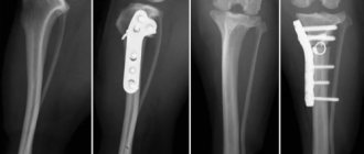
Tomographic examination can be performed for people with hip and knee joint replacement. An important rule for passing screening is the material from which the structure is made. It can be metal or ceramic, but their magnetic susceptibility should be minimal. Such material will avoid displacement of the structure or its overheating during diagnostics.
MRI is performed on patients with hernia meshes, dental prostheses, implants built into the chest and joints. All these elements are created from a material that does not in any way affect or interact with magnetic vibrations. All this allows us to make the tomography process safe for humans. It is important to consult with your doctor and diagnostician before scanning. Doctors will assess the likely risks and recommend precautions.
results
The study is interpreted by a diagnostician - a radiologist. The conclusion is issued on the day of the examination, but a description of the images may take the doctor several hours.
The doctor examines the obtained images and compares them with normal values:
- size and location of organs, symmetry;
- presence/absence of abnormal formations;
- presence/absence of an aneurysm, free fluid, blood clots in the blood stream;
- signs of infection and inflammation;
- stagnation in the ducts;
- traumatic injuries.
What are endoprostheses made of in the 21st century?
All plates with pins used in traumatology and orthopedic departments consist of various alloys. Different implants contain different amounts of paramagnetic and ferromagnetic materials. Its properties depend on the composition of the product. Not all prosthetics are created only from metal. Most are made of ceramic or polyethylene. The latter does not interact in any way with magnetic vibrations, which means it is 100% safe for MRI. However, there is one point. Ceramics usually contain aluminum oxide, but it does have some magnetic susceptibility. Therefore, it is necessary to notify the doctor about the presence of any prostheses in the body.
There are several possible combinations of materials in built-in structures:
- ceramics;
- metal;
- ceramics with polyethylene;
- metal together with polyethylene;
- ceramics with metal alloy.
Artificial compositions of joint additions include:
- titanium;
- zirconium;
- cobalt;
- chromium;
- tantalum.
Having studied the component parts of the implant, it will become clear exactly how it will respond to electromagnetic resonance.
The magnetic abilities of endoprostheses are determined not only by the material used to make the product, but also by its shape and dimensions. Pins and steels, the length of which reaches more than 20 cm, can heat up above the permissible values.
Interaction of different metals with a magnetic field
Different metals tend to interact with magnets differently. Some of them are attracted to it, others are repelled, and others do not react at all. All three types of metals are used for the manufacture of endoprostheses.
Table 1. Metal classes.
| Class | Representatives | Description |
| Diamagnets | Copper Zirconium Silver Zinc | They have negative magnetic susceptibility. This means that when interacting with a magnetic field, they repel rather than attract. |
| Paramagnets | Titanium Tungsten Aluminum Tantalum Chrome Molybdenum | These metals are characterized by the presence of low magnetic susceptibility, independent of the magnetic field strength. Paramagnetic prostheses usually tolerate the MRI procedure well and do not move or heat up. |
| Ferromagnets | Iron Nickel Cobalt Steel | They have high magnetic susceptibility, depending on the magnetic field strength. Implants containing large amounts of these metals may become dislodged or become hot during an MRI scan. |
Contraindications for MRI
When prostheses with plates are securely attached to the bones and do not move, then implants of a different location can easily move under the influence of a magnetic field. Therefore, under such circumstances, MRI is not performed - it is strictly prohibited. The main constructions for which screening cannot be done are:
- artificial heart valves;
- clips on vessels of different locations;
- ear implants;
- electronic pacemakers;
- artificial eye lens;
- Ilizarov device;
- insulin pump;
- impressive sized metal structures.
For consultation and clarification of all questions regarding diagnostics, call the hotline. A representative of the support center will help you choose a clinic for tomography, tell you about the restrictions, and will be able to book a place for an appointment with a doctor with a bonus of up to 1,000 rubles.
How does MRI affect implants?
Since the study is based on the use of magnetic vibrations, most patients have a question: “Is it possible to undergo tomography with dentures?” Innovations regularly occur in the dental field, prosthetic technology develops and new materials appear to create a beautiful smile. Previously, dental crowns were made only from copper or gold, but today patients have access to a large range of materials, for example, metal-ceramics or titanium. What's the difference?
Past methods that used copper and gold did not allow MRI patients to obtain detailed and clear images. The results were simply distorted, but there was no negative impact on health. Now dentists are massively using titanium alloys with admixtures of other particles, which has made dentures more durable and lighter. By nature, this substance is inert, so it does not:
- undergoes oxidation;
- generates toxic elements;
- responds to the influence of a magnetic field.
Because of this, people with titanium crowns can receive clear, detailed images after a CT scan. All this applies not only to dentures, but also to structures installed in bones and joints. Metal-ceramics during electromagnetic examination also does not affect the patient’s health or the screening results.
However, in addition to the built-in structure itself, the body contains pins, screws and plates with which it is secured. It is in them that a problem can arise. Implant fastener parts are made from various ferromagnets - iron alloys that have a certain magnetic susceptibility. The influence of resonance on them during an MRI scan can distort information and affect the patient’s well-being during diagnostic manipulation. All side effects can be avoided if you notify your doctor in advance about the presence of a prosthesis or other object in the body.
Myths about MRI
Our team has collected for you the top 3 common false theories about magnetic resonance scanning:
- During an MRI, the implants heat up and move. The specific gravity of the metal fastening elements of the structures is minimal, which means there are no defects in the examination results and no discomfort for the patient. But it is imperative to notify doctors about the material from which the product is made. Safe prostheses are considered to be those made of zirconium dioxide, ceramics or other expensive alloys. If you have a metal-ceramic structure, you need to clarify which metals and impurities were used in the creation.
- MRI cannot be performed during pregnancy. This is only half a lie. Due to the fact that in the first trimester the formation of tissues and organs in the unborn child begins, any effect on the fetus (chemical, physical or biological) must be excluded. In late pregnancy, tomography allows you to determine the position of the baby and determine the size of the mother’s pelvis. The results of the examination allow the obstetrician to prepare for childbirth without consequences for the child and the pregnant woman.
- MRI with prostheses is painful. Complete lie. Tomographic examination is one of the most painless and safe diagnostic techniques. MRI is allowed even with titanium inserts. Scanning can only be denied if the structure is implanted with steel, since it can heat up and move under the influence of magnetic vibrations. Only such a process can lead to injury to the subject.
If you have implants or not, but want to undergo an MRI, use. Our platform is designed to quickly search and select a clinic for diagnostics. The service presents dozens of medical centers, collects current prices for tomography and real reviews from visitors. To make an appointment, just call the hotline. When you reserve a place with a doctor using our resource, you will receive up to 1,000 rubles as a gift!
Bibliography:
- Konovalov R.N., Krotenkova M.V., Kremneva E.I. Functional magnetic resonance imaging [Electronic resource] // Journal “Annals of Clinical and Experimental Neurology”, 2011. https://cyberleninka.ru/article/n/funktsionalnaya-magnitno-rezonansnaya-tomografiya
- Kukhtevich I.I., Goryunova T.I., Goryunova V.V. Practice and safety of MRI studies [Electronic resource] // Journal “International Student Scientific Bulletin” - 2022. https://eduherald.ru/ru/article/view?id=18294
- Ternovoy S.K., Sinitsyn V.E., Belichenko O.I., Stukalova O.V. Clinical application of magnetic resonance imaging [Electronic resource] // Regular issues of “RMZh” No. 7, 1996. https://www.rmj.ru/articles/obshchie-stati/KLINIChESKOE_PRIMENENIE_MAGNITNO-REZONANSNOY_TOMOGRAFII/