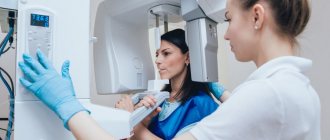
A dental visiograph is equipment with which you can quickly obtain accurate images of teeth to monitor treatment or evaluate tissue. Unlike an x-ray, the doctor immediately receives the image on the monitor, meaning little time is spent on the examination. The picture is clear; to improve the quality, special tools are used to increase picture brightness, clarity and contrast.
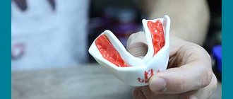
The radiovisiograph is compact in size and easy to use. To perform the procedure, a small sensor is placed in the Patient’s oral cavity, which transmits the image to the screen. No special preparation is needed; the patient does not feel pain or discomfort.
The device includes the following components:
- a base in the form of a high-frequency device, with the help of which particles acquire the desired direction of movement;
- wireless or wired sensor (depending on model);
- analog-digital system for converting images into electronic form;
- laptop or PC screen for viewing and image correction.
The radiation exposure during the procedure is minimal - 2-3 μSv, which is much less than during a standard examination. The resulting images can be stored electronically, accessing them if necessary to monitor treatment or the achieved result. The resulting visiograms can be used in the following cases:
- identification of foci of infectious lesions;
- search for pathologies invisible using other research methods;
- assessment of the course of the disease, including caries, periodontitis or pulpitis.
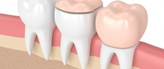
Diagnostics also makes it possible to detect foreign bodies in the tooth canal and cavities remaining after filling. Cysts, granulomas, and inflammations are painted black in the resulting picture; they stand out against the background of light healthy tissue. This improves the quality of the analysis and allows the doctor to immediately localize the pathology and the depth of the lesion. The ease of use of the device makes it easier to control therapy, for example, when filling cavities or dental canals.
What does a visiograph look like and when is it used?
Dental radiovisiographs are small in size. To take an image, a small sensor of the device is placed in the patient’s oral cavity and transmits the result of the image directly to the computer.
The visiograph consists of several components:
- The basis is the high-frequency apparatus in the photo above, which sets the desired direction of particle movement;
- Small wired or wireless sensor;
- Analogue-digital system providing image digitization;
- A PC or laptop that allows you to view, store, and send images via email.
The wired visiograph is equipped with a special sensor that is applied to the tooth. Every radiologist should know how to take targeted photographs of teeth using such equipment. Using a wired connection, the image is transferred to the computer screen. A wireless radiovisiograph has a scanner capable of reading an image and displaying it on the screen.
The radiation dose during the examination is small and amounts to 2-3 μSv. Compared to this device, fluorography creates an exposure of 500-800 μSv.
The resulting image can be stored in an archive and on a hard drive. A visiograph belongs to the category of diagnostic and treatment dental equipment and allows you to obtain a clear image of a diseased tooth.
Requirements for a visiographer in private offices
Since the device reduces the level of radiation, the requirements for a visiograph in private offices are much lower than for analog X-ray equipment.
According to SanPiN 2.6.1.802-99, radiovisiographic equipment (radiovisiography) can be placed in a dental office, which is located in a residential building. The only condition is to ensure compliance with radiation safety requirements within the given office.
For a private office, if you do not have the opportunity to rent a large area, you can work with the device in the main reception room. This is very convenient if the picture is taken during treatment and the patient is sitting with an open tooth. For personal safety, a special apron is used, and the photograph is taken directly by the attending physician.
The requirements for a visiograph are quite simple to fulfill even for a small clinic and a small private dental office. The cost of the device is high, but the ease of operation and regular use allow you to recoup the costs within 1.5-2 years.
There are interesting safe models with a wireless visiograph from the French company Satelec Action Group
This is what this PSPIX 2 radiovisiograph system looks like for reading X-ray images with a preview screen and memory plates.
On the X-ray equipment market in terms of visiography, there are also interesting models from the giant MyRay brand, one of the leaders in the field of developments in radiology. Compact wireless imaging device XPod
allows the dentist to immediately see x-ray images on the display
without using a computer. The average lifespan of a visiograph is 400,000 images. Use a radiovisiograph in dentistry, because innovative and advanced technologies are the key to success. Order radiovisiographic equipment right now! Take your business to a new level of service.
What does a radiovisiographic image reveal?
A digital radiovisiograph allows you to identify any foci of infection, the location of which is difficult to determine visually. A visiogram is done in case of possible occurrence of the following diseases:
- Caries;
- Pulpitis, which causes inflammation of the tissues surrounding the tooth;
- Small neoplasms in the oral cavity;
- Periodontitis.
The image allows you to detect hidden foreign bodies that have entered the canal. Any foci of inflammation, granulomas and cysts are painted black, which stands out sharply against the gray background of bone and connective tissue. With the help of detailed visual images, the dentist will be able to make an accurate diagnosis and prescribe appropriate treatment.
Radiovisiography allows you to determine the depth of tissue damage and control the quality of filling.
Frequent breakdowns of radiovisiographs
| Damage to the sensor layer for recording radiation from the visiograph sensor |
| Malfunction of the radiovisiograph matrix |
| Damage to the electronic board |
| Damage to the connecting cable or connector for connecting the sensor to the PC (USB) |
| Software malfunctions, visualization errors |
| Intermediate unit faulty. |
| Damage to the body of the visiograph sensor, depressurization |
| Damage to controls (select models) |
| Loss of image clarity, noise, banding, artifacts (for no apparent reason) |
Indications and contraindications for the procedure
List of main indications for use of the equipment:
- Prevention of pulpitis, caries, periodontitis;
- Viewing the condition of a filled tooth, decayed gums, damaged bone tissue, or installed implant.
Radiovisiographic examination allows complete control of the treatment process. It will help avoid many complications. This method is safer than x-ray examination. It does not pose a health hazard.
Radiovisiography can be performed during pregnancy and lactation.
The list of relative contraindications includes:
- Mental illnesses;
- Neurological disorders.
Diagnostics at the clinic Dr. Martin
Visiography is recognized as the best method of modern diagnostics in dentistry when it is necessary to obtain a targeted image that allows one to examine in detail a separate area of the dentofacial structure and joints of the lower jaw.
Clinic Dr. Martin offers dental imaging at an affordable price. Diagnostics can be either a stage of treatment by our specialists or a separately provided service. The cost for the procedure and the image is indicated in the price list on the clinic’s website.
You can make an appointment with a specialist, a consultation or an examination by calling the contact number, or using the website service by ordering a call back from a consultant (“Make an Appointment”).
Difference from X-ray and computed tomography
Computed tomography allows you to obtain a three-dimensional image of the jaw to examine it in all planes. A visiographic device helps to examine each problematic tooth individually. A similar examination is carried out before the therapeutic procedure, and the method is also used to monitor the treatment process.
X-ray can only create flat images. Even interproximal radiography is not always able to determine the presence of tumors in the oral cavity. Using a radiovisiograph, you can see a tumor in the initial stage of its formation.
Radiovisiography is safer than radiography; the level of dangerous examination in the first case is relatively low. The signal from the visiograph passes to a special CCD sensor or microcircuit that is more sensitive than x-ray film.
Example of resulting images
Visiography of a tooth is
Dental visiography is the most informative method for diagnosing pathologies of the teeth and jaw, carried out using a special device that operates on the basis of radio frequency radiation.
The procedure is absolutely safe and not contraindicated even in childhood. The radiation exposure to the body is minimal (10 times lower than with classic x-rays). The procedure is also called “radiovisiography”. The main difference between the procedure and classical x-rays is the ability to instantly obtain an image of the area under study not on film, but on a monitor screen. In addition, experts confirm the high quality of the image and the possibility of obtaining a targeted image.
A dental radiovisiograph is used to identify any pathological processes in the teeth or jaw, including surrounding soft tissues and temporomandibular joints. As a result of diagnosis, a specialist can determine hidden processes (including hidden caries), inflammation, maxillofacial injuries, pathologies of the maxillary sinuses, and the condition of the jaw bones. Visiography is also used in dentistry to monitor the progress of treatment and its results.
Advantages and disadvantages of a visiograph
The main advantages of radiovisiography:
- compactness of the device;
- image accuracy;
- the ability to instantly view a diseased tooth on a computer screen with multiple magnification of the resulting image, storing the image on a PC and transmitting it via a local network and e-mail;
- safety of the procedure;
- speed of manipulation;
- editing a digital visiogram, using various filters to improve the visual perception of the picture.
Radiovisiography of the tooth is carried out in a fairly low spatial resolution. This is the main disadvantage of the procedure. The device's sensor does not clearly show the difference between different structures with unequal densities. But thanks to the ability to adjust the contrast and brightness of the image, details become more visible.
Despite the fact that doctors do not prohibit performing the procedure during pregnancy, the expectant mother should warn the attending physician about her situation.
How is the diagnosis carried out?
The visiograph is a modern diagnostic device consisting of three main parts: a high-precision sensor, an analog-to-digital converter and a cord connecting the parts of the device. The operation of the visiograph is ensured by a whole complex of parts, including x-ray equipment with a special guide (tube). The device itself has an external resemblance to a tomograph.
The sensor is a silicone chip (digital sensor) that records all the signals that come to it. It is the sensor that transmits the signal to the device, which is formatted into an image with 16-bit quality. The analog-to-digital converter is a monoblock equipped with a USB port for connecting to a computer and a special port for connecting to a sensor. The data received by the sensor passes through the connecting cable to the computer. All information is converted using a built-in program and displayed on a computer monitor.
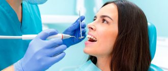
The diagnostic procedure takes on average 3–5 minutes. The patient sits down on a chair near the visiograph, the specialist adjusts the direction of the X-ray tube in the desired direction and places the sensor against the area being examined. Then the device turns on and a photo is taken.
How safe is the visiograph?
Thanks to a narrower, homogeneous beam and high sensitivity of the digital sensor, the radiation dose from visiographs is much less than from conventional X-ray units.
This explains the low level of X-ray radiation in the visiograph - it does no more harm than from an office scanner.
When diagnosing lower teeth, you will receive only 2 µSv in one procedure, and up to 5 µSv for upper teeth. For comparison, on modern X-ray machines the dose of radiation for a similar procedure is 7–14 μSv, and on outdated ones it can reach 80 μSv.
According to official standards, visiography can be carried out up to 60 times a year. But since the annual radiation dose is permissible within 1000 μSv, even a daily procedure will not cause harm to health.
Visiography is also recommended for children. In particular, it is safe during pregnancy and lactation, since there is no direct effect on the fetus during the procedure. However, in these cases, visiography is done as prescribed by a doctor, and the most favorable time for treating dental diseases in pregnant women is the second trimester.
Before examination with a visiograph, the patient wears a lead apron for additional protection.
How is a radiovisiograph repaired?
1. Consultation.
2. Diagnostics of the visiograph sensor.
3. Coordination of work.
4. Repair of the dental radiovisiograph sensor.
5. Connection and checking modes.
6. Transfer of equipment.
Modification differences
| Model | Xelia V8 | Xelia V7 |
| Sensor | CMOS sensor | CMOS sensor |
| Touch field | 32.6 x 20.5 mm | 32.6 x 20.5 mm |
| External dimensions of the sensor | 40x24 mm | 40x24 mm |
| Theoretical resolution | 22 line pairs/mm | 22 line pairs/mm |
| Actual Resolution | 14-16 line pairs/mm | 14 line pairs/mm |
| Sensor thickness | 6 mm | 6 mm |
| Gray level | 4096 | 4096 |
| Radiation dose reduction | by 80-90% | by 80-90% |
| Nutrition | USB 2.0 (5V) | USB 2.0 (5V) |
| Image transmission time | < 3 sec | < 3 sec |
| Available colors | red, green, blue, cyan, black | — |
| USB cable length | 2.5 /3/5 m (optional) | 2.5 /3/5 m (optional) |