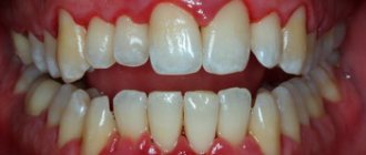
Filling is a complex of dental procedures, including the removal of tooth tissue destroyed by caries, disinfection and treatment of the resulting cavity with adhesive preparations, placement and grinding of a dental filling.
Normally, after performing the above procedures, the patient should not experience discomfort or pain. For this reason, the appearance of any unpleasant sensations in the area of a filled tooth is an absolute reason for a return visit to the dental office.
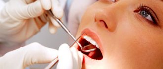
Causes of tooth pain after filling
Pulpitis
The appearance of acute, spontaneous pain that grows and intensifies at night in a filled tooth may be due to the development of chronic or acute pulpitis. If such symptoms are detected, it is necessary to contact a dentist as soon as possible to remove a previously placed filling, remove the inflamed pulp, fill the root canals and the carious cavity itself.
Development of secondary caries
The source of pain in the tooth after filling can also be secondary caries, which gradually develops under the filling. The most common causes of this pathology are:
Causes of secondary caries
- insufficient disinfection of the carious surface before placing a filling;
- displacement of the installed filling under the influence of external factors;
- shrinkage of the filling material, which contributes to the formation of gaps between the filling and dental tissues;
- injury resulting in damage to the filling;
- irregular oral hygiene;
- violation of filling technology.
Treatment of secondary caries
When a secondary caries process is detected, the dentist removes the previously placed filling, cleans the resulting cavity from destroyed tissue, disinfects it and re-fills the tooth.
Underdrying or overdrying of the cavity
The causes of spontaneous pain that increases with biting in a treated tooth may be under-drying or over-drying of the tissues before filling. Before applying adhesive preparations, the surface dental tissues must be dried to a “wet sand” state (that is, the surface must remain wet, but there cannot be drops of water on it).
Consequences of overdrying tissues
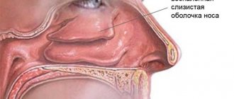
Overdrying of dental tissues leads to damage to the nerve endings located in the surface layers of dentin. In turn, the death of nerve fibers can cause inflammatory damage to the pulp and other serious complications. Mild pain caused by overdrying of the carious cavity most often goes away on its own within 5-13 days. In situations where pain intensifies or persists for two weeks, patients are advised to return to the dental clinic for help.
Consequences of not drying the cavity
Failure to dry out the treated carious cavity can also cause pain. Drops of moisture prevent the penetration of adhesive preparations into the dentinal tubules, therefore, when the filling material shrinks, the composite easily comes off from the cavity floor, thereby forming pathological sinuses in the dental tissues. The only way out of this situation can be to replace the previously installed filling.
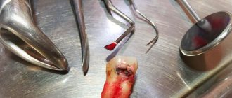
Granuloma or dental cyst
One of the likely causes of tooth pain after filling may be the formation of a granuloma (a localized area of inflamed tissue) or a root cyst (a pathological cavity in the gum tissue filled with bacteria, dead cells, blood and other physiological fluids). Granulomas and cysts are treated both conservatively (for example, using the depophoresis method) and through surgical intervention (cystectomy, hemisection, etc.).
Allergic reaction
In some cases, pain in a filled tooth can be the result of an allergic reaction to the components of the filling material. The clinical picture may be supplemented by urticaria, the appearance of an itchy rash and swelling on the patient’s skin and mucous membranes. If a patient is diagnosed with an allergy to the material used, the filling is immediately replaced.
Increased tooth wear - symptoms and treatment
Increased (pathological) tooth abrasion is an intense loss of hard tissues, which can lead to almost complete wear of the crown part of the teeth. It can be caused by congenital characteristics, somatic diseases, bad habits, abnormalities in the position of teeth, incorrect dental treatment and other factors. At the initial stage, the changes are almost unnoticeable, but as the disease progresses, the patient himself may notice a decrease in the length of the crowns of the teeth and a change in the shape of the chewing surfaces.
According to Alekseev, the author of scientific works and the book “Pathological abrasion of teeth,” increased abrasion of incisors, molars and premolars is detected in 4% of people aged 25–30 years. At the age of 30–40 years, almost 23% of patients seek orthopedic help due to increased tooth wear, at 40–45 years old - 35%, at 50–60 years old - 26%, after 60 years this figure decreases to 12% [2][ 13].
Physiological abrasion of the cusps of premolars and molars, the cutting surface of the front teeth in the process of chewing food is a natural process that is observed in every person. With age, the enamel wears off more intensively, and due to the natural mobility of teeth, facets are formed on the chewing surfaces, which are manifested by a decrease in the volume of hard tissues. The speed of this process depends on the strength of the enamel, bite and eating habits. Age-related changes in the temporomandibular joint and periodontal tissue can also cause tooth wear. Reducing the size of crowns serves as a compensatory reaction, allowing, under conditions of slowing metabolic processes, to maintain hard tissues in a normal state [1].
Causes of increased (pathological) tooth wear
The reasons can be general (pathological habits, work hazards, genetic abnormalities, chronic diseases) and local (bite pathology, tooth loss, bruxism, diet high in acids).
In some patients, pathological tooth wear is caused by harmful or professional habits: gnawing seeds or nuts, holding tools in the teeth, biting off threads. The characteristics of the enamel in these people correspond to the norm, but excess load leads to premature wear of the dental crowns. Even the strongest teeth need to be protected at a young age.
Other causes of pathological abrasion of enamel:
- malocclusion;
- bruxism (teeth grinding);
- loss of several teeth;
- wearing incorrectly made dentures, the shape of which does not correspond to the anatomical features of the patient’s oral cavity and does not ensure normal contact between the chewing surfaces;
- constant exposure of teeth to sand, soot and other solid particles;
- fluorosis (destruction of enamel associated with excess fluoride);
- amelogenesis imperfecta (hereditary disorder of the formation of tooth enamel);
- acid necrosis (destruction of enamel in patients who excessively consume acidic foods, as well as those in contact with acids in hazardous industries, as a rule, can be combined with enamel erosion);
- thyroid diseases;
- pathology of the pituitary gland;
- improper brushing of teeth [3].
Various malocclusions lead to a certain pattern of tooth wear:
- with a straight bite, the chewing surface of the molars and the cutting part of the incisors are intensively worn away;
- with a deep bite, the tissue of the front teeth decreases especially quickly, the lateral movements of the jaw in such people are limited, therefore the molars and premolars retain masticatory cusps even in adulthood;
- with an oblique bite or an anomaly in the position of part of the teeth, the process occurs asymmetrically with the abrasion of individual teeth.
When teeth are lost, the load during chewing is distributed unevenly and the remaining teeth take it upon themselves. This leads to their premature wear. If a person chews food on one side of the jaw due to dental disease, the wear of the crowns will be asymmetrical.
Another reason for premature enamel wear is improper installation of removable dentures . If the teeth, which are the supporting points for clasps (hooks for fixing the prosthesis), are not protected by artificial crowns, they will quickly wear out.
Increased tooth wear is observed in representatives of some professions:
- in persons engaged in physical labor, the pathology is associated with strong clenching of the jaws when lifting heavy objects;
- among foundry workers - with dust and gas contamination in the air;
- Chemical industry workers who come into contact with acids develop acid necrosis of the enamel.
Abnormal abrasion of enamel may be associated with diseases such as thyrotoxicosis , chronic cholecystitis, urolithiasis , and endemic fluorosis. They lead to the fact that the enamel loses its natural strength and quickly wears off even with minor mechanical stress. Diseases of the nervous system , one of the symptoms of which is bruxism, also have a negative impact
Pathological abrasion can be caused by hereditary anomalies, one of them is amelogenesis imperfecta . With this pathology, the quantity and mineralization of tooth enamel is disrupted and its density decreases. Accordingly, it will wear off faster than in people with healthy teeth [5].
With pathological tooth wear, it is first of all important to identify the cause; some patients need not only dental care, but also treatment of the underlying disease or lifestyle changes.
Recommendations for eliminating the causes of pain in a filled tooth
If you experience discomfort or pain in a filled tooth, it is recommended:
- eliminate the load on the diseased tooth;
- seek professional dental care immediately;
- stop using traditional medicine.
Procedures related to filling replacement, removal of granulomas and cysts are performed on an outpatient basis and do not require much time.
At the same time, timely seeking dental care at a 24-hour dentistry allows you not only to get rid of unpleasant symptoms in the shortest possible time, but also to prevent the development of serious complications.
What are the causes of pathology
In accordance with generally accepted sanitary standards, the maximum concentration of fluorine compounds in water should not exceed 1.5 mg/l. But it should be remembered that fluoride also enters the body during breathing and through food. An excess of this microelement negatively affects tooth enamel and begins to destroy it. If you do not consult a doctor in time, bone tissue pathologies may develop - osteoporosis or osteosclerosis.
Important! The occurrence of the disease is associated with the individual characteristics of the body. Sometimes fluorosis develops at lower concentrations of fluoride.
Mainly permanent teeth are affected, in rare cases – milk teeth in children. This is due to the fact that the mineralization of the baby’s first teeth begins and ends during the period of intrauterine development. At this time, the placenta protects the child’s body from an excess of fluoride. But if a pregnant woman lives in an area where the content of fluoride compounds in the water greatly exceeds the norm, then the disease can also affect the child’s baby teeth.
Children aged 3–4 years are at risk if they have consumed water with a high content of fluoride compounds for 3 or more years. This disease is also diagnosed in adults who work in industries with high levels of fluoride in the air.
Classification of the disease
Depending on the severity of the pathology, the following forms are distinguished:
- Streaked – characterized by the appearance of small stripes or streaks on the surface of the tooth. The upper jaw is most often affected by this form. The stripes are faint and cannot always be seen independently. Over time, they merge into one spot, in which strokes can also be distinguished.
- Spotted - this form is characterized by the presence of multiple chalky spots. They are well defined and located on the entire surface of the tooth. When drained, large spots form.
- A chalky-altered form - it is characterized by the presence of an affected light brown area, which turns into healthy enamel. This lesion is most often observed on the upper and lower incisors.
- Chalky-mottled - characterized by the presence of clear pigment spots. Sometimes there may be a variant of the presence of multiple dots on the enamel of a yellowish color. With this form, rapid thinning of the enamel is observed.
- Erosive - in addition to stains on the enamel, erosive defects occur. They contribute to the destruction of not only enamel, but also dentin.
- Destructive is the most severe form, in which the shape of the tooth crown is disrupted. This occurs due to thinning of the enamel and destruction of hard tooth tissue. The teeth themselves are quite fragile, prone to various damages (chips, breaks).
The disease can occur in mild, moderate and severe degrees. This depends on the number of teeth affected, as well as the depth of the pathological process. In severe cases, the patient experiences damage to more than 80% of the teeth. In this case, dentin is affected and crown deformation is observed. Fluorosis can also cause pathological disorders of skeletal bones.