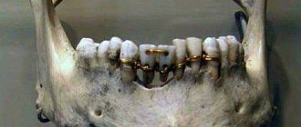
There are several types of malocclusion. Rare teeth are the most common. It is characterized by the presence of very wide spaces between dental units. At the same time, the chewing structures are externally similar to a comb with sparse teeth. This pathological condition cannot be ignored, as it can lead to the development of dangerous complications. Currently, there are several methods for treating rare teeth (a photo of the defect is presented below). The feasibility of using a particular method is assessed by the orthodontist.
Causes and consequences
The “culprit” of the pathology is a genetic factor. Large distances between chewing units are noticeable already at the stage of eruption of milk teeth. In extremely rare cases, pathology develops against the background of progression of dental diseases.
Rare teeth in humans are not only unsightly, but also dangerous to health. Initially, psycho-emotional instability appears. It is caused by complexes that have arisen and low self-esteem.
In addition, sparse teeth cause the following pathological conditions:
- Disorders of the gastrointestinal tract. Wide spaces between dental units do not allow for proper chewing of food. As a result, food enters the stomach in an insufficiently processed form. The natural consequence is indigestion.
- Tooth loss. Chewing units located far from each other are characterized by increased fragility. In addition, they are extremely vulnerable to any external influences.
- Inflammatory processes in the oral cavity. When there are large gaps between teeth, soft tissues also become more vulnerable to external influences. They are easily injured, and when pathogenic microorganisms penetrate the oral cavity, an inflammatory process develops.
To prevent the development of complications and make your smile perfect, you need to contact an orthodontist. Only a doctor can provide information on how to correct rare teeth in each specific case.
Reasons causing anomalies
Anomalies of the dental system are very diverse and include both anomalies of dental units and incorrect formation of the dentition, as well as abnormalities in the development of the jaws. The most frequently diagnosed abnormalities of dental units. They can be caused by a wide variety of reasons, including both exogenous (caused by external factors) and endogenous (formed as a result of the physiological state of the patient).
Endogenous causes
Endogenous anomalies are formed as a result of genetic and endocrine disorders.
- Many structural features of the dental system, leading to improper development of teeth, are genetically determined. This can occur through direct inheritance (abnormal number and shape of teeth, edentia, diastema), through inheritance of a mismatch in the size of the jaw bones, through inheritance of a mismatch in the size of the jaw and teeth (crowding of teeth or sparse arrangement of teeth). Dental anomalies of this type include disorders associated with hereditary and congenital pathologies (clefts of the soft and hard palate, cleft lip, Down syndrome, Vaanderburg syndrome, Seckel syndrome, Shershevsky-Turner syndrome).
- Another part of the endogenous causes of dental anomalies are problems of the endocrine system, such as hypothyroidism (in later stages), hyperparathyroidism, hypocortisolism. Their frequent symptoms may be delays in the eruption and replacement of teeth, and disturbances in the formation of the enamel layer.
Exogenous causes
Exogenous (external) causes of dental anomalies are, for the most part, external unfavorable factors that affect the baby’s body during the formation of tooth germs. There are prenatal (prenatal), postnatal (postpartum) and intranatal (associated with childbirth) periods.
- An example of a prenatal factor is a pregnant woman living in poor environmental conditions. Various developmental disorders of the fetus can also occur as a result of the mother’s poor lifestyle.
- Factors in the intranatal period may include complicated labor, birth injuries, consequences of asphyxia and oligohydramnios.
- In the postnatal period, dental anomalies develop as a complication of pathologies in early childhood. These include childhood infections, rickets, hypovitaminosis, and micronutrient deficiency.
All these reasons are of a common nature. Local factors that affect dental development include everything related to improper oral care, feeding errors or bad habits. Among them are feeding too soft food, prolonged sucking of a pacifier or finger in early childhood.
Childhood injuries and caries with complications lead to dental anomalies.
The death of tooth germs or the development of supernumerary teeth can occur due to osteomyelitis.
Diagnostics
After the examination, the orthodontist will refer the patient for a comprehensive examination. Based on its results, the doctor will be able to make a decision on what to do with the rare teeth of the person who contacts him.
Diagnosis may include the following studies:
- Orthopantomogram. This is a three-dimensional shot of the jaws.
- X-ray examination.
- Computer diagnostics. The method is considered the most accurate and informative. It allows you to evaluate both the structure of the jaw and the location of the teeth.
Based on the diagnostic results, the orthodontist will select the most effective method of treating rare teeth. It is important to understand that it is impossible to correct the genetic factor, but doctors are able to disguise the defect so that the patient’s smile is perfect. Contrary to popular belief, if adults have sparse teeth, braces are powerless. This method of correcting the bite is not used if a person has large distances between the teeth.
Preparing for treatment
Elimination of the defect requires a comprehensive examination of the oral cavity and selection of optimal treatment. In particular, the doctor assesses the size of the pathological holes. An x-ray examination is also prescribed, which allows you to identify the presence of hidden injuries. Regardless of the chosen scheme, before starting treatment of rare teeth, therapy for caries and other pathologies of the oral cavity is prescribed.
Self-medication is dangerous with complications!
Attention
Despite the fact that our articles are based on trusted sources and have been tested by practicing doctors, the same symptoms can be signs of different diseases, and the disease may not proceed according to the textbook.
Pros of seeing a doctor:
- Only a specialist will prescribe suitable medications.
- Recovery will be easier and faster.
- The doctor will monitor the course of the disease and help avoid complications.
find a doctor
Do not try to treat yourself - consult a specialist.
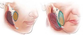
The most difficult operation to correct a malocclusion is implantation. Before it is performed, a panoramic photograph of the jaw is taken, which allows you to select the appropriate pin size.
Implantation is contraindicated in many cases. It cannot be used in the presence of oncological pathologies, tuberculosis, a number of chronic diseases and allergies to anesthesia. Implantation takes several hours. During the procedure, tooth extension is carried out in three projections: width, height and length.
After the operation, the patient should follow certain procedures for several months to prevent the development of complications.
Dental implantation
The technique is the most radical. The essence of the procedure is to replace rare dental units with prostheses. The latter are screwed into the jaw using a titanium pin that imitates a tooth root.
The main advantage of implants is that they are as similar as possible to natural chewing units and fully perform the functions of the latter. As a result, food is chewed efficiently and the load between the teeth is distributed evenly. As a result, the process of forming a correct bite begins.
In addition, implanted teeth are permanent. In other words, they do not need to be removed and cared for separately.
Dental implantation is contraindicated if the patient has type II diabetes mellitus, HIV, hepatitis and severe heart pathologies.
Anomalies of hard tissue structure
An altered shape, abnormal size or color of enamel is formed against the background of an abnormality in the structure of the hard tissues of the dental unit. Among such anomalies are:
- Hypoplasia (underdevelopment of tissue). The initial degree of the disorder is manifested by the presence of chalky spots on the enamel and areas where tissue deficiency is observed. Subsequently, all kinds of pits, grooves, and recesses appear on the surface of the enamel. The defect affects all teeth in the dentition.
- Hyperplasia (excessive tissue formation). Pathology also affects all teeth at the same time. It is characterized by areas where tissue growth is observed - tubercles, sagging enamel. The consequence of the pathology is a change in the shape and size of the teeth, a violation of the occlusion (the line of contact of the upper and lower jaws).
- Anomalies of amelogenesis (enamel formation) are expressed in the presence of brown or yellow spots on the surface of the teeth. Areas where the natural composition of the enamel is disrupted become especially sensitive. Microdentia develops against the background of pathology. The cause of the development of pathology is a deficiency of microelements that take part in the formation of dental tissue. Treatment consists of replenishing this deficiency. Additionally, local remineralizing therapy and physiotherapy are performed.
- Disturbance of dentinogenesis consists of dysfunction of the mechanism of dentin formation. The main symptoms of the disease are as follows: teeth become yellow-brown or grayish in color. The fragile dentin in these areas quickly wears off, causing tooth decay and then tooth loss. The disease occurs in genetically predisposed patients. The problem can only be solved by replacing the units destroyed by the disease with prosthetics.
Installation of a bridge
The use of fixed orthodontic structures is a modern way to fill wide gaps in the dentition.
In order for the prosthesis to be securely fixed, it is necessary to have supporting dental units. It is on them that the entire structure is attached.
The best option is to make a bridge from metal ceramics. The service life of this design is 7 years or more. At the same time, the bridge retains its original shade and does not differ from adjacent natural teeth.
Contraindications to installing a prosthesis on rare teeth:
- Acute periodontitis.
- Parafunctions of the masticatory muscles.
- Insufficient height of supporting teeth.
If there are absolute contraindications, the doctor makes a decision regarding the advisability of using a different treatment method.
Shape anomalies
There are pathologies in the development of dental units, due to which they acquire an unnatural shape. These disorders are named after the scientists who first described them. The following types of dental shape anomalies are found: spiny teeth, Pflueger teeth, Fournier teeth, Hutchinson teeth.
- Spiked or awl-shaped teeth. With this pathology, the teeth acquire a cone-shaped shape. Wide at the base, they gradually narrow and, towards the cutting edge, become sharpened into a spike shape. This problem is combined with microdentia. There are irregularities and stains on the surface of the teeth. The disease affects the front and lateral incisors. The disease occurs in childhood and is caused by genetic factors in combination with external factors. Among them are vitamin D deficiency and endocrine system problems.
- Hutchinson's teeth are characterized by a modified crown shape of the incisors. Externally, they have the shape of a barrel, since the neck is significantly thickened. The cutting edge of the teeth acquires an arched notch. The enamel layer also suffers, which is present only on the sides and absent in the center.
- Fournier's teeth are a form of systemic hypoplasia of dental units associated with metabolic disorders at the stage of intrauterine development of the fetus. The barrel-shaped shape of the teeth is preserved, but the notch of the incisal edge is absent. The color of the enamel is not disturbed, but the enamel layer is underdeveloped, which can be seen during microscopic examination.
- Pflueger's teeth are a disorder that affects permanent dental units. Their crowns take on a conical shape. Thickening develops in the cervical region, and the chewing surface is significantly underdeveloped. The chewing function of the teeth is completely preserved.
- Turner anomaly. With this pathology, there is no enamel on the teeth. They have an abnormally lumpy surface, and replacement tissue, dentin, forms in the exposed areas.
Systemic hypoplasia with a violation of the shape of the teeth has three degrees of development. The third degree is the most dangerous, in which the crown is severely deformed and the enamel layer decreases. In this case, the defective teeth are removed and replaced with dentures. It is also possible to restore affected teeth using reflective components.
Sealing
Currently, it is one of the most affordable ways to treat rare teeth. Its wide distribution is due to its low cost and ease of implementation.
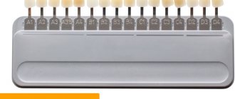
The essence of the method is as follows: fillings of the same color as the natural chewing units are applied to the teeth. The procedure is considered a jewelry procedure, since the doctor must literally mold new bone structures. As a result, the teeth become wider and the gaps between them disappear. Only a specialist can visually distinguish fillings from natural dental units.
During the procedure, orthodontists use only high-quality materials. These are ceramics and porcelain.
After filling rare teeth, the functioning of the gastrointestinal tract is normalized and a correct bite begins to form.
Installation of veneers
This is a highly effective correction method. Veneers are thin plates made of ceramic or porcelain. They are placed on top of natural teeth.
The main advantage of the method is that the plates cover and protect the enamel coating of the dental units. In other words, there is no need to grind or file down adjacent teeth.
Contraindications for installing veneers:
- Severe destruction of dental units from the tongue.
- The presence of large fillings.
- Grinding of teeth.
- Having a bad habit of opening bottles with your teeth, cracking nuts with them, etc.
In addition, veneers cannot be installed if six or more chewing units are missing from the mouth.
Installation of crowns
These are non-removable structures that can be used to eliminate dental defects. The installation of crowns is also indicated in case of severe destruction of dental units. The essence of the method is to insert an artificial tooth into the gum using a pin made of metal.
Installing crowns requires some preparation. The patient's natural teeth are prepared and, if necessary, pulp removal is performed. Crowns take several days to make. During this period, the patient is provided with temporary structures. The finished crowns are completely identical to the patient’s teeth. They differ only in width, which makes it possible to hide large gaps between chewing units.
The following conditions are contraindications: loose teeth, allergic reaction to the components used, periodontal pathologies. In addition, crowns are not placed on anyone under 16 years of age.
Formulas for calculating proportions
The normality of the crown part of the teeth is determined not by their size, but by the width and length of the dental arches, which are considered normal for a specific group of patients.
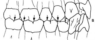
If the length and width of the arch correspond to the norm, and all the teeth on it are located without crowding or gaps, then their sizes are considered normal.
Many different techniques have been proposed to determine the normality of the jaw arches, taking into account the age, facial type and nationality of the patient. Three of them are discussed below - the Gerlach, Pon-Linder-Hart and Corkhouse methods.
Gerlach method
According to the Gerlach method, the dentition is divided into 3 segments:
- anterior, consisting of 4 incisors;
- two lateral ones, consisting of canines, premolars and the first molar.
Next, the calculation is made according to the following algorithm:
- The length of each segment is measured. In this case, the value of the length of the lower frontal segment is obtained by multiplying the length of the upper frontal segment by the Tone coefficient (1, 35).
- Compare the obtained lengths of the segments with each other and the tabular data compiled by Gerlach. With normal jaw sizes, they must be in certain proportions to each other.
If it turns out that the dimensions of the arches are normal, and there is no crowding or gaps, then the dimensions of the clinical crowns are normal.
Reference. All methods used involve measurements of dental arches at strictly defined points and according to certain methods. Because of their relative complexity and length, they are not included here. But they can be easily found in numerous sources.
Pon-Linder-Hart method
This technique is intended to determine the normal width of the dental arches in children with variable and permanent dentition. In this case, the formula developed by Pont is used and has the following form:
A ∶ 80 × 100 = distance between first premolars
A ∶ 64 × 100 = distance between first molars, where:
- A – total width of 4 incisors;
- 80 and 64 – Pon coefficients (indices).
Linder and Hart found that these coefficients for children of German nationality should be 85 and 65, respectively.
The algorithm for determining the parameters of the upper jaw according to Pon-Linder-Hart is as follows:
- The total width of all incisors along their cutting edge is measured.
- The obtained data is substituted into the Pon formula, and the distance between the first painters and premolars is calculated.
- The actual width of the dental arch between premolars and molars is measured.
- The measured value is compared with the results determined using the Pon formula.
- A conclusion is made about the compliance of the width of the arches and the size of the crowns with the norm.
Based on the data obtained, a treatment plan is developed. In particular, when the row in the area of premolars and molars is narrowed by more than 6 mm, it is recommended to remove some units or widen the jaw.
Corkhouse method
The Corkhouse method is based on the relationship that exists between the total width of the maxillary incisors and the length of the anterior segment of the jaw arch, measured according to a certain scheme.
The following algorithm of the Corkhouse technique is used:
- The total width of the 4 maxillary incisors is measured.
- The length of the anterior segment of the jaw arch is measured. This is done using a technique developed by Corkhouse by connecting certain points on the occlusal surface.
- The actual size of the arc segment is compared with the desired one.
Based on the data obtained, a conclusion is made that the size of the teeth and arches corresponds to the norm, and a treatment plan is developed.
Rare teeth in a child
Radical treatment methods are not used for children. In addition, a child under 5 years old just needs to be monitored. This is due to the fact that after teething, the jaw continues to grow, so there must be gaps between the dental units. This is a normal physiological process. It is necessary to consult a doctor only if the space between the teeth remains very wide after the eruption of all milk teeth.
Children may have braces. In most cases, after such an adjustment, the teeth will take the required position, and the distance between them will decrease.
If the cosmetic defect remains pronounced even after removing the braces, the doctor may recommend filling or installation of veneers. The advantage of these methods is that there is no need to prepare natural teeth. It is recommended to give preference to the installation of veneers. Thin plates can not only hide empty areas, but also protect the enamel from the harmful effects of external factors.