Composition of tooth enamel
Dental enamel is the most durable, wear-resistant and hard component in the human body. It represents the outer or surface layer of the teeth, completely covers its coronal and partially cervical part, and also performs protective functions. The main characteristics of enamel include:
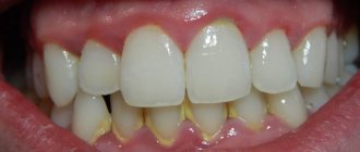
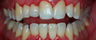
- The color of tooth enamel varies from white to yellowish, and it can change shade (color) depending on a person’s dietary habits or bad habits.
- The thickest areas of enamel cover the chewing molars in the area of their anatomical tubercles; their thickness varies between 2.3-3.5 mm;
- The thinnest areas of tooth enamel are localized in paroxysmal areas (places of contact in their lateral projection), here the thickness of the protective layer reaches approximately 1.3 mm;
- The enamel that covers all the teeth in the human oral cavity is not capable of regeneration, because there are no living cellular structures in the tissues of this protective layer;
- Depending on the characteristics of each person’s body, up to 95% of the chemical composition of tooth enamel is presented in the form of mineral compounds. The remaining percentage is divided between water and organic matter in approximately a 2:1 ratio, respectively. In addition, depending on the percentage of mineral content of tooth enamel, it can be more or less transparent (the higher the percentage of minerals, the more transparent the enamel becomes).
Microelements of dental plaque
The content of microelements in dental plaque is extremely variable.
The amount of microelements in dental plaque is comparable to their content in enamel and dentin. It can be noted that the enamel surface contains high concentrations of precisely those elements that are found in the greatest quantities in dental plaque (for example, iron, fluorine, zinc). One of the important components that actively influences the biochemistry of dental plaque is fluoride. Using colorimetric and enzymatic research methods, as well as using fluoride electrodes, it was possible to quite accurately determine the concentration of fluorine and the types of its compounds. It has been established that dental plaque can contain tens and hundreds of times more fluoride than saliva. The average concentration of fluoride in dental plaque, according to Hargreaves and Manly (1956), is 6 mg/kg, and according to Hardwick and Leach (1963) - 66.9 mg/kg in terms of wet weight.
The maximum amount of fluoride in dental plaque can reach 180 mg/kg. Only a small part of fluorine (2-3%) is ionized. The concentration of fluoride in dental plaque is largely influenced by its content in drinking water. There are three possible options for combining fluorine in dental plaque: a) the formation of inorganic crystals (fluorapatite, calcium fluoride); b) formation of a complex with inorganic substances (plaque matrix protein); c) penetration of bacteria.
Of the organic components in dental plaque, protein, carbohydrates, and enzymes were identified. The amino acid composition of dental plaque differs from the amino acid composition of mucin and pellicle, as well as saliva, while the composition of pellicle and salivary mucoprotein hydrolysates is identical. This casts doubt on the origin of dental plaque from saliva.
Of the carbohydrate components of dental plaque, glycogen, acidic mucopolysaccharides, and glucoproteins were discovered using histochemical reactions on decalcified sections of human teeth with plaque residues. In contrast to soft dental plaque, no histochemical reactions to detect the above components occurred in the pellicle. Several proteolytic enzymes of relatively low activity were found in dental plaque.
Other components in the enamel
In addition to the already mentioned main components of the enamel layer of the tooth, its chemical composition also contains a set of other components:
- Neonatal line - present exclusively on baby teeth, it looks like a dark-colored stripe (almost black). This line is located in the area of contact between two types of enamel, the first of which was formed before the baby was born, and the second after.
- Bundles and plates of dental enamel are special enamel formations containing prisms of a hypomineralized type, between which the interprismatic substance consists of the same material. It is noteworthy that the molecular structure of this material involves a large number of protein compounds. Many dentists are of the opinion that through the mentioned bundles and plates, various microorganisms penetrate into the enamel from the oral cavity, making their way to deeper dental tissues, causing caries, etc.
- Gunter-Schräger stripes are lines that stand out on the tooth enamel in a darker or lighter shade, the width of which does not exceed 100 microns. They are located perpendicular to the surface of the enamel layer and are formed as a result of opening its prisms.
- Retzius lines - in shape they resemble arches offset from the central one, located symmetrically in relation to each other. When cut across a tooth , these formations resemble rings inside a tree trunk. The formation of Retzius lines corresponds to different periods of mineralization of the enamel layer.
Symptoms, how to determine the presence of deposits
The formation of dental plaque cannot be tracked, since this process occurs at the cellular level. But its quantity must be controlled and not allowed to turn into hard stone, since this is fraught with the emergence of many problems. The following are symptoms that will help detect the presence of deposits in the oral cavity:
- yellow or white plaque that is located on the surface of the teeth, between them, as well as along the entire length of the gingival margin,
- teeth become rough if you run your tongue over them,
- there is an unpleasant odor from the mouth,
- As plaque turns into stone, bleeding gums appear, mucous membranes can move away from the surface of the teeth, change their color to bright scarlet or even bluish.
There are two types of dental plaque in terms of their localization, i.e. locations:
- superficial, that is, supragingival: visible to the naked eye, has a white, yellow or even brown color,
- subgingival: can be detected during an examination by a dentist, because is located under the mucous membranes and often the patient does not see or feel it (especially if the amount is small). It has a darker shade, even brown. It is impossible to remove it at home.
Features of the structure of the enamel of baby teeth
The main distinguishing feature of the enamel layer of children's teeth is that it is less durable and also much thinner than the enamel of permanent teeth. This is explained by the lower content of mineral compounds in teeth in relation to water and organic substances. Considering these features, if you examine baby teeth and their enamel layer under a microscope, you will notice the following differences:
- Due to the fact that the service life, as well as periods of mineralization and the tendency towards this process are shorter, the Retzius lines are much less pronounced in the structure of primary dental units.
- If in permanent teeth enamel prisms are located apically, then in milk teeth their direction is completely different, they are located horizontally.
- In children's primary teeth, the final enamel layer is much less pronounced; prisms are clearly visible on its surface, while its structure is much more porous, with microscopic cracks present.
Under the influence of each of these features, tooth enamel in children is more susceptible to wear and damage. For this reason, children develop caries much more often and it progresses faster, which is why it is important to visit the dentist regularly and have their teeth treated in a timely manner.
Types of damage to tooth enamel
Over the course of life, even if you provide your teeth with proper care and follow the rules of oral hygiene prescribed by dentists, the enamel layer gradually wears out and is destroyed. This contributes to the occurrence of various diseases of the oral cavity; it is influenced by the food a person eats, etc.
Among the main causes of damage and destruction of tooth enamel, dentists identify:
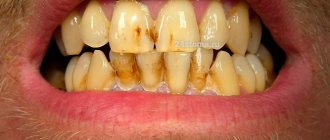
- Erosion is damage to the enamel layer, and then dentin, which is not associated with carious lesions of the teeth . The essence of this pathological process lies in disorders of mineral metabolism. As a result, disturbances occur in the crystalline structure of the enamel, which is manifested by its focal thinning and destruction. Externally, erosions look like local darkening on a round or oval tooth. The occurrence of erosions is provoked by the consumption of foods with high acidity levels, pathologies of the gastrointestinal tract, the use of certain medications, and the use of aggressive tooth powder or paste.
- Excessive sensitivity of tooth enamel - this disorder is especially pronounced in the form of painful sensations when the teeth cold or hot food, drinks, and even as a result of contact with cool air. The sensitivity of tooth enamel develops due to its thinning under the influence of the factors already described above. A thinned enamel layer puts teeth at increased risk of caries and other dental pathologies.
- Necrosis - this term characterizes multiple lesions of the hard tissues of the tooth, especially the enamel layer and dentin. The pathological process is initially expressed in the appearance of small light spots on the surface of the tooth , which subsequently darken and deepen. The progression of pathology threatens tooth destruction and is accompanied by a number of other oral diseases. The main reasons for the development of necrosis include gastrointestinal diseases, hormonal imbalances, metabolic disorders in the body, and work in hazardous industries.
- Caries is a carious lesion that threatens teeth , primarily affecting the enamel layer of the structure , gradually destroying it and spreading to deeper tissues. There are many reasons for the development of caries, from non-compliance with the rules of oral hygiene and irregular tooth brushing, to pathologies of the structures of the oral cavity, diseases of the gastrointestinal tract and metabolic disorders. If you start caries treatment at the stage when the lesions have affected only the enamel layer, you can only get by installing a filling or even restoring the enamel. But progressive caries is dangerous for teeth due to destruction, which may lead to the need for its complete removal.
- Mechanical damage - due to the fact that the main function of the enamel layer is to provide protection to the teeth , it primarily suffers from the effects of external adverse factors. Mechanical damage to the enamel includes cracks and other violations of its integrity due to blows, bruises, eating too hard food, etc. If the enamel of at least one tooth has been subjected to aggressive mechanical action, you should consult a doctor for an examination and, if necessary, subsequent treatment.
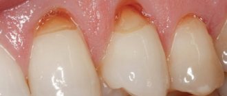
- Wedge-shaped defect – this term characterizes the pathological process in which the area of the dental neck is exposed. In such cases, the thinnest and most vulnerable areas of the enamel layer, located at the base of the teeth, are negatively affected. In addition to visible receding gums, damage to the enamel is indicated by a change in its color, as well as an acute reaction to hot and cold.
Physiological and pathological formations
Surface films of a physiological nature include the cuticle and pellicle.
Pathological deposits are dental plaque, tartar, plaque of various types.
Formations of a pathological nature are formed due to:
- poor oral care;
- the predominance of too soft foods in the diet;
- malocclusion;
- inflammatory diseases of the oral cavity;
- damage to the enamel structure.
Since the enamel, even under normal conditions, has a rough structure and is covered with microscopic cracks, external factors contribute to its uneven expansion. This creates conditions for the accumulation of minerals on the teeth, which over time form a dense structure.
Such deposits provoke the destruction of enamel, gum disease, caries, and bad breath. In addition, because of them, teeth do not look aesthetically pleasing.
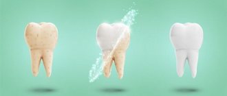
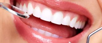
Strengthening tooth enamel
Today in dental practice there are many effective ways to strengthen the enamel, which allows you to maintain its integrity and prevent destruction and diseases of the dentition. At the same time, methods of strengthening and protecting the enamel layer are divided into two groups, the first are intended for adults, the second for children.
Strengthening baby teeth enamel
As was said earlier in relation to baby teeth, their enamel is more vulnerable. To protect it, saving the child from premature loss of dental units and problems in the future, doctors perform the following actions to provide temporary protection:
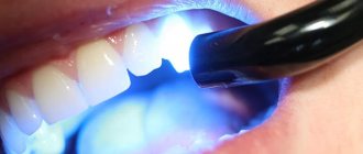
- Fluoridation involves treating teeth with special fluoride-based compounds; it is recommended to repeat this procedure 2-3 times a year.
- Fissure sealing - the dentist performs the procedure of filling the recesses and grooves of chewing teeth with temporary filling material, protecting the dental structures from the negative effects of harmful microorganisms and other unfavorable factors.
- Application gels and preventive mouth guards for teeth - the method is based on enriching the enamel layer with useful components (fluorine, calcium, vitamins) through the use of special products.
Standard method of brushing teeth Pakhomova G.N.
This method is recommended by most Russian dentists. It was invented by G.N. Pakhomov - professor of dentistry.
- Cleaning begins from the upper jaw in the following order - from the right molars to the front ones, then finishes on the left molars.
- On the lower jaw, the left major ones are cleaned first, then the anterior ones, and lastly the right molars.
- The front and back surfaces are cleaned with sweeping movements from bottom to top, with the brush head at an angle of 45° to the gum.
- It is important to make 10 movements for each tooth.
- Contaminants from the chewing side are removed by moving the head back and forth.
- To clean the front teeth, the brush is placed vertically and moved in a sweeping motion.
The Pakhomov method helps to effectively clean the oral cavity, but for the interdental spaces additional means are needed - irrigators, threads and a tongue scraper.
Strengthening the enamel of molars
There are more methods to preserve molars and maintain the condition of their enamel layer. Firstly, this is due to fewer contraindications for adults. Secondly, molars require long-term strengthening.
The main methods of strengthening the enamel of permanent teeth include:
- Drug therapy is based on the use of vitamin complexes containing vitamins of groups B6, B12, D. In addition, the patient is selected drugs that promote better absorption of calcium and fluoride by the body.
- Special gels and oral hygiene products – this technique uses specialized toothpastes and gels containing components necessary for teeth to strengthen and maintain the condition of the enamel layer. Also, teeth are subjected to unimaginative cleaning in a dental office.
- Mineralization and preventive cleaning – mineralization is performed using special means to increase the strength of the enamel and reduce its susceptibility to a number of negative factors. As for cleaning, such procedures are performed by dentists in the clinic using special equipment. During cleaning, plaque and tartar are eliminated , pathogenic bacteria and microorganisms that can harm the enamel layer are removed.
- Home prevention - to maintain healthy teeth and enamel, patients are advised to perform a light massage of the gums, enrich the diet with fresh vegetables and fruits rich in vitamins.
Author: Zhukov M.A.
Pellicle of the tooth
The leading role in the development of inflammatory periodontal diseases is played by microbial infection located in hard and soft dental plaque, especially strains of periodontal pathogenic anaerobic microorganisms (Prevotella intermedia, Porphyromonas gingivalis, Actinobacillus actinomycetemcomitans, etc.).
Dental plaque can be divided into 2 groups:
- Non-mineralized dental plaque
Pellicle Dental plaque, biofilm Soft plaque Food debris
- Mineralized dental deposits
Supragingival calculus Subgingival calculus
An unstructured cell-free film is formed on the cleaned surface of the tooth - pellicle . It consists of salivary proteins linked by electrostatic bonds.
The pellicle performs both a protective function and promotes the attachment of microorganisms.
Within a few hours, gram-positive cocci and actinomycetes attach to the pellicle Gradually, the thickness of the plaque increases due to the division and accumulation of bacteria. Poor oral hygiene results in the formation of dental plaque .
A mature dental plaque consists of a dense layer of bacteria that forms its matrix. The initially formed plaque contains aerobic microorganisms. Over time, anaerobic flora begins to predominate.
In recent years, researchers have introduced the concept of " biofilm ". Biofilm is a well-organized, interacting community of microorganisms.
Microorganisms are grouped into microcolonies, surrounded by an enveloping intermicrobial matrix, which have their own microenvironments that differ in pH levels, nutrient absorption, and oxygen concentrations. Bacteria in a biofilm communicate with each other through chemical stimuli (signals).
The main features of a biofilm are the formation of microcolonies by microorganisms surrounded by a protective matrix and the interaction between different types of microorganisms. Microorganisms in the biofilm are resistant to antibiotics and the host response.
The matrix surrounding the microcolonies also serves as a barrier that protects the biofilm from antimicrobial agents, both general and applied locally. This also explains the need for mechanical removal of plaque and the leading role of individual cavity hygiene in the treatment of periodontal diseases.