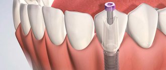
A sinus lift is aimed at forming a sufficient amount of bone tissue for further placement of a dental implant. Surgical intervention is performed exclusively on the upper jaw in order to raise the bottom of the maxillary sinus to create the required amount of hard tissue for fixation.
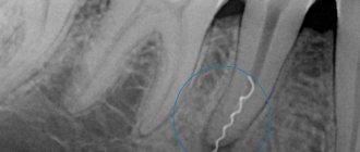
The development of a fistula occurs in the first 5 to 14 days after the operation. Its formation is associated with the introduction of pathogenic microflora during the procedure itself or in the first days after it, due to non-compliance with personal hygiene rules. The fistula after sinus lifting and bone grafting can have a diameter from several millimeters to 1 - 1.5 cm, creating communications between the oral cavity and the maxillary sinus.
Features of the maxillary sinus
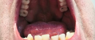
The maxillary sinus (its other name is the maxillary sinus) is located in the thickness of the bone tissue of the upper jaw. It is separated from the oral cavity by the alveolar process of the upper jaw, which forms its bottom. The volume of such a sinus is quite large, and in adults it can reach 10 cubic centimeters.
This sinus, or sinus, is not airtight. It communicates with the nasal cavity through a narrow slit.
Typically, perforation of the maxillary sinus occurs in the area of its bottom. Some of its features contribute to this:
- Close proximity of the roots of molars and premolars. In some cases, the thickness of the bone layer between the tooth roots and the bottom of the maxillary sinus can be relatively large - up to 1 cm, but in some people the bone border between these formations is very thin - no more than 1 mm.
- Sometimes the roots of the first and second molars are located in the sinus cavity itself, separated from it by just a layer of mucous membrane.
- Rapid thinning of the bone layer in the presence of acute or chronic inflammatory diseases: periodontitis, periodontitis, cysts.
- Relatively thin bony trabeculae in the tissue of the upper jaw.
All this predisposes to the occurrence of perforation during dental procedures, even if the treatment technique was not violated and the doctor did not apply significant traumatic force.
Causes of perforation
The physiological location of the upper teeth and associated pathologies are a risk factor that can cause perforation of the maxillary sinus. Complications can be characterized by dental operations. In particular, this is the sanitation of dental tissues in case of deep caries, the removal of part of the tooth or its root. It is important for the dentist to carry out surgical intervention extremely carefully without fragmenting or causing fractures of the tooth roots, which is why mandatory x-ray monitoring is required.
When carrying out dental treatment associated with the removal of an upper wisdom tooth, perforation can occur when manipulating the root of the tooth, which is often removed in fragments. Pathology also occurs during manipulations in the root of the tooth during the treatment of caries and when intensive canal filling is done.
Perforation may occur during root resection and installation of implants in the upper jaw. The dentist must act with extreme caution when removing the upper tooth. First, an image of the upper jaw is taken, which allows one to assess the location of the roots relative to the maxillary sinus, and then the doctor determines the subsequent tactics of therapeutic or surgical treatment.
Article on the topic: Open and closed sinus lift surgery
Causes of perforations of the maxillary sinus floor
The etiology of perforations of the maxillary sinus is always associated with any dental procedures. Perforation can occur:
- when removing teeth;
- during endodontic treatment;
- during dental implantation;
- during root resection.
When teeth are removed, damage to the bottom of the maxillary sinus can be a consequence of either rough actions by the dentist or failure to comply with treatment tactics, or as a result of the anatomical characteristics of the patient himself (for example, when the tooth roots are located directly in the sinus cavity).
When endodontic treatment is carried out, one of the complications is perforation of the tooth root, which is often combined with damage and perforation of the bottom of the maxillary sinus. This happens when the root canals are excessively expanded, when brute force is used when inserting pin elements or compacting filling cement. With this type of perforation of the maxillary sinus, filling material or root fragments almost always penetrate into its cavity.
If perforation occurs at the time of insertion of a dental implant (it can be an implant of any brand, for example, Mis, Nobel, Xive, etc.) or during root canal filling or insertion of pins into the tooth root, then it is always a therapeutic error doctor's tactics.
Damage to the bottom of the maxillary sinus is a serious complication when implanting artificial roots into bone tissue during prosthetics. This is explained by the fact that after tooth extraction, bone tissue very quickly undergoes degeneration processes. As a result, the height of the alveolar process of the jaw decreases. If the doctor does not take this point into account and incorrectly carries out preparations before implantation, and also incorrectly selects the size of the implant, then the risk of sinus perforation is very high.
Tooth root resection is a treatment method for the presence of cysts in the area of its apex. If the patient is underexamined, when the doctor does not know the exact size of the bone plate separating the bottom of the sinus from the wall of the cyst, and also if it is necessary to remove a large volume of jaw bone, then perforation of the maxillary sinus is not a rare phenomenon.
Causes and mechanisms of fistula development
The oral cavity, due to the peculiarities of its anatomical structure and physiological load (food intake and salivation), is aseptic. The presence of microorganisms creates conditions in which the penetration of infection into all kinds of wounds and surgical incisions is almost inevitable. Despite the fact that during bone grafting surgery the most sterile conditions are created to prevent bacteria from entering the wound, their infection cannot be ruled out both during the intervention and in the postoperative period. Most inflammatory processes after sinus lifting develop due to the patient’s violation of the dentist’s recommendations, namely: ignoring disinfectant rinses, antibacterial ointments and oral baths, eating on the side of the bone graft.
Once in tissues that have recently undergone surgery, bacteria find themselves in ideal conditions for reproduction. The presence of minor blood clots, tissue detritus, pieces of food, if hygiene requirements are not followed and the optimal temperature is present, create a breeding ground for them. In the process of life, microorganisms destroy the structures surrounding them, and the release of leukocytes from the bloodstream to destroy hostile microflora leads to the release of pus and inhibition of healing processes.
The fistula after sinus lifting and bone grafting is not able to heal on its own due to the presence of a defect in the bone tissue. The development of such a pathological course leads to the appearance of a focus of chronic infection, the penetration of bacteria into the maxillary sinus with the formation of sinusitis and other complications. In this case, the fistula after an open sinus lift can be located not only at the implant site, but also on the lateral surface of the gum. The appearance of a fistula tract more often occurs after closed bone grafting due to worse fixation of the denture.
Among other things, the formation of a fistula can occur when the tooth root is not completely removed during a sinus lift.
Symptoms of perforation
If perforation of the nasal sinus occurred at the time of tooth extraction, then its symptoms will be quite specific:
- The appearance of small air bubbles in the blood released from the tooth socket, the number of which increases with a sharp forced exhalation through the nose.
- The appearance of bloody discharge from the nose on the side of the perforated maxillary sinus.
- Changes in the timbre of the patient’s voice, the appearance of “nasality”.
Sometimes the patient begins to complain about the passage of air through the hole after tooth extraction, as well as a feeling of heaviness or pressure in the projection of the maxillary sinus.
If perforation of the maxillary sinus occurs during implantation or endodontic treatment, the doctor may suspect it by:
- characteristic failure of the instrument or implanted element after applying some force to advance it;
- changing the position of the instrument in the wound;
- the appearance of small air bubbles in the blood.
If perforation of the maxillary sinus for any reason was not diagnosed and treated immediately, then its cavity becomes infected with the development of acute sinusitis or sinusitis, which is characterized by symptoms such as:
- severe acute pain in the maxillary sinus area;
- swelling of the nasal mucosa on the corresponding side with difficulty breathing through the nose;
- the appearance of purulent nasal discharge.
The appearance of general symptoms of intoxication is also characteristic: headaches, chills, high fever, weakness.
Dial-Dent specialists who performed this work:
- ENT doctor A.V. Arkhandeev – endoscopic diagnostics, diagnostics using computed tomography of the sinuses, surgery on the maxillary sinus, treatment of chronic sinusitis.
- Implant surgeon V.P. Alaverdov – tooth extraction, elimination of a fistula in the maxillary sinus.
- Anesthesiologist D.V. Sidorov - sedation, monitoring the patient’s condition during surgery.
- Dentist-endodontist T.I. Matienko – analysis of the condition of teeth, development of treatment tactics.
- Periodontist surgeon N.V. Nekrasova – consultation on the treatment of periodontitis and the choice of management tactics for the patient.
- Assistant dentists M. Erkimbekova, E. Sheiko, A. Arzumanyan.
- Assistant to ENT doctor E. Shilova.
See other examples of treatment from Dial-Dent specialists here.
Make an appointment for a consultation by phone +7-499-110-18-04 or through the form on the website. You can ask questions about dental treatment to the chief doctor of the clinic, Sergei Vladimirovich Tsukor, at
Diagnostics
Diagnosis of perforation of the floor of the maxillary sinus during tooth extraction is based on a typical clinical picture. In doubtful cases, as well as when such a complication is suspected during implantation or endodontic manipulations, it is necessary to use instrumental diagnostic methods:
- Probing the socket of an extracted tooth or perforated canal with a thin probe . This allows us to determine that there is no bone bottom in the wound. In this case, the instrument passes freely through soft tissues and does not encounter obstacles along its path.
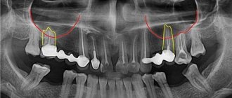
- X-ray of the sinus area . In this case, the pictures can reveal both darkening of the cavity due to the accumulation of blood in it, as well as fragments of dental roots, implants or filling material. Sometimes it is advisable to conduct radiography with contrast, when a contrast agent is introduced into the cavity through a perforation fistula.
- Computed tomography , which allows you to determine perforations and the presence of foreign bodies in the sinus with maximum accuracy.
- If old perforations are suspected, general clinical blood tests , the result of which may indicate the presence of an active source of infection in the body.
Diagnosis
At the time of a dental operation that led to perforation of the maxillary sinus, the doctor draws a conclusion about a new problem based on the signs described above. If perforation is questionable, the dentist's findings are rechecked using the following methods:
- probing with a thin probe;
- radiography - shows dark accumulations of blood, tooth root fragments and filling cement located inside the maxillary sinus, can be done with the preliminary administration of a contrast agent;
- CT scan;
- general clinical blood test - necessary when identifying an old problem to make sure there is an infection in the body.
Treatment
Treatment of perforations of the maxillary sinus floor depends on what changes are present in the sinus cavity itself.
Treatment without surgery is possible only in cases where perforation occurred during tooth extraction and was detected immediately, and according to radiography there are no signs of infection of the sinus cavity or the presence of even minor foreign bodies in it. With this option, the doctor’s tactics are to preserve the blood clot formed in the socket as carefully as possible, as well as to prevent its infection. To do this, a small gauze swab soaked in iodine solution is inserted into the lower part of the hole. Usually it is tightly fixed in the wound cavity on its own, but sometimes sutures are required on the gum. This treatment with iodine continues for at least 6-7 days until full granulations are formed and the defect is closed. In this case, the tampon is not removed from the hole so as not to damage the blood clot.
It is also possible to temporarily close the defect with a small plastic plate, which is fixed to adjacent teeth with clasps. It separates the oral cavity and sinuses, which promotes healing of the perforation.
At the same time, a course of preventive measures is prescribed, aimed at preventing the development of inflammatory complications. It includes taking antibiotics, anti-inflammatory drugs, drops with a vasoconstrictor effect. This course is carried out on an outpatient basis or at home.
If, during perforation, foreign bodies penetrate into the sinus (implant, filling material, fragment of a tooth root), then treatment is carried out only in a hospital setting. In this case, an operation is indicated to open the cavity of the maxillary sinus, remove the foreign body and non-viable tissue, followed by plastic closure of the perforated defect.
»
How to treat
The fistula after sinus lifting and bone grafting is initially subjected to antibacterial therapy. In the case of a “fresh” fistula, broad-spectrum antibiotics are used: Lincomycin, Erythromycin, Summed.
If the pathological course persists for a long time, then a test is performed to determine the sensitivity of bacteria to antibiotics, after which the necessary drug is selected. The use of rinses and baths should be strictly limited due to the risk of penetration of the contents of the fistula canal from the oral cavity into the maxillary sinus. It is possible to use ointments with an antibacterial effect. In the presence of purulent discharge, punctures of the maxillary sinuses and fistula are performed in order to create outflow paths for pus.
After eliminating the pathogenic microflora, surgical intervention is performed to close the oroantral tract. The operation can be performed under local or general anesthesia - it all depends on the scope of the intervention and the patient’s wishes. It is possible to make a special mouthguard in advance to protect the operated area from microflora. The operation takes place in several stages:
- An additional hole is formed for insertion of the endoscopic probe. Using a probe, the maxillary sinus is inspected and the fistula tract on its side is closed.
- Dead tissue is excised and the fistula tract is closed using one's own tissue or artificial substitutes.
- The external opening of the fistula is closed with a mucoperiosteal flap on a vascular pedicle.
The postoperative period requires special attention to prevent re-infection. If the outcome is favorable, a repeat sinus lift and bone grafting may be performed after 1 to 2 months. After closing the fistula tract, the bone defect is not eliminated, which is felt by the patient as a “sinking” of tissue when pressing on the site of the existing fistula. The lack of bone tissue creates discomfort when eating.
Old perforations
If the perforation of the maxillary sinus was not promptly identified and eliminated, then after 2-4 weeks the stage of acute manifestations will subside, and a fistula will form in the area of the defect, connecting the sinus cavity with the surface of the gum.
This process is simultaneously accompanied by symptoms of chronic sinusitis:
- constant dull pain in the sinus area radiating to the orbit and temple;
- nasal congestion on the affected side;
- purulent discharge from the nasal cavity, as well as from the fistula;
- Sometimes patients have swelling of the cheek on the side of the damaged sinus.
Most patients also complain of a sensation of air moving through the fistula when talking or sneezing, difficulty pronouncing certain sounds, and liquid food entering the nasal cavity from the mouth.
Treatment of such chronic perforations with fistulas presents some difficulties, since the presence of a chronic focus of inflammation in the maxillary sinus significantly reduces the effectiveness of therapy and quite often leads to relapse and re-formation of the fistula canal.
Such patients are indicated for surgical intervention, which includes opening the maxillary sinus with removal of all non-viable tissues and foreign bodies from its cavity, excision of the fistula and plastic closure of the defect. Antibiotics after removal of the fistula are prescribed for a course lasting 10-14 days with the simultaneous use of anti-inflammatory and antihistamine drugs, and the use of physiotherapeutic methods of treatment.
Symptoms of the disease
Some of the first manifestations of a fistula may be pain in the operated area when tilting and turning the head, a feeling of loosening of the implant, and an increase in temperature to 37.0 - 37.5 ° C.
The rate of progression of the disease is individual; over time, the denture may fall out (during its installation) or a hole may directly appear in the alveolar process. At this stage, there may be discharge of pus and mucus from the fistula, and a sensation of air vibrations in the oral cavity during breathing. When microorganisms or pieces of food enter the maxillary sinus, sinusitis develops, which aggravates the patient’s general condition. There may be a feeling of “softness” of the palate and alveolar process. If the fistula tract is 1 - 3 mm in diameter, then all kinds of discharge may be absent.
Eating becomes difficult due to severe pain. When coughing or sneezing, mucus may be thrown into the oral cavity, and there may be a sensation of air moving from the fistula.
One of the common signs of the oroantral tract is the development of odontogenic sinusitis. This disease can lead the patient to an otolaryngologist, directing the diagnostic algorithm and treatment in the wrong direction. This type of sinusitis is characterized by a long course with no response to treatment and antibiotics used. Puncture of the maxillary cave will also not have the desired effect.
Consequences of perforation
Perforation of the maxillary sinus is a fairly serious pathology that often has to be treated in a hospital. Attempts to independently treat it with folk remedies at home without medical assistance can lead to the development of serious and dangerous consequences:
- The development of a pronounced inflammatory reaction in the sinus cavity with the spread of infection to the surrounding bone tissue and the formation of foci of osteomyelitis of the upper jaw.
- Spread of inflammation to other sinuses of the skull (frontal, sphenoid and ethmoid).
- Loss of healthy teeth located in the area of untreated perforation.
- Formation of purulent foci (abscesses, phlegmons).
Due to the close location of the maxillary sinus and the brain, after perforation, infection may spread to the meninges with the development of meningitis or meningoencephalitis, which threatens the patient’s life.
Why shouldn’t you treat perforation of the maxillary sinus yourself?
To date, there are no effective treatments for perforation other than surgery. An attempt to cure yourself at home using “traditional medicine” means that time will be lost and the situation will become neglected. You can start the problem before complications arise:
- the sinus cavity becomes inflamed, the infection spreads to the bone tissue, the patient begins to suffer from osteomyelitis of the upper jaw;
- inflammation penetrates into other intracranial sinuses, and there are more foci of infection;
- next to the untreated perforation, the alveolar process is weakened, as a result of which healthy teeth may fall out;
- foci of suppuration develop.
The maxillary sinus is also located in close proximity to the brain. The lack of a timely response and the development of suppuration inside the sinus is fraught with meningitis and meningoencephalitis - these are diagnoses that directly threaten life.
Preventive actions
Prevention of perforations of the floor of the maxillary sinus consists of:
- in a full examination of the patient before complex dental procedures;
- in the correct assessment of the anatomical and topographical characteristics of each person;
- in strict adherence to the technology of therapeutic manipulations.
Timely detection of signs of perforation and its adequate treatment is the key to a favorable outcome for the patient. Incorrect therapeutic tactics or self-medication can aggravate the course of such a complication and cause the development of severe negative consequences.