Gingivitis
Acute catarrhal gingivitis is an inflammatory process with a predominance of exudation. This disease is characterized by complaints of pain, itching, bleeding gums, and refusal to eat. Upon examination, hyperemia and swelling of the papillae, gingival margin, and sometimes the alveolar gum are determined, which leads to an increase in size and a change in the shape of the gingival papillae (not sharp, but rounded). When the alveolar gum is involved, its granularity (symptom of lemon peel), characteristic of healthy gums, disappears. Bleeding gums are detected when you touch them slightly. This form of gingivitis is observed relatively rarely, mainly during teething, as a result of acute trauma (mechanical, chemical, thermal), in acute childhood infectious diseases, and also as a manifestation of allergies, a symptom of acute herpetic stomatitis.
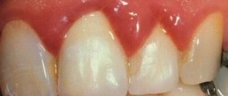
Chronic catarrhal gingivitis is the most common periodontal pathology in children. There may be no complaints; sometimes children notice bleeding gums when brushing their teeth. Upon examination, congestive hyperemia, pastosity, bleeding of the papillae and gingival margin are determined. The marginal gum is thickened like a roller (stretches like a rubber balloon), the surface becomes shiny. The granularity of the alveolar gum disappears.
With catarrhal gingivitis (both acute and chronic), with significant swelling of the gums, a gingival pocket, that is, a false periodontal pocket, can occur. The radiograph shows no changes in the alveolar bone.
Hypertrophic gingivitis is a chronic inflammatory process characterized by an increase in gingival papillae and marginal gums due to the proliferation of fibrous connective tissue and the basal layer of the epithelium. Most often, hypertrophic gingivitis is observed with close position of the teeth, open bite, small vestibule of the oral cavity, short frenulum of the lips, with improper orthodontic treatment, as well as during puberty (in girls with a late onset of menstruation or a long menstrual cycle, when the effect of estrogens predominates) , when taking antiepileptic drugs and blood diseases.
Hypertrophic gingivitis, caused by local factors (crowding of teeth, dental anomalies), is limited in nature, located mainly in the frontal area. Hypertrophic gingivitis, caused by endogenous causes (endocrine pathology, gastrointestinal diseases, etc.), is generalized, characterized by rapid development, a tendency to relapse and resistance to local treatment.
There are inflammatory (granulating) and fibrous forms of hypertrophic gingivitis. The inflammatory form is characterized by: hyperplasia of the gingival papillae, which are covered with granulations; their hyperemia with pronounced cyanosis and swelling; loose tissue and severe bleeding; pain on palpation; thickening of the gingival margin, its separation from the necks of the teeth, chronic catarrhal inflammation of the alveolar gum. False periodontal pockets are identified due to swelling and hyperplasia of the gums. There is a deposition of plaque and tartar. Hypertrophied marginal gum covers one or another part of the tooth crowns.
In the fibrous form, the gingival papillae are enlarged, dense, pale, do not bleed, and are painless.
According to the intensity of productive inflammation, there are 3 degrees of hypertrophic gingivitis:
— 1st degree — hypertrophy of gingival papillae;
— 2nd degree — hypertrophy of the gingival papillae and marginal gums;
— 3rd degree — hypertrophy of the marginal and alveolar gums.
Desquamative gingivitis is clinically characterized by swelling of the marginal gum, sometimes extending to the attached gum, pronounced hyperemia, severe bleeding and pain. A feature of this form of gingivitis is the constant desquamation of the surface layers of the gum epithelium, not only marginal, but also alveolar, as a result of which almost the entire mucous membrane becomes eroded and easily vulnerable. May occur against the background of hypertrophic gingivitis. Children complain of pain when brushing their teeth and eating, the presence of blood in saliva, the specific smell and taste of blood.
Desquamative gingivitis in children develops rarely, mainly during puberty (in girls, usually during a short menstrual cycle, when the action of progesterone predominates).
Periodontal diseases in children
Key words: periodontal diseases in children, gingivitis, periodontitis, periodontal disease, percentage
Introduction: The significance of periodontal diseases among the child population is determined by their steady growth, widespread distribution and the negative impact of periodontal foci of infection on the body as a whole. The severity of these changes depends both on the general condition of the body and on the age-related characteristics of the structure of periodontal tissues. During the period of growth and development of the dental system, and periodontal disease in particular, long-term pathological processes lead to disruption of tissue formation and early destruction of the periodontal complex, development of mobility and tooth loss. The prevalence of periodontal disease in children is increasing. Thus, according to A. M. Politun, in schoolchildren it is 39%; periodontitis is more common at puberty (13–15 years) and is 7.7% and 11.3% in 16–18 years. The issue of the prevalence of periodontal diseases in children has not been sufficiently studied, and therefore we decided to carry out this work.
Purpose of the study: To calculate the percentage of patients with various periodontal diseases among the child population of Vladikavkaz.
Materials and methods: The studies were conducted at the SOGMA dental clinic. The study involved 136 patients aged 4 to 16 years living in Vladikavkaz. All subjects were divided into 3 groups by age:
Group 1 – 4–7 years (39 people)
Group 2 – 8–12 years old (51 people)
Group 3 – 13–16 years old (46 people)
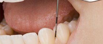
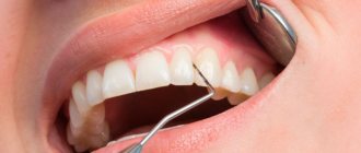
During the work, the data from the medical record (F.043/u) was taken into account. The study was carried out in two stages. At the first stage, a survey was conducted (identification of complaints and careful collection of anamnesis), examination of the oral cavity, percussion, and palpation. At the second stage, special research methods were used: Schiller-Pisarev test, x-ray, determination of Fedorov-Volodkina indices, PI and PMA. The presence of deposits of supragingival tartar, plaque (plaque), and subgingival tartar was assessed. Inflammatory phenomena in the gums were assessed according to the following signs: hyperemia, swelling, bleeding, desquamation, ulceration. In addition to these signs, atrophic and hyperplastic processes were noted. In the presence of a pathological process, its localization, quantity, symmetry, staining intensity, size, and quantity were taken into account.
Research results:
When examining the first group of patients (4–7 years):
− In 16 patients (41%), mild to moderate catarrhal gingivitis was found, which is characterized by hyperemia of the marginal periodontium, edema, bleeding from the apexes of the papillae when pressed at their base, and the presence of dental plaque. The general condition is not disturbed.
− 11 patients (28%) had hypertrophic gingivitis. Of these, 7 (18%) had an edematous form and 4 (10%) had a fibrous form. Clinically, this process manifests itself in the form of significant growth of the gums in the area of the front teeth. Swelling and hyperemia are weakly expressed.
- Hypertrophic gingivitis.
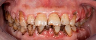
− Only 7 patients (18%) had ulcerative gingivitis. It is characterized by ulceration of the gingival margin, the ulcerative surface is covered with fibrinous plaque, which, when removed, causes bleeding.
− Localized periodontitis was observed in only 8 patients (21%), and generalized periodontitis (mild) in 15 patients (59%) and was objectively manifested by bleeding, deposits of subgingival tartar and plaque, mobility of teeth of varying degrees, pain, purulent discharge from pocket
− No periodontal disease was detected.
When examining the second group of patients (8–12 years old), we found:
− Catarrhal gingivitis with generalized symmetrical lesions in 19 patients (37%);
− Hypertrophic gingivitis was present in 21 patients (41%). Of these, the edematous form is in 15 (29%) people, the fibrous form is in 6 (12%) people.
− Ulcerative gingivitis, accompanied by fever, headache, insomnia and enlarged and painful regional lymph nodes, was observed in 10 patients (20%).
− Localized and generalized periodontitis was found in 11 (22%) and 17 (33%) patients, respectively.
− Periodontal disease was observed in 1 (2%) patient. In this form there are no inflammatory phenomena and periodontal pockets. It is characterized by good fixation of teeth, gum retraction, exposure of tooth necks and wedge-shaped defects.
When examining the third group of patients (13–16 years old), they received:
− Catarrhal gingivitis in 26 patients (57%), hypertrophic - in 23 patients (50%) and ulcerative - in 18 patients (39%). Localized periodontitis was observed in 15 patients (33%), and generalized in 10 patients (22%). Periodontal disease was observed in 3 (7%) patients.
Conclusions: Periodontal disease in children is one of the first morbidity problems in pediatric dentistry; it is necessary to devote more time to the prevention of periodontal disease in order to avoid morbidity, as well as to reduce the percentage of periodontal disease in childhood. It is necessary to talk with parents about the dangers of bad habits, proper nutrition and the consequences of abnormalities in the position of teeth and bite.
Literature:
- E. V. Borovsky, Yu. M. Maksimovsky // Periodontal diseases // Therapeutic dentistry 2001 - P.366–422.
- E. G. Denisova // Periodontal diseases in children // Textbook for dental interns, 2008 - pp. 55–75.