Anatomy of the human upper and lower jaws. Their structure and functions.
The jaws are part of the human facial skeleton. The upper and lower jaws are not connected to each other at the skeletal level and are connected only by soft tissues.
Schoolchildren often make the mistake of claiming that the upper and lower jaw are united by a joint. But in reality, the lower jaw is connected by a joint to the temporal bone , and this joint is called the temporomandibular joint .
Jaw structure: temporomandibular joint
The human lower jaw is the only movable bone in the human skull. And the joint that provides this mobility is special; it provides movement in three directions at once: vertical movements (opening and closing the mouth), horizontal movements (moving the lower jaw from side to side), and pushing the lower jaw forward.
The human upper jaw , anatomically speaking, is much larger in the area where the upper teeth grow. The upper jaw ends only in the area of the bridge of the nose, where it is fixedly connected to the frontal bone. It turns out that the bones of the face that surround the nose up to the bridge of the nose and the inner corners of the eyes are all the upper jaw. Scientifically speaking, the upper jaw has four processes: the alveolar (where the teeth grow), the frontal, the zygomatic and the palatine, which forms our hard palate.
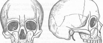
Human skull bones, upper jaw highlighted in pink
The human upper jaw is considered an air-bearing bone, since it contains the maxillary sinuses , anatomically these are cavities in the bone lined with epithelium. This epithelium is ciliated, that is, it ensures the movement of air inside the sinus; it also contains glands that secrete mucus.
It is interesting that scientists are still not completely clear why a person needs the maxillary sinuses; some say that they are needed to warm and humidify the air, others hypothesize that they house a whole internal laboratory of the body, where it studies microbes that enter them from the environment.
Maxillary sinus or sinus
Scheme
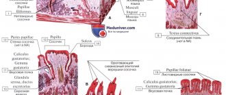
Human teeth consist of three elements:
- Crown . The uppermost visible part, which fully or partially protrudes above the alveolus after eruption;
- Cervix . A narrower area located in the gum between the main crown and the root.
- Root . The lowest part, located in the alveolus. The pulp of the root contains intertwining nerves and blood vessels. With the help of the periosteum, the roots are tightly fixed in the alveolar socket. Depending on the functions performed by the tooth and the anatomical features of the person, the number of roots can vary from 1 to 4 units.
The main substance in the structure of a tooth is dentin, which makes up most of its mass. From a chemical point of view, dentin is collagen impregnated with various salts, phosphorus and other minerals.
Photo: diagram of the structure of human teeth and jaw
The crown is covered with enamel on top. Due to the fact that the crown is based on inorganic compounds, its strength is close to that of diamond . Metabolic processes take place only in a thin skin that tightly envelops the surface of intact enamel.
The tooth is fixed using “special cement” covering the root. In its structure, cement is very close to the structure of bone tissue. Blood flow occurs through the branches of the external carotid artery, tightly intertwined. The outflow of venous blood occurs through vessels directly connected to the blood circulation of the brain.
Such blood circulation, in turn, carries a danger: if the initial infection is localized in the oral cavity, through these vessels it can enter the dura mater of the brain and cause a number of serious diseases.
In an adult, the row consists of two arches, each of which contains from fourteen to sixteen teeth. For children under twelve years of age, the row looks a little different - they, as a rule, only have twenty dairy products.
The external similarity of the structure of the upper and lower jaws does not indicate their identity, so you should familiarize yourself with their structure and distinctive features.
In the following video you can clearly see all of the above:
Blood circulation and sensitivity in human jaws
In the human lower jaw there is a maxillary canal, an elongated narrow cavity through which the mandibular artery, vein and mandibular nerve pass. The upper jaw also has a maxillary artery, vein and nerve, but their structure, as can be seen from the figure below, is more branched.
These vessels and nerves provide nutrition and sensitivity to the teeth, but not only to them, they are also responsible for the sensitivity of the soft tissues of the face and partially provide blood circulation to them.
Blood circulation in the upper and lower jaw, as well as the facial nerve
If you look closely at the drawing, it becomes clear why half of the jaw, and sometimes even half of the face, can hurt because of one bad tooth. All these areas are united by the trigeminal nerve .
Nature has made sure that the facial nerve is reliably protected by bone tissue. But damage to it is dangerous because it can lead to loss of sensitivity and immobility of part of the face.
Structure of the lower jaw
Through the mental foramen , a bundle of nerves and blood vessels exits the body of the jaw into the soft tissues. Typically, the mental foramen is located between the 4th and 5th teeth, but its location may vary slightly from person to person.
About the blood circulation in the human jaws, we can say that the upper and lower jaw arteries are branches of the carotid artery. And venous blood from the jaws subsequently ends up in the jugular vein.
Blood supply to human jaws
Upper jaw
The central incisor is characterized by the presence of a flat shape, a beveled cutting edge and a single root. The front part of the incisor is convex and contains three small tubercles.
The appearance of the lateral incisor is identical to the central one. But due to the fact that the central tubercle is large and stands out much more strongly, the cutting edge itself takes on a convex, streamlined shape.
Find out the reasons for the formation of a yellow coating on the tongue, and ways to prevent this disease.
In this article you will find photos with symptoms of oral leukoplakia.
Here: https://www.vash-dentist.ru/lechenie/zubyi/kakie-simptomyi-vospaleniya-troynichnogo-nerva.html - you can find out why the trigeminal nerve becomes inflamed and how it is treated.
Fang is an element inherited by a person from predatory representatives of the fauna. There is only one voluminous tubercle on the canine crown. With the help of a groove running along the inside, the fang is divided into two parts.
Small molars (called premolars ). Unlike the frontal ones, premolars are characterized by a more square shape. The roots, although flattened, are already beginning to bifurcate.
Large molars (aka molars) are the largest in the entire row and are responsible for the direct grinding of food. The first molar has a rectangular shape with four cusps, which allows you to chew food as efficiently as possible. The second molar is somewhat smaller in size, but in terms of functionality and root structure it is practically no different from its predecessor.
The third molar, also called the wisdom tooth, grows much later than the others. Sometimes it may not erupt at all, which is not very scary, since it does not perform any important functions and is largely a vestigial organ.
Structure and functions of the upper and lower jaw: how many teeth does a person have?
How many teeth does a person have? By nature, an adult should have from 28 to 32 teeth (including wisdom teeth). It is interesting that not everyone has 8 teeth or wisdom teeth; some people only grow 3 or 2 wisdom teeth, and the rest do not grow at all. In addition, wisdom teeth are often underdeveloped, with one root, and with weak enamel.
Scientists consider wisdom teeth to be an atavism, they say that their presence is not necessary, and over time, fewer and fewer people grow wisdom teeth. There is also a tendency towards a decrease in the size of the human jaws themselves. For example, Neanderthals had a much smaller brain and much larger jaws than modern humans; the trend towards changes in the size of these organs continues to this day.
Jaw size of humans and monkeys
By nature, a person should have 28 teeth, but the sad statistics are that the presence of such a number of teeth is the exception rather than the rule.
INTERESTING: According to statistics, 14% of the Russian population are completely toothless. And the average resident of the Russian Federation who is over 65 years old has only 12 teeth.
Statistics also prove a direct relationship between the income level of citizens and the number of teeth they have in old age. With time and the development of dentistry, people are increasingly able to preserve their teeth into old age. So, according to Danish sociologists, in 1975 in their country 25% of the adult population was completely toothless, and now this figure is 5 times lower.
Lower jaw
The name and number of teeth in the upper and lower jaws are the same, but they have differences in structure and functional features.
The front incisors are significantly smaller than their counterparts on top. The outer surface has two edges: sharp and blunt. The roots are shallow and not large.
The lower fangs are practically no different from those located above, they just have narrower edges.
The molars and premolars of the lower jaw have a different number of tubercles for chewing food, as well as roots and canals in them. Unlike upper molars, lower molars have one less root.
Useful information for patients
The jaws are the basis of the facial skeleton. Not only the beauty of the profile depends on their anatomical structure, but also functional capabilities that are important for life - chewing, swallowing, breathing, speech, the formation of cavities for the sensory organs and much more.
SYMPTOMS OF CORRECT BITE:
- There are no gaps between teeth.
- Chewing is not difficult.
- There is clear pronunciation.
- There are no cracks, chips or stains on the enamel.
- The teeth have the correct shape.
- There is no crowding of teeth.
- There is no pronounced inclination of the teeth in or out.
- The center of the incisors is located right in the middle of the face.
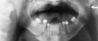
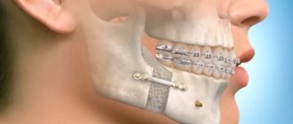
With improper development of bone tissue and the alveolar process, deformations such as microgenia, prognathia, overbite and others occur. In adults, the shape can also change, and this is mainly due to factors such as:
- Severe mechanical injuries.
- Consequence of surgical interventions.
- Various pathological processes (osteomyelitis, ankylosis, purulent inflammation and others).
- Incorrect orthodontic treatment or relapses after it.
- Partial or complete adentia (senile jaw).
Changes in structure, various inflammatory and tumor processes lead to severe diseases of the jaw. The most common pathologies are osteitis, periostitis, osteomyelitis. At the first signs, such as swelling, redness, pain, fever, you should contact a dentist or surgeon.
ANATOMY OF THE JAW
UPPER JAW
The upper jaw is a fixed paired bone of the facial skeleton. It has an air cavity (sinus), 4 surfaces (orbital, anterior, posterior, internal), as well as 4 processes (frontal, zygomatic, palatine and alveolar).
The dentition of the upper jaw is deviated slightly forward and outward. The crowns of the teeth increase in size from the incisor to the first molar, but the second molar is smaller than the first, the third is smaller than the second. The height of the crowns decreases successively from the incisor to the third molar, with the exception of the canines. The large roots of the teeth provide significant stability to the dentition of the upper jaw and to each tooth individually.
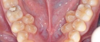
LOWER JAW
The lower jaw is a movable horseshoe-shaped bone of the facial skeleton. A large number of muscles are attached to it. The dentition of the lower jaw has a parabolic shape.
The teeth of the lower jaw are characterized by the fact that the incisors and canines are located perpendicular to the alveolar process, the chewing teeth are slightly inclined towards the tongue. The pattern of changes in the size and height of tooth crowns on the lower jaw is the same as on the upper jaw.
JAW DEVELOPMENT
The growth and formation of the jaw is closely related to the development of tooth germs and alveolar process. This process begins as early as the 7th week of intrauterine life. It is influenced by various endogenous factors (maternal toxicosis, hormonal imbalances, levels of vitamins and minerals), and after the birth of a child - also exogenous (external).
The jaw of a newborn is not yet completely ossified and fused; the lower part is shifted back in relation to the upper.
When a permanent dentition is formed (from the age of 6), intensive growth occurs due to the eruption of molars and incisors. Also, growth spurts are observed at 11–13 years of age; in boys, this usually occurs later. By the age of 18, the formation of bone tissue is completely completed.
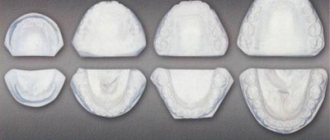
CHANGES IN THE SHAPE OF THE LOWER JAW WITH AGE
| Jaw of a newborn |
| Jaw of a 4 year old child |
| Jaw of a 6 year old child |
| Jaw of an adult |
| Elderly man's jaw |
For patients who want to get a free consultation with a dentist in Krasnodar , we inform you that pre-registration for an appointment is being made at Dentistry on Gagarin. Call the numbers indicated on the website and sign up for a free consultation with a dentist with a diagnosis (consultation duration up to 15 minutes).
Anatomy of molars and premolars
Molars in dentistry are divided into large ones - molars, and small ones - premolars. And their structure in humans is very different from the anterior ones.
Premolars
A person has two small molars on the left and right sides. In the first premolar, the central part of the chewing surface has a long shape, while the distal part is shorter and larger.
The second premolar retains all the features of the first, but it is more massive. The upper premolar is slightly smaller in size than its lower counterpart.
Molars
Depending on individual anatomical features, the number of molars in a person can vary from eight to twelve. Due to the structural features of the jaw, the molars gradually become smaller from the center to the edges.
The crowns of molars are large, with a pronounced square or even triangular closure surface. From three to five chewing tubercles are located on top, allowing the molars to fully perform their functional duties - the primary processing of food.
Upper molars are characterized by the presence of three roots, two of which are directed to the cheek, and one to the tongue. The lower molars have only two roots: posterior and anterior. In the outer molars, the roots sometimes grow together. Third molars also have a very unpredictable crown shape, which depends on the structure of the skull and jaw.
We'll tell you what antibiotics are usually prescribed for gumboils and how to take them correctly.
How malocclusion is corrected with braces - see our “Before” and “After” photos.
Here: https://www.vash-dentist.ru/lechenie/desnyi/parodontoz/sposobyi-narodnyimi-sredstvami.html - you will learn about the causes of periodontal disease and traditional methods of treating it.
Incisors and canines
Dentists divide human front teeth into canines and incisors.
Incisors
The incisors include two teeth located in the upper and lower jaw arches. The crown has a narrow, flattened shape with a sharp edge, as it is intended for cutting pieces of food, which are subsequently chewed by molars and premolars.
The incisors of the upper jaw are much wider and more massive, while the lower ones are almost half as large. The roots are single and flat, especially for the incisors located below. The upper part of the roots deviates to the side.
Fangs
The canines are located directly behind the incisors in the upper and lower jaw arches. Their distinctive feature is that both cutting edges converge at an angle at one point, forming such a recognizable shape. The canines have one long root with grooves on the side.
The upper canine is larger and more massive, while the lower one is less pronounced. The fangs located below have a shorter and smoother cutting edge and narrow longitudinal ridges. The roots are noticeably shorter than the upper ones and have pronounced grooves.
Functions of the human upper and lower jaw
- Support function. The upper and lower jaw of a person serve as a support for the facial and chewing muscles. And there are many of these muscles; according to some reference books, a person has 57 separate muscles on the face alone.
- Protective function . The jaw bones protect the sensory organs and the upper digestive system.
- Digestion. Jaws are needed to bite and chew food. In addition, some muscles necessary for sucking and swallowing movements are attached to them.
- Breathing . The jaws serve as a container and support for the upper respiratory tract, in which air is filtered, humidified and heated.
- Speech. The jaws are involved in the process of articulation. Regulation of the opening of the mouth by the temporomandibular joint, the presence of teeth, the work of the muscles attached to the jaws - all this is necessary to make our speech intelligible.
- Facial expressions and communication. A movable lower jaw and facial muscles are necessary to reproduce emotions that are understandable to other people and communicate with the outside world.
INTERESTING: The mouth is the most developed part of the baby's body. This is where the most sensitive nerve endings are located, and therefore small children, in order to explore an object, pull it into their mouth.
Wisdom teeth
a — vestibular surface; b — mesial surface; c - lingual surface; d — figure-of-eight cut; d — mesiodistal section; 1, 2, 3 - cross section: in the plane of the crown, in the middle and upper part of the root
Wisdom teeth, or third molars as they are correctly called, can erupt at any age, and not necessarily all of them. But at the same time, even if they never appeared, remaining in their infancy, this is not a deviation from the norm.
Third molars are among the most problematic teeth in humans. They are located at the end of the row on both sides, and there are four of them in total. The structure of the third molars is no different from the structure of the other large molars. But at the same time, it also has its own characteristics:
- the wisdom tooth is located last in the row and is not sandwiched between its neighbors;
- at the location of the third molar, children do not have milk teeth that prepare the ground for its eruption, which makes this process more unpleasant and painful;
- the roots of third molars often grow together into one large one, which may have an irregular cone shape;
- the crown does not necessarily erupt completely and has different shapes.
Typically, third molars grow in between the ages of eighteen and twenty-five. But sometimes they can appear much later, or even not appear at all. Such unerupted teeth are called impacted or semi-impacted if the crown has only partially appeared.
Problems with the growth of wisdom teeth are caused by evolutionary changes in the skull. In the jaw of a modern person, they are a rudimentary organ, and often there is simply no space left for their normal development.
Kinds
The main characteristic of a correct bite is the central occlusion (closing) of both jaws.
Based on the analysis of the nature of closure, five main types of physiological occlusion were derived, which differ slightly from each other:
- Orthognathic . In orthodontics, this type is considered close to ideal. The main characteristic is that the top row, when closed, leaves 2/3 of the bottom row in the visible zone. They also pay attention to the density of contact: ideally, the surfaces fit tightly to each other, and there are no gaps between the cutters. With this type, the load on the temporomandibular joint is distributed evenly.
- Progenic. It is determined by a slight protrusion of the lower jaw forward. But, since this extension is weakly expressed, the closure is not disturbed, and the load on the jaw joint remains uniform.
- Straight . With this type, the dental arches close together with their cutting surfaces, without overlapping each other. This variety is correct, but has one drawback - high jaw abrasion. This phenomenon is observed due to the pronounced load on the cutting surfaces.
- Biprognathic . Both arches move slightly towards the lips, but the closure remains normal.
- Opisthognathic. Slight displacement of the arches into the oral cavity. At the same time, the teeth look perfectly straight from the outside.
Types of correct bite according to Angle's classification
Baby teeth
Their formation in a child begins to occur in the womb at the twelfth week . As a rule, the first to appear in a child are the incisors and canines, and only at the very end the molars.
The timing of this process is purely individual and can vary, but in most cases, the formation of the primary bite begins to occur at the age of seven months and ends at three to four years. By this time, the child should have twenty baby teeth.
Compared to permanent teeth, milk teeth have their own characteristics:
- smaller sizes;
- fewer chewing tubercles;
- the roots spread out to the sides.
Despite this, primary and permanent teeth have the same number of roots.
The deciduous row in the jaw consists of ten teeth: four molars, four incisors and two canines. At the age of six or seven years, baby teeth begin to fall out and are replaced by permanent ones.
First of all, the large molar is replaced, and the final formation of the row ends by the age of twelve to fourteen, with the exception of the third molar.
If you find an error, please select a piece of text and press Ctrl+Enter.
Tags: tooth structure
Did you like the article? stay tuned
Previous article
Photos of the smiles of stars and ordinary patients BEFORE and AFTER installation of veneers
Next article
What to do if a wisdom tooth grows into the cheek?