How does salivary gland cancer develop?
Oncology begins with the appearance in the body of just one altered cell, different from healthy ones. It becomes like this due to various diseases and exposure to certain chemicals, multiplies, creates many copies of itself and creates a tumor. Most of these cells detect and destroy the immune system, but some manage to evade or resist our natural defenses. In addition, they have special properties - they are able to move throughout the body using the circulatory and lymphatic systems. The lymphatic system complements the cardiovascular system. The lymph circulating in it - the intercellular fluid - washes all the cells of the body and delivers the necessary substances to them, taking away waste. In the lymph nodes, which act as “filters,” dangerous substances are neutralized and removed from the body. systems Blood and lymph transport them to other organs, where they take hold and create metastases - additional foci of the disease. Such neoplasms gradually increase in size, destroy all tissues located next to them and disrupt their functioning.
What are salivary glands and how do they get affected by cancer?
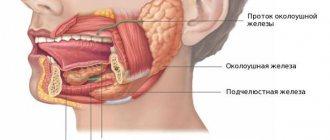
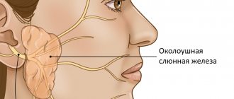
These important organs produce saliva - a liquid that not only moisturizes our mucous membranes, but also contains enzymes - proteins that start the process of digesting food. In addition, they contain special substances that help prevent the development of infections in the mouth and throat. There are 3 sets of large glands on each side of our face:
- The largest: parotid
- located in front of the ears. Up to 7 out of 10 tumors develop in them, most of which are benign, that is, not cancerous - they do not grow into nearby tissues and do not create metastases - additional foci of the disease in other tissues. - Submandibular
- located under the lower jaw. They secrete saliva under the tongue. Approximately 1-2 out of 10 neoplasms arise in them, about half of which are malignant. Malignant tumors are called tumors whose cells multiply uncontrollably, destroy surrounding tissues and create metastases - additional neoplasms in other organs located far from the main focus of the disease. - The smallest: sublingual
- located under the oral cavity on both sides of the tongue. In them, oncology begins quite rarely. - In addition to the above, there are several hundred small salivary glands, which can only be seen under a microscope. They are located under the mucous membrane of the lips and tongue, in the palate, inside the cheeks, nose and larynx. Tumors in them are not common, but the vast majority of them are cancerous.
How to prepare for the procedure
Ultrasound of the salivary glands is an absolutely safe and painless manipulation, for which the patient does not require special preparation. Patients of almost any age tolerate the procedure well, without showing any anxiety before and during its implementation, and therefore it does not require premedication (pre-administration of sedative medications) even in children.
On the eve of the visit to the specialist (about 3-4 hours in advance), the patient is advised to refrain from eating, and immediately before the examination he needs to thoroughly clean the oral cavity.
Types of salivary gland tumors
Almost all neoplasms arising in these organs are benign
, that is, they are not related to oncology and do not spread to other parts of the body. Most of them are treated surgically and are extremely rarely life-threatening - as a rule, their degeneration occurs with prolonged absence of therapy or re-growth after partial removal. The mechanism and principles of this transformation are not known to doctors today.
A minority of tumors are malignant
, capable of destroying tissues into which they grow and creating metastases - additional foci of disease in other organs:
- Their most common type is mucoepidermoid cancer
, most often starting in the parotid glands. It is generally slow growing and responds well to treatment. - Adenoid cystic carcinoma
: usually does not develop quickly, but can recur - appear again after therapy, including many years later. Recovery prognosis largely depends on the size of the tumor - the smaller it is, the better the prospects. - Many subtypes of adenocarcinoma
: formed in gland cells that secrete various substances. They grow slowly, but appear at a younger age than other types of cancer.
Rare types of malignant tumors:
- Squamous cell carcinoma
: occurs mainly in older men. The prognosis for its owners is unfavorable. - Epithelial-myoepithelial carcinoma
: usually grows slowly, but can recur - come back after treatment, and spread to other parts of the body. - Anaplastic small cell carcinoma
: Most often develops in the minor salivary glands, grows rapidly, and its cells are similar to those that make up nerves. - Undifferentiated carcinomas
: often spread quickly and are difficult to treat, and the outlook for patients with this diagnosis is generally poor.
In addition to the above, other types of oncology can develop in these organs:
- Non-Hodgkin's lymphomas
: formed in the cells of the immune system of the glands, behave and are treated differently from other types of tumors. - Sarcomas
: Occur in cells of blood vessels, muscles and connective tissue. - Secondary cancer
: begins in other organs and spreads to the salivary glands.
Treatment
The patient is treated by a team of doctors, which may include: a clinical oncologist, an ENT doctor, an oncologist-surgeon, an oral and maxillofacial surgeon, a chemotherapist, a radiotherapist, etc. The treatment program is determined by the stage of cancer, the histological type of the tumor, its location (which gland is affected) , age, general condition and concomitant diseases of the patient.
Surgery
If the tumor has not grown much into the surrounding tissue, then it is resectable, that is, it can be removed surgically. The surgeon’s task is to excise the tumor while capturing the surrounding tissue so that there are no cancer cells left on the cut line, that is, to ensure a negative resection margin. If tumor cells have spread to the lymph nodes, or a biopsy reveals aggressive cancer, the lymph nodes are also removed.
For parotid salivary gland cancer, surgery presents certain difficulties, because the facial nerve passes through the gland, which controls the work of facial muscles. If the tumor affects only the superficial lobe of the gland, you can remove it separately - perform a superficial parotidectomy. There is no risk of damaging the facial nerve. In some cases, it is necessary to remove the entire gland, and if the tumor has grown into the facial nerve, then it too.
For cancer of the sublingual and submandibular gland, the surgeon removes the gland itself and some of the tissue located around it, including, possibly, bone tissue. In some cases, it is necessary to excise the nerves that control sensitivity, movements in the lower part of the face, in the tongue, and the sense of taste.
For cancer of small glands, the affected gland and part of the surrounding tissue are removed. The extent of the operation depends on the size and location of the tumor.
Radiation therapy
Indications for the use of radiation therapy for malignant tumors of the salivary glands:
- To combat malignant tumors that cannot be removed surgically due to their location or size. Sometimes radiation is supplemented with courses of chemotherapy.
- After surgical treatment. This type of radiation therapy is called adjuvant and is sometimes combined with chemotherapy. Radiation after surgery helps destroy remaining cancer cells and prevent recurrence.
- For advanced cancer. In this case, radiation therapy is aimed at combating pain, difficulty swallowing, bleeding and other symptoms.
Radiation is typically given five days a week for 6–7 weeks. If radiation therapy is used for palliative purposes, the course will be shorter.
Chemotherapy
Chemotherapy is used quite rarely for malignant neoplasms of the salivary glands. Anticancer drugs can reduce the size of the tumor, but are not able to completely destroy it. They are most often prescribed for advanced cancer as palliative treatment or in addition to radiation therapy.
Depending on the type and other characteristics of the cancer, the doctor may prescribe combinations of different chemotherapy drugs: carboplatin, cisplatin, 4-fluorouracil, doxorubicin, paclitaxel, cyclophosphamide, vinorelbine, docetaxel, methotrexate. Chemotherapy for cancer is always given in cycles. The patient is administered the drug, then takes a “break” for several days. The course of treatment may consist of several cycles.
Rehabilitation
After treatment, some problems associated with nerve damage may persist: dysfunction of the facial muscles, speech disorders, swallowing, and cosmetic defects. Some side effects of chemotherapy and radiation therapy go away after treatment is completed, while others persist for a long time. In such cases, rehabilitation courses are indicated. The doctor draws up a rehabilitation treatment program individually, depending on the severity and nature of the disorders.
Causes of salivary gland cancer
Doctors and scientists do not know the exact causes of this disease - they only know about the factors that can lead to the occurrence of this event.
- These include exposure to radiation
, including that received in the workplace or as a result of radiation therapy in the treatment of other types of tumors. - Age
: The older a person is, the higher their risks. - Gender
: this type of dangerous neoplasm occurs more often in men than in women. - Family history
: blood relatives of those who already have a similar diagnosis are more likely to become a patient of an oncologist.
Some studies have shown that frequent contact with silica and asbestos, bad habits such as alcohol and smoking, and a diet low in vegetables and high in animal fats can trigger the development of cancer. In addition, according to incompletely confirmed data, workers in woodworking and rubber industries, as well as people interacting with certain metals, including nickel alloy dust, are more likely to get sick. This information cannot be called absolutely accurate, since dangerous tumors of the salivary glands are quite rare, which makes them difficult to study.
Signs and symptoms of salivary gland cancer
Symptoms of the disease are often caused by damage to important nerves and other structures that pass through or near the salivary glands. Possible signs of cancer include:
- Bumps, lumps, or lumps in the mouth, cheek, jaw, or neck.
- Numbness of part of the face.
- Persistent pain in the mouth, cheek, jaw, ear, or neck.
- Weakness of the muscles on one side of the face.
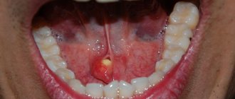
- Non-healing mouth ulcers.
- Pain when trying to open your mouth wide.
- Outflow of fluid from the ear.
- Changing the size or shape of the left or right sides of the face and neck.
- In the later stages of the disease, problems with swallowing often appear.
Many of the above symptoms may be caused by benign tumors—those that are not life-threatening and do not spread throughout the body—or other conditions. However, if any of them appear, it is necessary to consult a doctor as soon as possible, who will determine the cause of this condition and prescribe treatment.
In the human body during normal development there are three pairs of salivary glands
The parotid gland is most often affected by various diseases. There are a number of diseases in which the sublingual and submandibular salivary glands become inflamed. If you do not start treatment on time or undergo inappropriate therapy, then serious complications may occur after such diseases, such as encephalitis, orchitis, meningitis, nephritis, neuritis and pancreatitis. However, do not worry, treatment often gives positive results. To avoid inflammation of the salivary glands, you just need to follow a few recommendations.
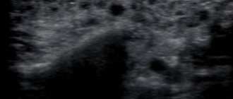
Diagnosis of salivary gland cancer
An examination is a very important moment for any patient, since during it doctors do not just detect a disease. They find out how far the altered cells have spread throughout the body, what tissues have been damaged, and what treatment is best for each individual person.
Lapino-2 specialists carry out full diagnostics and any treatment for salivary gland cancer without queues or delays. We have a whole team of professionals with extensive experience in identifying and combating oncology. We carry out all the necessary studies with high quality and analyze their results, and our patients do not have to waste time visiting various medical organizations and retaking tests.
The examination begins with a survey about well-being, symptoms, the time of their onset, as well as risk factors - the presence of relatives with cancer or work in hazardous industries. Then a thorough examination of the mouth, face, neck and jaw is carried out, after which a number of studies are prescribed:
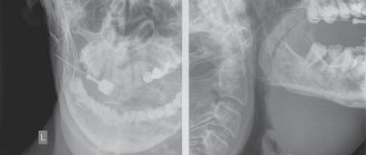
- X-ray
: allows you to see damage to the jaw, teeth and lungs. - Computed tomography
- creates a clear image of internal tissues to assess the size, shape and location of the tumor, as well as search for changed cells in the lymph nodes. - Magnetic resonance imaging, MRI
- obtaining a detailed “picture” of tissue using radio waves and strong magnets. The method helps determine the location and exact dimensions of the tumor, and identify damage to the lymph nodes or other organs. - Positron emission tomography, PET
: used to detect clusters of cancer cells throughout the body. Before the test, a person takes a special substance – radioactive sugar, which accumulates in the altered cells. A PET scan does not create the very clear images of tissue obtained during a CT or MRI, but thanks to it, a specialist can notice all the foci of cancer in the body. This procedure is usually prescribed when there is a suspicion of the presence of metastases - additional tumors in other tissues located far from the salivary glands. - Symptoms and examination results can strongly indicate the presence of a disease, but the actual diagnosis is made only by the results of a biopsy
- the removal of a small amount of altered tissue. As a rule, samples are collected using a thin needle, which is inserted directly into the tumor after preliminary anesthesia. If this method does not produce enough cells, the surgeon makes a small cut with a scalpel and removes a tiny area of the tumor. The material is sent to the laboratory, where a specialist carefully examines the cells and determines whether they are cancerous. Unfortunately, even a biopsy cannot always give a clear answer as to whether a patient has cancer. In such cases, the doctor can perform an operation to completely remove the suspicious lesion and confirm or refute the correctness of his conclusions after the intervention. - Blood, urine
and
stool
: are prescribed not to identify life-threatening neoplasms, but to assess the condition of the patient’s body and the quality of the functioning of its organs.
During a laboratory study, the degree of differentiation of the tumor
– the difference between its cells and healthy ones.
Such data allows specialists to imagine the possible speed of development of a neoplasm and the complexity of its treatment: G1
is highly differentiated: its cells are very similar to healthy ones, they grow slowly and respond well to therapy.
G2
– moderately differentiated: they are noticeably different from normal.
G3
– poorly differentiated: cells do not look like healthy ones, multiply quickly, spread throughout the body and respond poorly to treatment.
Complications and relapses
Even if the treatment is successful and the examination results show no signs of the presence of cancer cells in the patient’s body, a relapse may occur in the future. Therefore, you need to regularly see an oncologist, come for examinations, undergo various studies and take tests.
Typically, the doctor prescribes examinations once every few months for several years, then less frequently.
- If cancer recurs, treatment options may vary:
- If the tumor can be removed, surgery is performed followed by a course of radiation therapy.
- If the tumor cannot be removed surgically, the doctor prescribes radiation therapy in combination with chemotherapy.
- If there are distant metastases, chemotherapy becomes the main treatment method. Radiation therapy and surgery can be used to control some symptoms.
With advanced cancer with metastases, achieving remission becomes extremely unlikely. In this case, treatment will be aimed at slowing the progression of cancer, combating symptoms, and prolonging the patient’s life.
Euroonco doctors undertake cancer treatment at any stage. For us there are no hopeless patients. You can always help, and we know how to do it correctly, we have all the necessary technologies, the latest generation drugs.
Stages of salivary gland cancer
After identifying cancer, doctors determine its stage - find out how far the changed cells have spread throughout the body and what tissues have been damaged. Such information is necessary for specialists to understand the patient's approximate prognosis and draw up an individual treatment plan that is best suited in each specific case.
Staging is carried out according to the international TNM system, each letter of which has its own meaning:
- " T
" describes the size of the main tumor and its growth into the tissues closest to it; - using “ N
” indicate the number of lymph nodes affected by cancer; - “ M
” is used to indicate the presence (M1) or absence (M0) of metastases - additional foci of disease in other organs located far from the salivary glands.
Stage 0
, carcinoma in situ: altered cells are present only in the tissue lining the salivary ducts, through which the secretions of the glands exit into the oral cavity.
Stage I
: tumor size does not exceed 2 centimeters.
Stage II
: neoplasm with a diameter of 2 to 4 cm.
Other organs are healthy. Stage III
: the tumor is larger than 4 cm and/or it has managed to damage the soft tissue located next to it;
or a lesion of any size, and its cells have damaged 1 lymph node located on the same side of the head or neck. Stage IV
:
- a neoplasm of any size has grown into the nearest structures - jaw bone, skin, ear canal or facial nerve, and has damaged 1 lymph node, increasing to no more than 3 cm;
- or tumors up to 6 cm in size were found in 1 or more lymph nodes.
IVB stage
:
- 1 or more lymph nodes larger than 6 cm in size have been affected by cancer, or the tumor has spread beyond their limits;
- or the disease has spread to other structures, such as the base of the skull and other nearby bones, or has grown around the carotid artery, a vessel that supplies blood flow to the brain.
IVС stage
: A tumor of any size has created metastases - additional foci of disease in organs or tissues located far from the salivary glands - for example, in the lungs.
Stages of the disease
Salivary gland cancer, like other malignant tumors, is classified into stages according to the generally accepted TNM system. The letter T in the abbreviation denotes the characteristics of the primary tumor: the size and degree of its growth into surrounding tissues, N - spread to the lymph nodes, M - the presence of distant metastases. Depending on these indicators, the following stages are distinguished during salivary gland cancer:
- Stage 0 is “cancer in situ” (carcinoma in situ). The tumor is located within the layer of cells that form the salivary gland and does not grow into neighboring tissues.
- Stage I is a tumor that is located within the salivary gland and measures no more than 2 cm.
- Stage II - the tumor reaches a size of more than 2 cm, but not more than 4 cm.
- Stage III - a tumor that reaches a size of more than 4 cm and/or spreads into surrounding tissues, or a tumor of any size that has grown into surrounding tissues, has spread to one cervical lymph node on the same side, and the focus in the lymph node is no more than 3 cm and does not extend beyond its borders.
- Stage IV includes substages IVA, IVB or IVC. The first two are characterized by varying degrees of spread of the malignant tumor to the anatomical structures of the head, neck, and lymph nodes. If stage IVC is diagnosed, it means that there are distant metastases.
In addition to stages, there are three degrees of malignancy of salivary gland cancer:
- Grade I - low degree of malignancy. Such tumors are called highly differentiated. Tumor tissue is as similar as possible to normal salivary gland tissue. It grows slowly, and the prognosis for such patients is most favorable.
- Grade II are moderately differentiated tumors. Tumor tissue differs more significantly from normal tissue. This cancer is more aggressive and has a poorer prognosis.
- III degree - poorly differentiated tumors. Cancer cells almost completely lose the features of normal ones. Such tumors behave the most aggressively.
Determining the degree of malignancy helps the doctor predict how the cancer will behave and plan treatment correctly.
Treatment of salivary gland cancer
Fighting a life-threatening tumor is not an easy task. To solve it, the participation of many different specialists is necessary: oncologist, surgeon, chemotherapist, radiologist and others. It is extremely important that they all work together and exchange information in a timely manner, since only this approach allows taking into account all the characteristics of the patient.
The oncology department has a whole team of professionals with extensive experience in the diagnosis and treatment of any type of cancer. Our doctors are candidates and doctors of medical sciences, constantly improving their knowledge and skills. We use only the most modern equipment and original drugs that give predictable results. Our clinic’s specialists offer each patient individual treatment and draw up a clear plan, following which you can get the best possible result.
Several methods are used to combat this type of tumor. Surgery is often the main one.
. With the help of surgery, it is possible to completely remove tumors that have not seriously damaged nearby structures. The success of the procedure is largely determined by the experience and skills of the doctor, including because the nerves responsible for tongue movements, control of facial muscles and taste sensations pass near the salivary glands. During the procedure, the organ and surrounding healthy tissue are removed so that not a single altered cell remains in the body. In some cases, lymph nodes also have to be removed.
Radiation therapy
– destruction of cancer cells using radiation. The method can be applied:
- Alone or in combination with chemotherapy for some forms of tumors that cannot be treated with surgery alone.
- After surgery, possibly simultaneously with “chemistry” - to destroy any remaining changed cells and reduce the likelihood of recurrence of the disease.
- As palliative treatment in the final stages to relieve symptoms - reduce pain, eliminate bleeding or difficulty swallowing.
Chemotherapy
– destruction of abnormal cells with special drugs that are injected into blood vessels or taken like regular tablets. They enter the blood and spread throughout the body, thanks to which they work against all foci of the disease. Most often, such drugs are prescribed for patients with metastases - additional tumors in distant organs. Chemistry can reduce the tumor, but it cannot cure this type of cancer on its own. Some substances increase the effectiveness of radiation therapy and make it easier for radiation to destroy damaged cells. Such therapy is prescribed in courses, which are necessarily followed by a period of rest, allowing the body to recover. Typically, such cycles last from 3 to 4 weeks. To combat salivary gland cancer, drugs such as Cisplatin, Carboplatin, Doxorubicin, 5-fluorouracil, Cyclophosphamide, Paclitaxel, Docetaxel, Vinorelbine and Methotrexate are usually used.
Acute mumps
Mumps is an infectious disease caused by a filter virus. Most often occurs in childhood.
Symptoms of mumps
Often, in the prodromal period, the first symptom of mumps is stomatitis. Then there is swelling of the gland, pain, protruding earlobe, decreased salivation, temperature up to 39 C. Fever lasts 5-7 days.
The patient may complain of increased pain when chewing, opening the mouth, and dry mouth. In the oral cavity, you can see swelling and hyperemia of the mucous membrane of the pharynx and around the opening of the excretory duct. The swelling of the gland lasts for 2-4 weeks.
Acute mumps can be accompanied by damage to the nervous system (meningitis, meningoencephalitis, neuritis), the digestive system (dyspepsia), the cardiovascular system (pain in the heart, shortness of breath), the organs of vision, and hearing.
Differential diagnosis of mumps
Differential diagnosis of mumps is carried out with false mumps, parenchymal mumps, and Mikulicz's disease.
False parotitis is a unilateral acute serous lymphadenitis of the intraglandular lymph nodes of the parotid region. Develops as a complication of difficult eruption of third molars. With this disease, salivation remains normal.
Parenchymal parotitis is characterized by a long chronic course with periods of exacerbation.
With Mikulicz's disease, all salivary and lacrimal glands are affected.
Treatment of mumps
Treatment of mumps is aimed at eliminating symptoms and preventing complications, since there is currently no specific therapy. Patients are prescribed bed rest for 7-15 days, pureed food, warm compresses, and rinsing the mouth with antiseptics. Antibiotics are prescribed for prophylactic purposes.
In case of suppuration, surgical treatment is performed. Incisions are made in the direction of the branches of the facial nerve, the skin and subcutaneous fat are dissected with a scalpel, and the capsule and gland are separated with a hemostatic clamp. The wound and excretory duct are washed with antibiotics.
Prevention of mumps
Prevention of mumps involves isolating the patient for the duration of the illness and 14 days after all symptoms disappear.
Vaccination with mumps-measles and mumps live vaccines is also carried out. Included in the mandatory vaccination calendar. Vaccination is carried out at 12 months and revaccination at 6 years.