Causes
The main causes of damage to the NAS are:
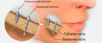
- implantation (doctor’s errors, lack of a full preliminary examination);
- removal of dystopic “eights” on the lower jaw;
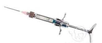
- errors when performing conduction anesthesia;
- exit of the filling material beyond the root apex into the nerve canal;
- infectious lesion of the periapical region of the lower row.
But the most common reason is the first reason - damage as a result of implant installation, usually in the chewing area.
First signs of damage
The first symptoms of nerve damage are discomfort in the gums, cheeks, and lower lip. Manifestations of the problem are:
- paresthesia, that is, a change in the level of sensitivity without pain;
- dysesthesia with pain in the affected area, a feeling of “pins and needles”, changes in the general sensitivity of the area;
- anesthesia - complete loss of sensation in a certain area.
In some cases, the lingual nerve, which runs from the side of the tongue into the gum tissue, may be affected. This is usually observed as a result of the removal of "eights" (in approximately 2.1% of all cases). When implanted, this nerve is affected less frequently. If this situation occurs, the following symptoms appear:
- salivation becomes profuse;
- involuntary biting of the tip of the tongue appears;
- diction disorders;
- burning sensation, numbness in the tongue;
- loss, change in taste;
- swallowing is impaired.
In 90% of cases, the problems go away on their own within seven to ten weeks; no special treatment is required.
Mandibular nerve
Mandibular nerve, n. mandibularis , mixed; it represents the most powerful branch of the trigeminal nerve. The mandibular nerve is formed by a sensitive branch extending from the trigeminal ganglion, to which the motor root of the trigeminal nerve joins.
The mandibular nerve exits the skull down to its base through the foramen ovale and is divided into two main branches - the anterior, predominantly motor, and the posterior, predominantly sensory.
Even before dividing into these branches, a thin meningeal branch of the mandibular nerve, r, departs from the mandibular nerve. meningeus n. mandibularis , which returns through the foramen spinosum into the cranial cavity, innervating the dura mater of the middle cranial fossa. From the posterior surface of the mandibular nerve, 3-4 short trunks extend to the ear ganglion, ganglion oticum.
Meningeal branch of the mandibular nerve
A number of nerves arise from the anterior branch:
1. Masseteric nerve, n. massetericus , goes outward and gives 1-2 thin branches to the temporomandibular joint, then passes through the notch of the mandible to the inner surface of the masticatory muscle and innervates it.
2. Deep temporal nerves, nn. temporales profundi , usually two nerves (smaller - posterior and larger - anterior), are directed laterally into the gap between the upper edge of the lateral pterygoid muscle and the infratemporal crest of the sphenoid bone and, turning upward onto the inner surface of the temporal muscle, branch in its thickness.
Nerves of the temporal muscle, right. (Inner surface of the muscle.) 1 - posterior intramuscular branches of the deep temporal nerve; 2—middle intramuscular branches; 3—anterior intramuscular branches; 4—temporalis tendon.
3. Lateral pterygoid nerve, n. pterygoideus lateralis, - short, often departs along with the buccal nerve, approaches the lateral pterygoid muscle from the inside, innervating it.
4. Buccal nerve, n. buccalis is a rather powerful nerve, the only sensitive one of this group. Most often it passes between the heads of the lateral pterygoid muscle, follows forward along the lateral surface of the buccal muscle and ends in the skin and mucous membrane of the cheek; also innervates the skin of the corner of the mouth. At the point of branching it has connecting branches with the branches of the facial nerve.
The following nerves arise from the posterior branch:
1. Medial pterygoid nerve, n. pterygoideus medialis, starts from the inner surface of the posterior branch, approaches the medial pterygoid muscle and innervates it.
At the level of the auricular ganglion, two small branches arise from the medial pterygoid nerve:
1) nerve of the tensor tympani muscle, n. musculi tensoris tympani, which is directed slightly upward and posteriorly, passes through the ear ganglion and innervates the indicated muscle;
2) nerve of the muscle that strains the velum palatine, n. musculi tensoris veli palatini, departs slightly above the previous nerve, less often from the mandibular nerve and, going down and anteriorly, innervates the corresponding muscle.
2. Auriculotemporal nerve, n. auriculotemporalis , mixed in composition. It contains sensory and secretory fibers approaching it from the ear ganglion. The nerve begins with two roots from the posterior surface of the trunk of the mandibular nerve, is directed posteriorly, covering the middle meningeal artery, passes along the inner surface of the condylar process of the lower jaw, is directed posteriorly and upward along the capsule of the temporomandibular joint, located under the parotid gland, in front of the auditory canal. Heading further upward, it ends in the skin of the temporal region.
Along its path, the auriculotemporal nerve gives off a number of branches:
1) parotid branches, rr. parotidei, arise from the auriculotemporal nerve where it passes under the parenchyma of the gland and connect to the temporal branch of the facial nerve. These branches mainly contain secretory fibers (from the ear ganglion);
2) nerve of the external auditory canal, n. meatus acustici externi , penetrates the wall of the external auditory canal at the border between its bone and cartilaginous parts and innervates the skin of the external auditory canal;
3) branches of the tympanic membrane, rr. membranae tympani , two or three thin branches, approach the outer surface of the eardrum, innervating its anteroinferior part;
4) anterior auricular nerves, nn. auriculares anteriores , usually there are two of them, go to the anterior part of the auricle, innervate the skin of the tragus and parts of the helix;
5) superficial temporal branches, rr. temporales superficiales, are the terminal branches of the auriculotemporal nerve. They branch in the skin of the temporal region, have connecting branches with the branches of the facial, frontal and greater occipital nerves;
6) connecting branches with the facial nerve, rr. communicantes (cum nervo faciali), join the latter behind the neck of the lower jaw.
3. Inferior alveolar nerve, n. alveolaris inferior , mixed in nature. It is a powerful trunk that runs down first along the medial surface of the lateral pterygoid muscle, and then, passing between the pterygoid muscles, along the lateral surface of the medial pterygoid muscle. Directing slightly anteriorly and entering the mandibular canal through the mandibular foramen, it follows it along with the artery and vein of the same name and emerges from the mental foramen onto the surface of the face.
Along its length, the inferior alveolar nerve gives off a number of branches:
1) mylohyoid nerve, n. mylohyoideus , departs from the inferior alveolar nerve at the place where the latter enters the mandibular foramen, goes forward and down, runs in the groove of the same name on the inner surface of the lower jaw. Then it approaches the mylohyoid muscle, branches in it and sends a small branch to the anterior belly of the digastric muscle;
2) inferior dental plexus , plexus dentalis inferior , is formed by the branches of the inferior alveolar nerve, extending from the main trunk along its entire length as it passes through the mandibular canal.
The branches connect with each other and form a plexus that sends out two types of branches:
a) lower gingival branches, rr.gingivales inferiores, innervating the gum of the lower jaw;
b) lower dental branches, rr. dentales inferiores, next to the teeth of the lower jaw.
4. Mental nerve, n. mentalis , is the terminal branch of the inferior alveolar nerve. Coming through the mental foramen, the mental nerve splits into a number of branches ending in the skin of the chin - the mental branches, rr. mentales, and lower lip - lower labial branches, rr. labiales inferiores; often sends one or two thin branches to the mucous membrane of the lower lip.
5. Lingual nerve, n. lingualis , mixed in nature, since it consists of fibers that perceive general sensitivity (touch and temperature) of the mucous membrane of the anterior 2/3 of the tongue, and fibers that make up the chorda tympani - a branch of the facial nerve involved in the taste sensations of the anterior part of the tongue.
Lingual nerve, n. lingualis
Separating from the anterior edge of the mandibular nerve, the lingual nerve, like the inferior alveolar nerve, first runs along the medial surface of the lateral pterygoid muscle, and somewhat lower penetrates the gap between it and the medial pterygoid muscle (anterior to the inferior alveolar nerve).
Here the lingual nerve receives the fibers of the chorda tympani (a branch of the facial nerve), which enters it from behind at an acute angle. Between the lingual nerve and the chorda tympani there are connecting branches with the chorda tympani, rr. communicantes (cum chorda tympani).
Further, the lingual nerve is directed in an arcuate manner down and forward along the inner surface of the lower jaw and, lying above the submandibular gland, approaches the lower surface of the body of the tongue, where it sends its terminal branches into its thickness.
Along its course, the lingual nerve gives off the following branches:
1) branches of the isthmus of the pharynx, rr. isthmi faucium, - several thin branches heading to the mucous membrane of the anterior arch of the pharynx and to the palatine tonsil;
2) nodal branches, rr. ganglionares , to the submandibular nerve ganglion, represented by two or three short trunks, which contain, in addition to their own sensory fibers, also secretory ones included in the chorda tympani;
3) hypoglossal nerve, n. sublingualis , originates from the anterior surface of the lingual nerve and innervates the sublingual gland, the mucous membrane of the floor of the mouth in the area of the sublingual fold and the anterior parts of the gums of the lower jaw;
4) connecting branches with the hypoglossal nerve, rr. communicantes (cum nervo hypoglosso), 2 or 3 branches in the form of arches, with their convexities facing forward, running along the outer surface of the hyoid muscle, joining the trunk of the hypoglossal nerve;
5) lingual branches, rr. linguales are the terminal branches of the lingual nerve. They approach the tongue from its lower surface, enter its thickness and, connecting with each other, follow upward, approach the mucous membrane and innervate its anterior two-thirds (the apex, edges and back of the tongue), giving thin branches to the filiform and mushroom-shaped papillae of the tongue . At the border of the root and body of the tongue, the lingual branches connect with the lingual branches of the glossopharyngeal nerve.
Classification
In accordance with Seddon's classification, the following types of NAS injuries are distinguished:
- Neuropraxia. This injury is reversible; the sheath of the nerve fibers does not suffer, that is, there is no development of degeneration. After treatment, the problem goes away, which usually takes a couple of weeks.
- Axonotmesis. A reversible type of lesion, for which long-term, intensive treatment is prescribed - from three to six months. The development of degeneration and fiber damage are observed.
- Neurotmesis. A serious disorder affecting all structures, including fibers and connective membranes. Scar tissue develops, and surgery is indicated to correct the problem.
In accordance with the generally accepted WHO classification, there are five categories of NAS injuries:
- compression, nerves are not damaged;
- swelling of tissues;
- partial rupture of fibers;
- complete ruptures of the NAS are observed;
- post-traumatic fibrosis develops.
Assessment methods
To determine the cause and extent of the disorder, diagnostics are required. For this purpose they prescribe:
- mechanoceptive methods, in which the response to mechanical irritation and stimulation is recorded;
- nociceptive, allowing to determine sensitivity to painful stimulation.
The first type of research includes tests of the following types:
- tests with a brush, during which the Patient’s lip is passed with a brush and asked to determine the direction of movement;
- two-point irritation using a probe.
The second diagnostic method includes:
- pin tests;
- Temperature tests for sensations of cold or heat.
Tests are also carried out for the presence of deficits in the sense of taste, and data from the right and left sides of the maxillofacial apparatus are compared. The accuracy of the research should be no more than 1 mm.
Treatment
The following regimens are used for therapy:
- observation of the Patient at intervals of 2, 3, 4, 6 months;
- drug therapy;
- unscrewing, removing the implant;
- microsurgical intervention.
There is no strict protocol; tactics are determined individually in each case. But there are limitations to surgical intervention - it will be effective only in the first year, after which this method is considered ineffective.
Drug therapy includes:
- taking painkillers;
- use of hydrogen pump blockers;
- prescription of anti-inflammatory drugs (glucocorticosteroids are prescribed);
- therapy for sensory impairment.
If symptoms appear in the first 1.-1.5 days after installation of the implant, its removal may be indicated. During this period, the prognosis is favorable, but over a longer period of time, removing the artificial root will no longer bring a noticeable improvement. The reason is irreversible changes in nerve fibers.
Surgery is indicated in cases where all previous measures have not brought the expected effect. To do this, the doctor must determine the location of the NAS and evaluate sensitivity disorders. The effectiveness of the operation is influenced by the following factors:
- time between injury and surgery;
- severity, type of lesion;
- blood supply;
- Preparation;
- tension zone;
- Patient's age, health status.
Predictions and prevention
Treatment has an uncertain prognosis; with microsurgical intervention, the positive result is in the range of 55-82%. If the damage is severe, complete recovery does not occur with any treatment method. According to some experts, complications such as pain persist even after the intervention. It is also worth noting that the success of treatment depends entirely on the extent of the lesion. It is impossible to guarantee a high result in any situation, that is, the forecasts are uncertain.
To avoid complications, prophylaxis is recommended. This is a mandatory full examination before the implantation procedure. The specialist will determine the exact location of the nerve, the topography of the nerve trunks and the maxillofacial system. This allows you to exclude the development of pathology and conduct conduction anesthesia correctly, without the risk of complications.
The main preventive measures include:
- competent selection of implantation system;
- diagnostics, including CT, production of a surgical template, computer modeling;
- planning the procedure for introducing an artificial root;
- determining the paths of nerve trunks, ensuring the distance between them and implants is not less than 2 mm;
- when drilling, it is necessary to avoid excessive force;
- When immersing a drill or implant, stoppers are used to limit the depth and ensure safety.
Treatment of damage to the mandibular nerve during implantation requires the doctor to have high qualifications and experience. But even under such conditions, the success rate does not exceed 82%. Some complications may remain, such as numbness or soreness.
Iatrogenic trigeminal neuropathy.
This nosological entity arose due to the fact that treatment of trigeminal neuralgia in most cases began with neurodestructive operations (alcohol-lidocaine blockades, neuroexeresis, destruction of the trigeminal ganglion). As a result, a significant number of patients experienced iatrogenic traumatic or toxic-traumatic neuropathies of the trigeminal nerve. The maxillary and mandibular nerves were most often affected.
In many textbooks on neurology, in case of trigeminal neuralgia, it is proposed to carry out alcohol-novocaine and alcohol-lidocaine blockades of its peripheral branches or node - the so-called alcoholization . The analgesic effect in this case is achieved on average after the second or third procedure, but it is explained by the fact that numbness occurs due to the development of destructive changes in the nerve trunk. Over time, toxic-traumatic neuropathy develops, practically resistant to treatment, so the patient must continue to undergo blockades, the effectiveness of which decreases in proportion to their number.
Thus, neurodestructive operations that are performed in the treatment of neuralgia lead to the development of toxic-traumatic neuropathy. This determines the nature of the pain syndrome.
Clinic.
The clinical picture is usually represented by the presence of constant aching, burning or dull neuropathic pain in the area of innervation of the affected nerve, against the background of which neuralgic paroxysms occur with irradiation of pain, respectively, to the segmental areas of the face (Zelder segments). Patients experience various types of paresthesia (numbness, crawling, burning) and sensitivity disorders (hypesthesia with symptoms of hyperpathia or hyperesthesia), which sometimes spread beyond the innervation of one of the branches of the trigeminal nerve.
In many cases, vegetative fibers are drawn into the process, which leads to trophic changes in the oral mucosa (gingivitis), the dental system (progressive periodontitis) and facial skin (pigmentation or depigmentation, dryness, peeling, soft tissue atrophy). In such cases, the pain becomes unbearably burning, tearing, boring, and is accompanied by vegetative reactions (redness and swelling of the facial skin, local increase in body temperature, lacrimation, salivation).
During neurodestructive manipulations on the mandibular nerve, painful contraction of the teeth may occur (patients are forced to eat through a straw and cannot speak or open their mouth). With each subsequent alcoholization, the nature of the pain syndrome changes: neuralgic paroxysms become longer, more frequent, a neuralgic status can form, and slightly pronounced trigger areas appear on the skin of the face. Pain is provoked by weather conditions (cold or heat), exacerbation of somatic pathology, food consumption, and physical activity. The exit points of the trigeminal ditch are painful during palpation in approximately 2/3 of patients.
The data presented convincingly indicate that neurodestructive manipulations are not the method of choice for the treatment of trigeminal neuralgia, since in most cases a short-term effect is achieved. However, such therapy leads to the development of toxic neuropathy, disease progression and the development of resistance to conservative treatment methods. Only when all the methods used to treat neuralgia are not effective, and the intensity of the pain syndrome remains severe, can neurodestructive operations recently developed by neurosurgeons be used.