Most patients at a dentist's appointment have long become accustomed to complex dental terms. Bridge, veneer, filling, premolar – these words are practically easy to understand when a doctor diagnoses a problem and prescribes treatment.
Most often, patients are confused by incomprehensible numbering . For example, when talking about wisdom teeth, the dentist can explain to the patient that the lower “eights” need treatment. Moreover, each tooth has its own numerical designation, which can reflect its type and exact location in the oral cavity.
Over the entire existence of dentistry as a science, several classifications of teeth have been invented, which are called dental formulas. Depending on the chosen numbering method, each tooth is assigned a specific number. In some systems, teeth are designated by a combination of letters, numbers and even symbols, which allows the dentition to be depicted in the form of an accurate diagram.
Functional properties of elements
Teeth perform several functions at once. Each element has a distinctive shape according to its purpose. The main role of the units is digestive. The quality of assimilation of useful elements by the body depends on the thoroughness of grinding the products. Only after chewing and moistening with saliva will the food be ready for subsequent processing by the digestive organs.
Chewing one piece lasts on average 10-20 minutes. The force exerted by the teeth on food sometimes reaches 80 kg. Incomplete dentition can lead to disturbances in the digestive system.
Another function of the elements is aesthetic. With malocclusion, a person may develop psychological problems, since the first thing others pay attention to during a conversation is a smile. Teeth also play a role in the formation of syllables. In the absence of one or more elements, the pronunciation of syllables changes: a lisp appears, speech is distorted.
Age-related changes in dentogingival tissues
Over time, the human dentofacial system undergoes the following changes:
- reduction in the height of the enamel layer due to abrasion of the enamel;
- the formation of microcracks, which provokes gradual staining of teeth yellow and brown;
- active formation of secondary dentin and, accordingly, a decrease in the volume of dental pulp;
- age-related thickening of the walls of blood vessels also causes a narrowing of the pulp cavity;
- an increase in the amount of cement, which is observed both with an increase in chewing load and without it;
- thickening of periodontal bundles in the area of teeth that serve as support for bridges.
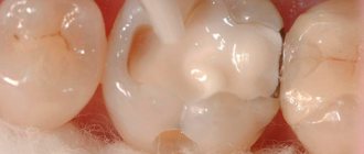
Age-related microcracks and darkening of enamel
Tooth anatomy
The structure of all units is the same, but each tooth has a distinctive shape and dimensions, depending on the functions it performs.
Internal structure of teeth in the photo
What does a tooth consist of? The top layer (enamel) is predominantly composed of minerals. It protects the inner layers of the tooth from mechanical, thermal and chemical damage. The rest of the solid surface of the elements consists of proteins and liquid involved in metabolic processes. This layer tends to wear out and wear off over time. Typically, caries first affects the surface of the tooth. For this reason, you should carefully monitor the condition of the oral cavity and carry out hygiene procedures in a timely manner.
If you examine a tooth in cross-section, you can also see dentin. It also consists of mineral substances and protects the pulp with the nerve fibers located in it. Many small channels are localized in dentin, with the help of which metabolic processes in bone tissue are carried out. Through these structures impulses are transmitted between nerve endings. For 1 sq. mm of dentin there are about 70,000 microscopic tubules.
The internal cavity of the elements is represented by pulp. It consists of nerve endings and lymph nodes.
The structure of the tooth should be considered depending on its location relative to the gum. The visible part of the element is the crown. The area of contact between the tooth and the gum is called the periodontium. The root is located in a special cavity, which dentists call the alveolus. The root of the tooth ends at an apex, which is supplied with blood vessels. Through these structures, the elements are fed.
No Ads
Fangs
Upper canine
The upper canine, one per quadrant, erupts at approximately 11-12 years of age and completes its formation after 3-4 years.
This is the longest tooth in the arch (about 27 mm or more).
The crown has a diamond shape with a peculiar sharp cusp, which on the vestibular side divides the mesial and distal sides of the crown. The crown is 10 mm high (length) and has a maximum mesial-distal diameter of 7.5 mm at the point of proximal contact and decreases to 5.5 mm cervically. The buccal-palatal diameter is wider (8 mm), and it remains up to 7 mm at the cervical level due to the presence of a prominent marginal ridge (Fig. 1.16).
Rice. 1.16. Upper canine: palatal view
The long root, like the crown, is narrow in the mesial-distal direction and more pronounced in the buccal-palatal direction. There is almost always only one root canal with lateral canals at different levels in 24% of cases. The apical third is straight in 40% of cases, and is often curved distally (32%) and/or vestibularly (13%) (Fig. 1.17).
Rice. 1.17 Upper canine: graphical representation of the access cavity
The pulp space at the level of the cervical third and the root middle third has an oval shape in the buccal-palatal direction; often two canals are present with a tendency to merge into one common round canal in the apical third (Fig. 1.18a, b).
Rice. 1.18 (a, b) Upper canine: buccal and proximal view
The endodontic access cavity is oval-shaped, extending from the cusp to the marginal ridge of the coronal cervical third to gain access to the radicular space without obstruction that could cause instruments to create steps or transposition of the apical foramen (see Fig. 1.19).
Rice. 1.19. Anatomical pictures from Hess and Keller: the endodontic space of the upper canine at the level of the cervical third of the canal is represented by one oval canal, and in the middle third it is divided into two different canals and often merges into one round canal in the apical third
Lower canine
The lower canine, one per quadrant, erupts approximately 8-10 months before the upper one and completes its formation after 3-4 years.
The crown has a length of 11 mm, and its mesiodistal diameter is narrower than the upper one (7 mm), and decreases to 5.5 mm at the cervical level. The maximum buccolingual diameter is 7.5 mm wide and decreases to a minimum at the cervical level. The cervical lingual marginal ridge is less pronounced than that of the upper canine (Fig. 1.20).
Rice. 1.20 Lower canine: lingual view of the crown
The lower canine is about 27 mm long, and has two separate roots in 6% of cases, or two canals in one root, connected by isthmuses, with a common or two separate apexes (Fig. 1.21). In 20% of cases, the apical third has a distal slope (Fig. 1.22).
Rice. 1.21 Lower canine: graphical representation of the access cavity
Rice. 1.22. Anatomical pictures from Hess and Keller: the endodontic space of the lower canine has two separate canals connected by an isthmus in the coronal third; the canals merge into one opening in the apical third
The endodontic space is oval in shape in the bucco-lingual direction for two-thirds of the root, and then gradually becomes rounded.
The endodontic access cavity should be oval in shape in a buccolingual direction, from the cusp to the marginal ridge (Fig. 1.23a, b).
Rice. 1.23 (a, b) Lower canine: buccal and proximal view
Kinds
What are the units? Teeth in the oral cavity are divided into 4 types: incisors, canines, molars and premolars. The human jaw is symmetrical and each part (right and left) has the same number of elements. If this does not happen, then they talk about malocclusion development, which requires immediate correction.
Incisors
Incisors are the elements located in the center of the jaw. There are 8 such units in total - 4 at the bottom and 4 at the top. The peculiarity of the anatomy of the incisors is that they have a flat crown with a sharp cutting surface. There are 3 tubercles on the cuts of the incisors, which wear off over time. The centrally located elements are usually larger than the lateral incisors, since they bear more load when chewing food.
The upper central teeth are most susceptible to damage in the form of cracks in the enamel and chips. It is in these areas that aesthetic defects associated with improper placement of elements in the oral cavity and trema are usually noted.
Crown – part of the tooth protruding above the gum
It should be noted the main functions of the incisors are the primary capture of food, cutting pieces and chewing them. The central elements are the first to begin to chop the food and pass it further. Due to the sharp cutting edge, units and twos crumble and crush food for further normal absorption. The incisors protect the remaining elements from severe damage during chewing loads. In addition, they protect the digestive organs from the development of chronic diseases associated with insufficient absorption of nutrients by the intestines.
On the upper jaw, the units have impressive dimensions - up to 23 mm including the root system. Unit width is from 4 to 6 mm. Thanks to the flat crown, the chewing load is distributed evenly between all areas of the tooth. The thinnest enamel of the incisors is in the area of contact with the gum. It is this part that is most vulnerable to carious processes.
Several features of incisors should be noted:
- rounded root shape;
- the presence of grooves on the surface of the root;
- chisel shape;
- greater curvature of the incisors located on the sides.
Fangs
A person has 2 fangs on each jaw. They resemble a triangle in shape and are easily recognized among other elements of the series. With the help of fangs, a person can bite off pieces of hard foods - apples, carrots, crackers. The elements have only one long root. The upper units are larger than the lower ones. Some patients resort to extension procedures to give greater effect to the canines on the upper jaw.
No ads 2
Premolars
The first premolar is also called a quadruple. The main function of the tooth is transitional and exciting. Due to the wider crown, the elements take part not only in biting off pieces of food, but also in grinding it to the desired state.
On the chewing surface of the element there are several types of chewing tubercles - flat and vestibular. The first tubercle is located closer to the cheek, and the last one is directed towards the oral cavity. In rare cases, three-tubercle or four-tubercle teeth are found. An anomaly is observed if the palatine tubercle passes into the median tubercle.
Due to the many grooves and fissures, pathogenic bacteria can accumulate on the surface of premolar teeth, especially if oral hygiene is insufficient. For this reason, these segments should be given the most attention when brushing your teeth. The second premolar of the upper and lower rows has larger dimensions, but is shaped like a quad.
Molars
Molars have the same structure as the rest of the series. But, despite this, several features can be identified that are characteristic only of sixes and sevens. The main function of molars is to grind and grind food during chewing. They play an important role in the formation of an anatomically correct bite.
Each half of the jaw has 4 molars below and above. Molars have several chewing surfaces. The buccal surface of the tooth is convex. There is a groove on it, which is a place where pathogenic flora accumulates. These areas also need to be given special attention when brushing your teeth, since the salivary glands are located near the buccal surface, which are affected during the development of carious processes.
There are 3 tubercles on the chewing surface: paracone, protocone and metacone. On the buccal side there is also the most important morphological structure of bone tissue - the Carabelli tubercle. The degree of expression of the structure is varied.
In some cases, the Carabelli tubercle may have its own process
The mesial border of the molars has a slightly convex shape. The root system on this side varies in shape among different patients. It can be cylindrical or barrel-shaped. The structure of the distal surface of the molars is similar to the mesial one. The only difference is inaccessibility during hygiene measures. For high-quality cleaning of the last elements, it is better to use floss or irrigator.
Wisdom tooth
Eight, or wisdom teeth, usually appear on the surface of the gums in adulthood. The shape of the element is similar to 6, but has more powerful roots. The number eight is considered the most problematic tooth: it is difficult to break through to the surface and quickly collapses. Dentists may have difficulty removing figure eights due to their abnormal position in the mouth and crooked roots, which can be diagnosed using x-rays. In such cases, the problem area is immediately extracted, otherwise purulent inflammation of the soft tissues may develop. Another serious complication of improperly positioned wisdom teeth is the destruction of the roots of adjacent units.
No ads 3
What other options exist for naming a person’s teeth by numbers?
There are three other systems common among dentists that can be used to designate and record dental units:
- A universal alphanumeric system developed by the American Dental Association.
- Zsigmondy-Palmer system. One of the oldest counting systems (it was created back in 1876), it is often used in their practice by maxillofacial surgeons and orthodontists.
- Haderup system.
All of them are convenient and easy to use in their own way, and in all such systems the arrangement of teeth by numbers in adults and children is indicated differently.
Alphanumeric system
It provides an indication of not only the number of the dental unit, but also its functional purpose. Thus, incisors are designated by the letter I, canines by the letter C, premolars by the letter P and molars by the letter M. As for the numbers, they indicate the serial number of a specific functional unit in the segment (whereas in other systems the functions of the dental unit are not taken into account, only its position is taken into account in a row). For example, if according to the Viola system the wisdom tooth is numbered 8, then according to the alphanumeric system it is also designated as M3, that is, the third molar in the segment. Additionally, digital segment designations are also used, which is why the serial number becomes two-digit. The same 48th tooth mentioned more than once with such a scheme will be designated as M43.
For milk teeth, lowercase (small) Latin letters are used in the recording, and instead of numbers, sometimes letter values from A to K are also indicated, counting dental units clockwise from the upper right incisor.
Zsigmondy-Palmer system
In old, and sometimes in new dental outpatient records, you can see a special plate for recording the condition of dental units, based on the Zsigmondy-Palmer square-numeric system. The order of growth of an adult’s teeth is indicated here by the familiar Arabic numerals from 1 to 8; for children, they use Roman numerals from I to V. The numbers of the jaw segments are not indicated here, the data is simply entered into the corresponding parts of the table diagram. The system is quite convenient and visual, but in an oral conversation it can cause difficulties when indicating a specific dental unit.
Classification of dental surfaces
To conveniently describe the localization of the pathological process, dentists use a term such as “crown surface”. There are 4 types of such surfaces:
Where is the eye tooth + photo
- Chewable. This part of the tooth faces the elements on the opposite jaw. In canines and incisors, this area is represented by the cutting edge.
- Vestibular. Located in the vestibule of the oral cavity. The vestibular surface of the anterior teeth has an area of contact with the lips. In the molars, this part is in contact with the inner shell of the cheek and is called the buccal surface.
- Lingual or lingual - the area of elements facing the tongue. It is in this area that the largest amount of plaque accumulates due to the difficulty of carrying out hygiene measures. The lingual side of the elements is not visible to others during a conversation, so many patients seek to install corrective systems on the inside of the tooth.
- Approximal. This type of surface is divided into medial and distal. In the first case, the area is located closer to the middle of the dentition, in the second case, closer to the edge of the arch.
Tooth surfaces
Upper incisors
Central upper incisor
The central upper incisor erupts (one per quadrant) between the ages of six and seven years, and the complete formation of the apical third occurs after 2 or 3 years. The average tooth length is 22-23 mm.
The crown has a triangular shape, about 10.5 mm long, the base extends in the mesial-distal direction, corresponding to the anterior edge of the tooth, up to 9 mm in size, and its buccal-palatal size is 7 mm (Fig. 1.9).
Rice. 1.9 Upper central incisors: palatal view of the crown
The root is usually straight (75%), but according to Ingle, a slight curvature may be present in a small percentage of cases. Lateral canals may be present in more than 20% of cases, and an apical delta is also common (35%).
The coronal pulp space also has a triangular shape, especially in the region of the cervical radicular third, with the base facing the vestibular wall and the apex located palatally, after which it gradually becomes a round canal until the apex.
The access cavity is triangular and follows the shape of the pulp space (Fig. 1.10).
Rice. 1.10 Upper central incisor: graphical representation of the access cavity
Lateral upper incisor
The eruption of the upper lateral incisor (one in the quadrant) occurs 1 year after the central one, and its complete formation takes approximately 3 years.
It is approximately 1 or 2 mm shorter than the central incisor, its mesial-distal size is smaller - about 7 mm, and the vestibular-palatal size is only 5.5-6 mm (Fig. 1.11).
Rice. 1.11 Upper lateral incisor: palatal view of the crown
Normally there is only one root canal, straight in 30% of cases, and often it is curved distally (53%), in a small percentage of cases there is curvature in other directions. The shape of the canal is ovoid in the cervical region, with a tendency to become more rounded in the apical region; the access cavity has a similar structure (Fig. 1.12).
Rice. 1.12 Upper lateral incisor: graphical representation of the access cavity
Features of milk elements
Milk units act as precursors of permanent elements. They usually appear in the first year of a baby's life. Usually the period of eruption of the first incisors is accompanied by painful symptoms for the baby. About 2-3 years may pass between the appearance of the first and the eruption of the last milk tooth. A 3-4 year old child has 20 fully formed baby teeth in his mouth.
The structure of teeth in children
The change of the primary occlusion to a permanent one is carried out from 6-7 years of age. The shape of the permanent elements will differ little from their predecessors. The only difference is the more massive crown part and dense enamel.
In children, the degree of mineralization of enamel and dentin is weaker than in adults. Therefore, their dental care should be carried out with special care. The milk tooth is designed in such a way that most of its volume is occupied by the pulp, so caries in children becomes more complicated faster.
Another difference between milk units and permanent ones is short roots. For this reason, tooth loss in children sometimes occurs unnoticed and painlessly.
Lower incisors
The lower incisors, two per quadrant, are very similar to each other, with some peculiarity retained.
Lower central incisors
The lower central incisors are the first permanent teeth to emerge in children's mouths, usually between 6 and 7 years of age, and are fully formed by 9 or 10 years of age. The lower central incisor is approximately 21.5 mm long. The apical third of the root is straight in 60% of cases and curves distally in 23% of cases.
The crown has a trapezoidal shape with a large base corresponding to the cutting edge and a smaller base that continues into the cervical third of the root. Its mesio-distal size is 5.5 mm at the greatest width of the incisal edge and gradually decreases to 3 mm at the cervical level (Fig. 1.13).
Rice. 1.13 Lower incisors: lingual view of crowns
The lower central incisor has a buccal inclination. In the root, it is possible to have a second canal located lingually in relation to the main canal in 18-23% of cases (Fig. 1.14).
Fig 1.14 Lower central incisor: graphical representation of the access cavity
Lower lateral incisor
The lower lateral incisor is similar to the central incisor, but slightly larger - about 1 mm, usually erupts 1 year after the lower central incisor and completes its formation after 3 years.
Root examination confirms the presence of two canals in 15% of cases.
The access cavity of the lower incisors is formed in a buccal-lingual direction to search for a second canal located lingually (Fig. 1.15a-c).
Rice. 1.15 (a - c) Lower central incisor: buccal, lingual and proximal views
Malocclusions
With a correct bite, the teeth of the upper jaw protrude onto the elements of the lower jaw by no more than 1/3. This position will make it easier for the jaws to chop food. Malocclusions are usually associated with the following conditions:
- The upper units overlap the lower ones by more than ½. This type of bite is usually called a deep bite.
- Certain groups of teeth may not fit together at all. In this case, they talk about an open bite.
- Distal bite. What it is? The problem is said to occur when the upper jaw is overdeveloped.
- If the lower jaw is pushed forward, then there is a mesial deviation.
- When the structures are displaced relative to each other, a cross anomaly occurs.
If not treated promptly, malocclusions can cause toothless jaws. For this reason, it is important to contact an orthodontist at the first sign of a problem.
Upper molars
Three molars in a quadrant without corresponding primary teeth.
Upper first molar
It erupts between 6 and 7 years behind the second primary molar. The formation of the apex is completed between the ages of 9 and 10 years.
The crown has four cusps on the occlusal surface and three roots, one palatal and two vestibular (Fig. 1.38).
Rice. 1.38 First upper molar: occlusal view
The length of the tooth is about 21-22 mm and there is one canal in the palatal and disto-buccal roots
The mesiobuccal root has two separate canals in 60-70% of cases (MB1 and MB2). The second canal in the distobuccal root (DB2) is quite rare (2-3%) (Figs. 1.39, 1.40 and 1.41).
The access cavity to the pulp chamber has a triangular shape with the base facing the buccal side and the apex facing the mesial-palatal tubercle; the cavity is always located mesial (in front) of the transverse ridge, which unites the mesiopalatine tubercle with the distal buccal tubercle. The mouths of the canals are located at the vertices of the triangle, which form the access cavity (Fig. 1.42); if we combine the orifice of the MB1 canal with the palatal orifice, we can find a second mesiobuccal root canal (MB2), which is often closed by calcification (Fig. 1.43).
Rice. 1.39 Anatomical pictures from Hess and Keller: the upper molar has a very complex canal anatomy
Rice. 1.40 Anatomical pictures from Hess and Keller: the upper molar has a very complex canal anatomy; MB1 and MB2 visible in the mesiobuccal root
Rice. 1.41 Anatomical pictures from Hess and Keller: the upper molar has a very complex canal anatomy; MB1 and MB2 visible in the mesiobuccal root
Rice. 1.42 Upper first molar: triangular access cavity with three canals
Rice. 1.43 Upper first molar: access cavity extended to the mesial ridge to locate the MB2 canal on the line connecting the mesiobuccal and palatal canal
Second upper molar
Eruption occurs at the age of 12 years with the completion of the apical third at approximately 14 years.
It is smaller and shorter than the first molar by about 1-2 mm, is about 19-20 mm high and has four occlusal cusps. There are three roots and three canals, just like the first molar, and the MB root also has a second canal (37-42%). The shape of the pulp chamber is rhomboidal rather than triangular (see Figs. 1.42 and 1.43).
Upper third molar
This tooth erupts after age 17. It has a very unique anatomical morphology; the crown has three or five tubercles (Fig. 144). The average height of the third molar is about 18 mm. There may be one root or more often than one - three or four roots, usually curved distally (Figs. 1.45, 1.46, 1.47 and 1.48).
Figure 1.44. The upper third molars have a very distinctive occlusal morphology with three or four cusps
Rice. 1.45. Upper single root third molar
Rice. 1.46. Upper third molar with four distally curved canals
Rice. 1.47. Upper third molar with atypical anatomy
Rice. 1.48. Upper third molar with strong 3D root curvature