1.General information
The word “ostitis” (not to be confused with otitis) in a broad sense means inflammation of the bone, or, more correctly, inflammation of bone tissue.
In fact, this is a group of heterogeneous diseases, pathological conditions and processes in relation to which much remains to be explored, clarified and clarified. In the current Tenth Revision of the International Classification of Diseases (ICD-10), such inflammatory processes are classified under the heading “Other bone diseases,” and the unifying characteristic is precisely the presence of inflammation.
A must read! Help with treatment and hospitalization!
2. Reasons
The main trigger (trigger factor) in the etiopathogenesis of osteitis is secondary infection during fractures and other injuries, which include surgical interventions on bone structures. The infection can be either nonspecific (streptococci, staphylococci and other widespread pathogenic microorganisms) or specific (mycobacterium tuberculosis, gonococcus, treponema pallidum).
Osteitis also includes Paget's osteitis deformans - a chronic, progressive disease of unknown etiology, in which the regeneration of bone tissue (normally, its renewal occurs constantly) is carried out due to the formation of tissues with an irregular, altered structure. Paget's disease usually affects the pelvic, femur, humerus bones, as well as the spinal column and skull bones.
Unfortunately, in isolated cases, the cause of the development of osteitis is non-compliance with the BCG vaccination procedure.
Visit our Traumatology and Orthopedics page
Indications for prophylactic antibiotics
Antibacterial drugs may not always be prescribed during implantation. For example, when installing artificial roots without tilting the mucoperiosteal flap, antibiotics are usually not prescribed. This is due to the low traumatic nature of the operation, in which even no stitches are applied. In other cases, antibiotics are mandatory for dental implantation. Among the indications:
- simultaneous installation of several root substitutes with the formation of a large flap;
- replanting of bone material simultaneously with implantation;
- ridge augmentation (hard tissue augmentation) using grafts or own bone blocks transplanted from another location;
- multiple simultaneous implantation with immediate loading of artificial roots with temporary or permanent crowns;
- the use of closed or open sinus lift when installing implants under the maxillary sinuses.
Antibiotics for all types of dental implantation can also be prescribed for unsatisfactory initial oral hygiene, installation of root substitutes against the background of periodontitis, and chronic diseases of the nasopharynx.
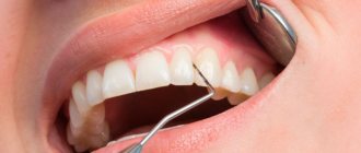
3. Symptoms and diagnosis
The clinical picture of osteitis in each specific case is determined by a number of individual factors, i.e. generally varies widely. Quite typical is an asymptomatic or low-symptomatic course in the initial stages. Over time, patients notice some swelling, pain discomfort and some degree of functional decline in the affected area (for example, limb mobility is limited, chewing becomes painful, etc.).
Under unfavorable conditions (hypothermia, abdominal surgery, exhaustion, hypovitaminosis and other factors that weaken the immune system), an exacerbation may occur during osteitis, including such severe complications as the formation of fistulas, phlegmon, osteomyelitis.
Chronic osteitis (especially fibrous) is characterized by an increased risk of fractures, due to the tendency for the affected bone tissue to soften and become brittle.
Diagnosis is based on the radiographic method; in cases of insufficient information content, radionuclide testing, computed tomography or magnetic resonance imaging is prescribed. According to indications, immunological and bacteriological laboratory tests are performed; in some cases, a biopsy is required for differential diagnosis.
About our clinic Chistye Prudy metro station Medintercom page!
Types and regimens of prescribing antibacterial drugs during implantation
The patient does not need to know what antibiotics are prescribed for dental implantation. It is enough to follow the instructions of the dentist: purchase prescribed antibacterial drugs at the pharmacy and take them according to the prescribed regimen.
The name of the drug, frequency and duration of its administration depend on the type and complexity of the operation performed. In some cases, for example, the doctor may prescribe a single dose of the antibiotic amoxicillin 60 minutes before installing the implants. Others require an additional dose of the drug six hours after surgery. When performing complex traumatic procedures, the dentist may prescribe a stronger antibiotic (Augmentin, Tsipran, Tsiprolet) several times a day for three or five days.
In general, it is useful for the patient to know that before any dental surgery you can independently reduce the number of microorganisms in the mouth. This can be done by rinsing the mouth with a 0.2% chlorhexidine solution. If you don't have it, you can use plain water. Rinsing even with it alone removes about 60-70% of bacteria.
4.Treatment
The therapeutic regimen is determined by the results of the diagnostic examination. Depending on the scale, localization and etiology of inflammation, antibacterial drugs, anti-inflammatory drugs, immunomodulators and immunostimulants are prescribed. Absolute indications for surgical intervention are often identified - in particular, to remove necrotic (dead) areas. Therapy with special proteolytic enzymes, as well as physiotherapeutic methods, are effective. Sanitation of all foci of chronic infection, relief of exacerbations of diseases concomitant and/or primary in relation to osteitis (for example, rheumatoid arthritis, syphilis, tuberculosis) are mandatory.
An important task is to prevent the inflammatory process from becoming chronic. In this regard, the time factor is crucial: at the first symptoms of trouble in any bone tissues or structures, contacting a doctor should be immediate. The earlier a reliable diagnosis is made and treatment is started, the more favorable the prognosis, i.e. higher chances of complete healing of osteitis.
Approaches to the treatment of purulent-inflammatory diseases of large joints
Dear visitors of our site! This article is devoted to the surgical treatment of purulent-inflammatory diseases of the joints, complicated by the destruction of bone tissue (osteomyelitis), and purulent complications of endoprosthetics.
This group of diseases is a serious, unresolved problem in modern orthopedics. There are no uniform standards for the treatment of this pathology. Treatment approaches are based on the personal experience of individual authors and clinics. We would like to highlight the fundamental aspects of diagnosing this group of diseases and share our personal experience of their treatment.
The incidence of septic arthritis ranges from 2 to 10 cases per 100,000 population. The predominant lesion is the knee joint (up to 55%), followed by the hip and ankle joints. Mostly children and the elderly suffer from such diseases.
What are the prerequisites for the development of purulent arthritis? These are injuries to the joint area, degenerative and rheumatoid diseases of the joints, injections in the joint area, arthroscopic interventions, a history of joint replacement, purulent-inflammatory diseases, trophic ulcers of the distal extremities, sexually transmitted infections, systemic administration of glucocorticoids.
Clinical picture of the disease. Manifestations of the disease are quite distinct when joints close to the skin are affected (elbow, knee, ankle). This is tissue swelling in the joint area, local redness and increased skin temperature. When large, deep-lying joints, such as the shoulder and hip, are affected, local changes may not be pronounced, and the patient will only be bothered by pain in the joint area. An increase in body temperature may also not appear immediately. The photograph below shows a typical clinical picture of arthritis of the knee joint: the contours of the right knee joint are smoothed, the joint is increased in volume.
Laboratory diagnosis of the disease. The examination of a patient with a suspected purulent process must necessarily include a clinical blood test with calculation of the leukocyte formula and erythrocyte sedimentation rate, and a blood test for C-reactive protein. Among the laboratory indicators, I would like to dwell specifically on the level of procalcitonin. The procalcitonin test is currently the most objective method for determining the generalization of a bacterial infectious process. A dynamic increase in this indicator in the patient’s blood tests is an indicator of the spread of the purulent process beyond the joint and the development of sepsis.
Instrumental diagnostics: - radiography of the affected joint; — spiral computed tomography of the joint; - joint puncture followed by microscopy and culture of the discharge to determine the infectious agent and its sensitivity to antibiotics. This manipulation must be performed before starting antibiotic therapy.
Treatment. In the treatment of purulent-inflammatory diseases of the joints, our task is to quickly stop the inflammatory process and, if possible, preserve the function of the joint. According to various authors, during a purulent process in the joint, the articular cartilage is affected 4–6 days from the onset of the disease and finally dies off in the 4th week of the disease, after which the effectiveness of joint-preserving operations sharply decreases. In the absence of signs of bone tissue damage, preference is currently given to operations that preserve the joint. If superficial joints are affected in the early stages of the disease, it is possible to use puncture methods and broad-spectrum antibiotic therapy. If puncture methods are ineffective, drainage of the joint is recommended within 48 hours from the onset of the disease. Ideally—most modern authors tend to do this—the use of arthroscopic techniques.
Our personal experience: treatment of septic arthritis of the ankle joint in a 19-year-old patient through 3 weeks of flow-through drainage. The flushing system was installed through accesses up to 2 cm. The result of treatment was complete relief of the inflammatory process and recovery of the patient. During two years of follow-up, no relapse of the disease was noted. When the purulent process spreads to the bone tissue, non-viable tissue is removed and the joint area is stabilized using an external fixation device. As a result, the inflammatory process is stopped and arthrodesis of the affected joint is formed, that is, fusion between the bones that form the joint, which subsequently ensures adequate supporting function of the limb.
Personal experience: An 81-year-old patient was admitted to the clinic with pain in the hip joint that had been bothering her for 4 months after falling from a height. During the week before admission to the clinic, the pain steadily increased, and the supporting function of the limb was impaired. She did not notice any increase in body temperature; she constantly took anti-inflammatory drugs (Movalis) in large doses.
X-ray upon admission.
Zones of destruction are identified in the body of the ilium and in the head of the femur.
Puncture of the hip joint reveals thick purulent contents.
The patient underwent emergency surgery: the joint was opened, the focus of osteomyelitis was sanitized, the head of the femur was removed, the joint was fixed with a rod-based extrafocal device. A flow-through drainage system has been installed. The patient was treated with broad-spectrum antibiotics for 6 weeks. During treatment, pain in the hip joint was relieved, and clinical and laboratory tests showed a decrease in the inflammatory response.
The external fixation device was removed 8 weeks after surgery. The hip joint is fixed with an orthopedic fixator. 3 months after surgery, the fixator was removed. The patient walks independently with full weight bearing on the operated leg. The inflammatory process has been stopped.
Results of spiral computed tomography of the patient’s pelvic bones 3 months after surgery.
Signs of the formation of a fusion between the femur and the body of the ilium are determined. This fusion ensures adequate support function of the limb in the future.
Currently, a serious problem in modern orthopedics is purulent complications of endoprosthetics . According to CITO data for 2012, 80,000 endoprosthetics of large joints were performed in Russia. The ratio of primary and revision endoprosthetics was 9:1. The complication rate for primary arthroplasty of large joints was 2.4%. Of these, 40.6% are infectious complications. The rate of complications during revision arthroplasty was 12.9%. Of these, 51.4% are infectious complications.
In case of early deep suppuration after hip replacement, it is possible to preserve the endoprosthesis . Chronic infection of the endoprosthesis area and destructive osteomyelitis in adults, which developed against the background of septic arthritis, are indications for radical surgical sanitation, removal of endoprosthesis components and necrotic areas of bone tissue.
Approaches to performing operations for suppuration of endoprostheses vary. In our practice, we use articulating spacers (temporary “substitutes” for the prosthesis) - individually made from bone cement with the addition of antibiotics. The use of these devices allows you to maintain limb length, ensure walking with support on the operated leg, improve the patient’s quality of life in the postoperative period, and facilitate subsequent re-endoprosthetics .
Clinical example: Patient 65 years old. History of total hip replacement performed abroad in 2002. No dynamic monitoring of the joint was carried out. Over the past two years, since 2012, I have been experiencing periodic pain in the hip joint. Since September 2013, over the course of 3 months, he has noticed a gradual increase in pain. Two weeks before admission to the clinic, massage of the joint area and acupuncture were performed at home. After a course of these procedures, a sharp increase in pain and a rise in body temperature to 39 degrees were noted.
Radiographs of the patient upon admission.
Signs of destruction of the acetabular component of the prosthesis, signs of destruction of bone tissue along the femoral component of the prosthesis are determined.
During puncture of the joint, thick purulent contents were obtained. The joint cavity was opened, the components of the endoprosthesis were removed, and the osteomyelitis lesion was radically treated. During the inspection, purulent melting of the proximal femur was revealed. An articulating spacer is installed.
X-ray of the patient after surgery.
The model of the proximal femur is formed from bone cement with antibiotics, reinforced with a proximal femoral rod.
The postoperative course is smooth. The inflammatory process has been stopped. Re-endoprosthetics is planned. This clinical example clearly demonstrates the need for timely contact with a specialist traumatologist - orthopedist and the inadmissibility of performing physiotherapeutic procedures without preliminary adequate diagnosis of the cause of pain in the joints.
The Orthocenter specialists are proficient in all methods of surgical treatment of purulent-inflammatory joint diseases and are ready to come to your aid.