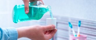
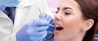
From this article you will learn:
- how to rinse your mouth if your gums are inflamed,
- comparison of the effectiveness of funds,
- The best mouthwash for sore gums.
The article was written by a dentist with more than 19 years of experience.
Mouth rinses for gum inflammation can be divided into – 1) antimicrobial, 2) anti-inflammatory, 3) combined action. Rinse solutions with an antimicrobial effect contain antiseptics or antibiotics, and these components act directly on the cause of gum inflammation - pathogenic bacteria. In dentistry, antiseptics such as chlorhexidine, hexitidine or cetylpyridine are usually used, and among antibiotics, triclosan.
In turn, rinses with anti-inflammatory effects may contain: methyl salicylate, phenyl salicylate, allantoin, bisabolol, thymol and eugenol, as well as medicinal plant extracts. All of them have the property of blocking the so-called “inflammatory mediators,” thereby stopping the inflammatory reaction in the tissues. Some mouth rinses have a combined composition, combining both antiseptics and anti-inflammatory components.
In this article, we will analyze the best mouthwashes –
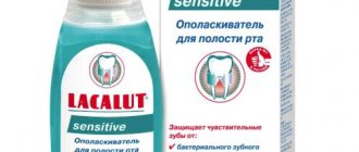
- chlorhexidine solution,
- LACALUT Active,
- PRESIDENT PROFI Antibacterial “Powerful protection against bacteria”,
- Hexoral (0.1% hexitidine solution),
- miramistin,
- PRESIDENT PROFI Active,
- Periodontocide,
- LISTERINE Expert “Gum protection”,
- Stomatophyte.
Combination of rinses and application gel –
The selection of rinse aid and gel is carried out in such a way that both antimicrobial and anti-inflammatory effects are equally realized.
Those. if you choose a rinse with antimicrobial components - such as chlorhexidine, hexitidine, cetylpyridine, triclosan, then in parallel you need to use a gum gel containing anti-inflammatory components (this can be choline salicylate, phenyl salicylate, methyl salicylate, etc.). But if you use a rinse primarily with anti-inflammatory components (for example, Parodontocid mouthwash), then you need to choose a gum gel that contains an antiseptic and/or antibiotic. Such gels include Perio-Aid, PRESIDENT Effect gel and others. For more information about anti-inflammatory gels for the treatment of gum inflammation and patterns of their use, read the article:
→ Rating of the best gels for gum inflammation
The importance of choosing the right medicine
It is impossible to completely destroy microorganisms living in the human mouth. But when treating some diseases and conditions, it is necessary to temporarily reduce their number for a short time. Modern antiseptics help a lot with this.
It should be understood that those solutions that are used to treat the skin should not be taken by mouth. Firstly, it is fraught with poisoning. All sanitizers for external use are not intended for oral use.
Secondly, such therapy is always pointless. This is explained by the fact that the human mouth is inhabited by bacteria. Unsuitable products are inactivated by salivary proteins. This means that they do not work on the mucous membranes of the tongue and gums.
Mouth rinses may be unsafe -
You may notice that the instructions for rinses with antiseptics and antibiotics (at least from some manufacturers) recommend using them for 3-4 weeks, and in some cases even up to 1-2 months. It is absolutely impossible to follow such recommendations, because... Long-term use of antiseptics radically changes the composition of the oral microflora, and not for the better. Pathogenic microflora tends to get used to antiseptics (as they say, the fittest survive), and therefore, after the end of the use of antiseptics, the rate of destruction of the attachment of teeth to the bone and gums can only increase. Therefore, the course of rinsing with antiseptics should not last more than 10-12 days.
In addition, clinical studies (source) revealed that after just 7 days of rinsing the mouth with chlorhexidine, the ratio of microflora in the oral cavity changed in such a way that strains of pathogenic bacteria that produce large amounts of lactic acid received an advantage. This leads to a shift in the pH of the oral fluid to the acidic side, to a decrease in the buffer capacity of saliva and a decrease in its ability to neutralize lactic acid, which is secreted by cariogenic bacteria. Thus, the risk of developing dental caries increases.
Important for patients with cardiovascular pathology -
Several reputable scientific studies have shown that chlorhexidine interferes with the ability of oral bacteria to convert nitrates (found in foods) into nitrites. This bacterial activity is called “nitrate-reducing” and is one of the mechanisms for maintaining normal blood pressure. Normally, nitrites entering the blood help lower blood pressure and, accordingly, a decrease in their production will lead to an increase in blood pressure.
This is primarily important for patients who already have chronic cardiovascular pathology. This information is confirmed by the clinical study “The increase in plasma nitrite after a dietary nitrate load is markedly attenuated by an antibacterial mouthwash. Nitric Oxide" (2008), authors – Mirco Govoni, Emmelie Å. Jansson, and others. If you wish, you can familiarize yourself with this study using the link above, for example, using a browser translator.
Indications for use
Each medicine has its own indications for use. Among the common diseases that are successfully treated with the described dental medications are:
- stomatitis;
- tonsillitis;
- angina;
- periodontal disease;
- periodontitis;
- gingivitis;
- ulcers;
- period of gum recovery after tooth extraction.
Medicines reduce the risk of infection and speed up healing if bacteria have entered the wound. But in order to get positive dynamics with their help, you must strictly follow all medical prescriptions and rinse regularly.
What you don't need to rinse your mouth with -
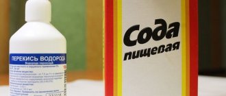
Many patients who try to be treated at home sometimes treat their gums with hydrogen peroxide.
Of course, hydrogen peroxide is a good antiseptic, and it is excellent in treating many infectious processes (for example, purulent wounds). It can also be used in dentistry, but not for rinsing the mouth, but for rinsing periodontal pockets with a syringe. When hydrogen peroxide comes into contact with any organic matter, it begins to foam, releasing atomic oxygen. When rinsing periodontal pockets, 3% peroxide is drawn into a syringe and the sharp edge of the needle is broken off (so as not to pierce the gum). Then the blunt end of the needle is inserted along the tooth root into the lumen of the periodontal pocket and washed under pressure. As a result, all infection and pus are washed out of the pockets.
But when rinsing the mouth, peroxide will react most quickly only with oral fluid, and other than a full mouth of foam, you will not get any effect. You won’t be able to rinse your pockets with a syringe at home yourself. For this you need a doctor, because... if you suddenly inject peroxide not into the lumen of the periodontal pocket, but directly into the soft tissue of the gums, you will receive a severe chemical burn with subsequent necrosis.
Gargling with infusions of medicinal herbs –
Many patients prepare their own rinse solutions by brewing medicinal herbs such as chamomile or oak bark. Such infusions actually have a good astringent effect, moderate anti-inflammatory, and also a weak antiseptic effect. And this can really help reduce swelling and bleeding of the gums, but such herbal infusions also have one big disadvantage.
The fact is that self-brewed herbal infusions contain a lot of pigments and tannins, which are very quickly deposited on the teeth in the form of pigment plaque. This plaque forms very quickly, and it will serve as a good basis for the already microbial plaque to attach to the surface of the teeth. But it is microbial plaque and tartar that cause gum inflammation. Therefore, if you use solutions with herbs, it is better in the form of ready-made rinses (since manufacturers remove from them all pigments that stick to tooth enamel).
Medicines for the treatment of diseases of the mucous membrane of the mouth, tongue and lips
Diagnosis, treatment and prevention of diseases of the mucous membrane (OM) of the mouth, tongue and lips remain an important problem in clinical medicine. Changes in the CO of the mouth, tongue and lips in many systemic diseases occur long before the appearance of general clinical symptoms. Therefore, the correct interpretation of such changes and the choice of appropriate medications are of great importance not only for dentists, but also for other specialists.
When presenting the material, we used the classification of diseases of the oral cavity by E.V. Borovsky and A.L. Mashkilleyson [7], which provides for the identification of 9 groups of this pathology, taking into account etiological and pathogenetic factors, which, due to the impossibility of citing them, will be omitted in this publication. We limited ourselves only to issues of medical tactics, drug treatment of major diseases of the oral mucosa, tongue and lips.
The authors have made every effort to ensure the accuracy of the information presented, including drug dosages. Aware of our responsibility associated with the preparation of the publication, and taking into account the constant changes occurring in medical science, we recommend that the dosage of medications be updated according to the appropriate instructions.
Basic principles of treatment of diseases of the mucous membrane of the mouth, lips and tongue:
- Rational treatment requires contact between the dentist and other dental and non-dental specialists.
- Treatment must be carried out in compliance with the principles of bioethics, these diseases must be considered from the standpoint of the state of the whole organism, therefore in most cases one cannot limit oneself to local influences only.
- An axiom for the dentist should be the elimination of all unfavorable irritating factors in the oral cavity in the patient, which can support and provoke the development of the pathological process. It is unacceptable to use so-called cauterizing agents and long-term use of the same mouthwashes.
- Treatment should begin only after at least a preliminary diagnosis has been established and meet the following requirements: be comprehensive; provide a pathogenetic approach; do not disturb the anatomical and physiological characteristics of the oral mucosa; eliminate the pain factor; promote rapid epithelization of lesions; provide for the active involvement of the patient in performing medical procedures at home.
Traumatic lesions
Medical tactics for traumatic lesions of the mucous membrane due to the action of mechanical factors, high and low temperatures, radiation, unfavorable meteorological factors, and chemicals boils down, first of all, to eliminating the effect of the traumatic factor, which not only facilitates differential diagnosis, but is also a therapeutic intervention.
Decubital ulcer
Heals, as a rule, 10-12 days after eliminating the mechanical cause. The surface of the ulcer is treated with oxygen-containing antiseptics (hydrogen peroxide, potassium permanganate, sodium hypochlorite), and after cleaning its surface from necrotic plaque, keratoplasty agents are used. It should be remembered that with prolonged existence (more than 2-3 months), especially in older people, a decubital ulcer can become malignant.
Chemical injury CO
It occurs as a result of its immediate immediate contact with concentrated solutions of acids, alkalis, some medications and agents sometimes used by dentists (silver nitrate, arsenic paste, resorcinol-formalin mixture). In case of acute chemical injury, the CO drug that caused the burn is immediately neutralized (in the absence of a neutralizer, the mouth is washed with water). Subsequently, the affected areas are treated with anesthetic substances, weak antiseptic solutions, proteolytic enzymes, and then keratoplasty agents are applied.
Physical trauma CO
It can also be acute and chronic. Painkillers, antiseptics, and drugs that promote tissue regeneration are used. In some cases, it is necessary to resort to excision of the affected area.
Leukoplakia
A fairly common disease of the oral mucosa and red border of the lips, pathomorphologically characterized by chronic inflammation with pronounced hyperplasia and keratinization of the epithelium.
The scope of treatment measures is determined both by the form of the disease and the size of the lesion and the speed of development of the process. Among the medications for flat form of leukoplakia, applications are made to the lesion and vitamin A is prescribed orally (3.4% oil solution of retinol acetate or 5.5% retinol palmitate, 10 drops 2-3 times a day for 1. 5-2 months). Courses are repeated 2-3 times a year. For verrucous and erosive forms, the same is done within one month, and in the absence of positive dynamics, the lesion is excised, followed by mandatory histological examination, which determines further medical tactics.
The manifestations and severity of changes in the mucous membranes of the mouth, tongue and lips during infectious diseases depend both on the type of pathogen and on the individual characteristics of the patient’s body, which determines the dentist’s tactics.
Adenoviral diseases
In case of influenza and adenoviral diseases, the lesions of the mucosa are not specific; after a few days from the onset of the disease, hyperemia with “granular” rashes on the mucous membrane is replaced by petechial rashes, and by the 7-9th day the pathological changes disappear.
Chronic recurrent herpes
It appears at any age in persons previously infected with the herpes simplex virus.
Medical tactics for viral infections include the use of drugs from several groups that affect various parts of the etiology and pathogenesis of diseases: antivirals, immunocorrectors and immunomodulators, vaccines, interferons and their inducers. The most pronounced therapeutic effect is observed with the combined use of 2-3 drugs of general and local action with different mechanisms of antiviral effect.
Ointment and cream are started to be used when the first signs of infection activation appear and continue until the erosions are epithelialized. Early treatment can prevent the development of vesicles. A large group of antiviral drugs includes nucleoside analogues, similar in structure to intermediate products of DNA and RNA biosynthesis (iododeoxyuridine, chlordeoxyuridine, fluorodeoxyuridine), effective against generalized forms of herpes and herpes zoster. An effective antiviral agent is acyclovir (Zovirax, Virolex). In addition to acyclovir, the group of abnormal nucleosides includes: valacyclovir (Valtrex) in tablets of 500 mg; take 2 times a day for 5-7 days; famciclovir (famvir) tablets 250 mg; take 3 times a day for 7 days.
It should be noted that the therapeutic effect of antiviral drugs is most effective when prescribed in the first hours and days of development of the lesion elements and in the prodromal period of the viral disease.
Local antiviral drugs include: adimal, bonafton, florenal, riodoxol in the form of ointments and liniments (gossypol). Ointment and cream are started to be used when the first signs of infection activation appear and continue until the erosions are epithelialized. Moreover, early treatment can prevent the development of vesicles.
The leading role in the development of antiherpetic immunity belongs to interferon. The antiviral drug "Infagel" (it includes recombinant interferon alpha-2) has a wide spectrum of antiviral activity, bacteriostatic and anti-inflammatory effects, as well as antitumor and immunomodulatory activity. It is used in the form of applications to the lesions 2 times a day with an interval of 12 hours and dried for 10-15 minutes to form a protective film [10].
Interferon inducers (poludan, larifan) and antiviral vaccines (in the form of a cycle of intradermal injections to prevent relapses and during remission of the disease) have found widespread use in dental practice. The therapeutic effectiveness of the vaccine increases significantly when combined with poludanum or other interferon inducers.
In addition to these drugs, salicylates, analgesics, antihistamines and antiseptic solutions, multivitamins, EF radiation and helium-neon laser radiation are used in the treatment of herpes infection. To stimulate the processes of regeneration of the mucous membrane of the mouth and the red border of the lips, applications of oil solutions of vitamins A, E, ointment and jelly of solcoseryl and Actovegin, aerosols “Livian”, “Spedian”, “Gipozol” are used. After eliminating the elements of the lesion, sanitation of foci of chronic infection is mandatory.
In case of herpes zoster, along with the measures mentioned above, the doctor’s task includes eliminating the pain syndrome and preventing postherpetic neuralgia. For this purpose, analgesics, ganglerone, and B vitamins are prescribed.
Vincent's ulcerative-necrotizing gingivostomatitis (YANGS)
This is an inflammatory disease of the mouth, characterized by necrotic decay of the affected areas. Medical tactics for YANGS in some cases (especially in severe cases) include hospitalization of the patient, local and general measures, and medical examination.
Local treatment is important and begins with the patient’s first visit to the doctor, and then is carried out daily and in stages:
- Pain relief (applications, oral baths).
- Treating the mouth with antiseptic solutions (more effective - those that release atomic oxygen and chlorine-containing preparations: hydrogen peroxide, potassium permanganate, hypochlorite).
- Mechanical (and after application of proteolytic enzyme solutions to the lesion) removal of necrotic tissue, removal of mineralized dental deposits and elimination of other local traumatic factors.
- After cleansing the lesions from necrotic plaque, it is possible and advisable to use (in the form of applications) keratoplasty agents and preparations that promote tissue regeneration (oxycort aerosol, methyluracil ointment, oil solutions of vitamins A and E).
- It is better to postpone the removal of damaged teeth and roots, treatment of chronic forms of pulpitis and periodontitis until the ulcers are completely epithelialized.
- Subsequently, final sanitation of the oral cavity (planned treatment of caries, its complications, periodontal and mucosal diseases).
General treatment (especially for severe disease) involves the use of antimicrobial drugs. Orally prescribed: metronidazole (Flagyl, Klion) 0.25 g 2 times a day for 7-10 days, in some cases it is necessary to use broad-spectrum antibiotics. General strengthening and stimulating therapy is also carried out: ascorutin, vitamin C in large doses (up to 3.0 g per day), and antihistamines are prescribed. The nutrition of patients with YANGS should be complete and balanced.
Fungal infections of CO
Fungal infections of the mouth (oral mycoses) are most often caused by fungi of the genus Candida albicans.
General treatment includes:
- Ingestion of antifungal drugs for 10 days: diflucan (capsules) 50-100 mg 1 time per day; the use of polyene antifungal drugs (nystatin, levorin, amphotericin B) is traditionally included in general treatment, although it is known [11] that polyenes are practically not absorbed in the gastrointestinal tract, thus exerting a local effect on oral mucosa, and therefore It is recommended to dissolve the tablets in the mouth after meals (nystatin 1 tablet 500,000 units every 6-8 hours, levorin 1 tablet 500,000 units every 8-12 hours); Amphotericin B preparations are used mainly for severe systemic mycoses due to their high toxicity [11].
- Vitamins C, PP, group B taken inside.
- Use of antihistamines.
- Treatment together with other specialists of concomitant and background diseases.
Local treatment includes:
- Antifungal drugs for application and lubrication of lesions: 0.5% decamine ointment, 1% ointment and clotrimazole solution (canesten); 1% pimafucin ointment; for yeast infections and cheilitis, it is advisable to use 5% levorin ointment.
- Aqueous solutions of aniline dyes: 1-2% solution of gentian violet, 2% solution of methylene blue.
- Agents that alkalize the oral cavity: rinse with a 2% sodium bicarbonate solution; applications and lubrication of lesions with a 20% solution of borax in glycerin.
- A decoction of wild rosemary for mouth rinsing and oral baths.
Lesions of the oral mucosa are observed in all periods of syphilis, and dentists with similar manifestations have become much more common. Treatment of patients with syphilis is carried out in specialized venereological institutions. Dental interventions are usually associated with the sanitation of the oral cavity and are carried out in strict compliance with the disinfection and sterilization regime in health care facilities.
Treatment of fixed and widespread allergic and toxic-allergic diseases includes the “exclusion” of the allergen, for example, a drug, the use of painkillers, antiseptics, desensitizing and keratoplasty agents, if necessary. Sanitation of odontogenic foci of chronic infection is mandatory.
Erythema multiforme exudative
A fairly common dermatosis affecting the oral mucosa, observed mainly in young and middle-aged people. Stevens-Johnson syndrome is a severe variant of exudative erythema multiforme (also called acute mucocutaneous-ocular syndrome).
Medical tactics depend on the severity of the disease. In some cases, patients need to be hospitalized.
General treatment is usually prescribed by other specialists (dermatologist, immunologist) and includes:
- Detoxification therapy: drinking plenty of fluids or infusion therapy if it is impossible to take liquids orally.
- Taking glucocorticoids: prednisolone in an initial dose of 20-60 mg per day orally for 5-7 days (the dose depends on the severity of the process and the form of the disease, with Stevens-Johnson syndrome higher dosages are required), then the dose of prednisolone is reduced by 5 mg every 2 —3 days before complete withdrawal of the drug.
- Immunomodulatory therapy: for example, Lavomax (Tilorone) is prescribed 125 mg (1 tablet) orally in the first two days, then 125 mg after 48 hours. The dose of the drug per course is 2.5 g [15].
- Prescription of antihistamines, B vitamins, ascorutin.
- Antibiotics are prescribed when there is a threat of purulent-inflammatory complications and the doctor is confident that there is no connection between the development of the disease and the use of these antibacterial agents.
Local treatment is aimed at relieving inflammation and accelerating epithelization of the affected areas of the mucous membrane of the mouth and lips. It includes:
- The use of painkillers (1-2% solutions of trimecaine, lidocaine, anesthetics in aerosols) before each manipulation in the oral cavity (baths, rinses, applications) and before meals.
- Antiseptic treatment of the mouth (solutions of hydrogen peroxide, potassium permanganate, chloramine).
- To improve the rejection of necrotic tissues, apply solutions of proteolytic enzymes (trypsin, chymotrypsin) to the lesions.
- After cleaning the eroded surfaces from purulent-fibrinous plaque, use keratoplasty preparations (sea buckthorn and rosehip oil, oil solutions of vitamins A and E, the drug “Gipozol”).
- EF irradiation of the oral mucosa.
After the elimination of acute phenomena, the foci of infection are sanitized. In the inter-relapse period - specific desensitizing therapy.
Chronic recurrent aphthous stomatitis
One of the most common diseases with an autoimmune component of pathogenesis, it is characterized by periods of remission and exacerbation with the formation of aphthae (a superficial painful defect of the mucous membrane of a round or oval shape, covered with fibrinous plaque and surrounded by a bright inflammatory rim).
General treatment is prescribed after consultation with an immunologist:
- Immunocorrective and immunomodulatory therapy with thymogen, levamisole.
- Metabolic drugs for 10 days (calcium pantothenate orally 0.1 g 4 times a day; potassium orotate 0.5 g 3 times a day an hour before meals, lipamide 0.025 g 3 times a day after meals).
- Sedatives and adaptogens.
- Exclusion from the diet of hot, spicy foods, prohibition of smoking and drinking alcohol.
- In the inter-relapse period - specific and nonspecific desensitizing therapy.
- Vitamins: C, group B.
Local treatment:
- If there are aphthae, apply painkillers to the aphthae.
- Injections under the base of the aphthae with a mixture of 0.1 ml of 0.1% atropine sulfate and 1 ml of 0.25-0.5% solution of novocaine or trimecaine.
- Applications to aphthae of biopolymer soluble films (for example, oblekol films).
- Applications to aphthae of oil solutions of vitamins A, E, Oblekol, “Aevita”.
Pemphigus
Among the pathological processes of the oral mucosa with the formation of blisters (vesical dermatoses), the central place is occupied by pemphigus - an autoimmune process in which intraepithelial blisters appear on the skin and mucous membranes.
Treatment is coordinated with a dermatologist and begins in the hospital. Glucocorticoid therapy (first in shock and then in minimal, maintenance doses) interrupts the progression of pemphigus and leads to the disappearance of clinical symptoms of the disease. It should be remembered that local use of glucocorticoid drugs is ineffective. Sometimes cytostatics (mainly methotrexate) are also used to treat pemphigus.
Local treatment is aimed at preventing secondary infection of erosions and ulcers. Non-irritating CO solutions of antiseptics and keratoplastics are used.
Lichen planus
Mucocutaneous reaction - lichen planus occurs in 1-2% of the population. When treating lichen planus, an integrated approach is required, the basis of which is psychotherapy, especially at the initial stage, supplemented by the prescription of tranquilizers, sedatives, adaptogens, desensitizing agents, vitamins (A, PP). An effect was detected when prescribing the anxiolytic “Tenoten” (from 1 to 4 tablets per day for 3-6 weeks) and the antioxidant “Kudesan” [9].
Smoking and taking allergenic products is prohibited. It is necessary to exclude dissimilar metals in orthopedic structures and replace metal fillings. With exudative-hyperemic and erosive-ulcerative forms, it is sometimes necessary to resort to steroids (prednisolone) and drugs such as delagil.
Local events include:
- Local anesthetics.
- Applications of 1% emulsion of dibunol, vitamins A, E, Solcoseryl ointment.
- Irradiation of lesions with a helium-neon laser in combination with oral administration of tigazone capsules (10 mg 3 times a day for a course of up to 6-8 weeks).
- Electrophoresis of vitamin PP from dimexide medium.
In his medical practice, the dentist must proceed from the fact that lichen planus can become malignant.
Changes in the mucosa of the mouth and the red border of the lips often occur in pathologies of various organs and systems of the body and metabolic disorders.
It should be noted that in most cases, the manifestation of systemic diseases in the oral cavity is not specific, however, some changes in the oral cavity clearly indicate one or another type of organ or systemic disorder and are of great clinical significance.
Vitamin A deficiency
Causes disturbances in the epithelial structures of the skin and oral mucosa. Retinol is prescribed (in the form of a concentrate or pill) up to 50,000-100,000 IU per day.
Vitamin B1 deficiency
Accompanied by hyperplasia of the fungiform papillae of the tongue, paresthesia and allergic reactions of the oral mucosa. For treatment, preparations of thiamine chloride and thiamine bromide are used in courses of up to 30 days. It is unacceptable to mix a solution of vitamin B1 with other B vitamins in one syringe or to administer them sequentially through one needle.
Vitamin B2 deficiency
It manifests itself as a peculiar change in the skin, red border of the lips, ulceration in the corners of the mouth (angular stomatitis), weeping, maceration of the epithelium. For medicinal purposes, riboflavin is prescribed orally at a dose of 0.01 g 3 times a day for 4-6 weeks.
Vitamin B6 deficiency
With a deficiency of vitamin B6, symptoms of disorders of the nervous system (polyneuritis) and gastrointestinal tract, angular stomatitis, cheilitis, and glossitis are observed. The daily therapeutic dose of pyridoxine for adults, administered primarily parenterally, is 0.05-0.1 g.
Vitamin B12 deficiency
Accompanied by neurological disorders, changes in hematopoiesis (B12-deficiency anemia). Hunter-Meller glossitis is characteristic. For therapeutic purposes, 1000 mcg of vitamin B12 is administered intramuscularly daily until hematological disorders are completely eliminated, during the same time (4-6 weeks) manifestations in the oral cavity are stopped. Subsequently, lifelong maintenance therapy is prescribed (one injection of vitamin B12 per month).
Vitamin PP deficiency
Severe PP (nicotinic acid) deficiency is described as “pellagra,” which is characterized by a triad of symptoms: dementia, dermatitis, diarrhea. Treatment consists of taking nicotinic acid orally after meals (0.1 g 2-3 times a day for a course of 2-3 weeks). At the same time, thiamine, riboflavin and pyridoxine are usually prescribed, since in most cases polyhypovitaminosis occurs.
Vitamin C deficiency
It manifests itself as hemorrhagic syndrome (petechial hemorrhages in various parts of the oral mucosa, swelling and bleeding of the gums) and complications caused by the addition of a secondary infection (ulcerative necrotizing gingivitis). For therapeutic purposes, ascorbic acid is prescribed up to 1.5 g per day (after meals) with simultaneous intake of rutin (50-100 mg 2-3 times a day).
Most diseases of the blood and hematopoietic organs have typical manifestations in the oral cavity: changes in the color of the mucous membrane, the phenomenon of hemorrhagic diathesis, paresthesia, ulcerative-necrotic processes. The mucous membrane of the mouth in acute leukemia is almost constantly affected. In more than half of the patients, changes in the mucosa are ulcerative-necrotic and hemorrhagic in nature.
The dentist’s task when treating such patients (usually in a hematology hospital) remains pain relief, proper professional antiseptic treatment of the oral cavity, careful removal of necrotic plaque (mechanically and with the help of proteolytic enzymes), and stimulation of epithelization of lesions.
Salivary dysfunction
Disorders of this kind are of great clinical significance, especially associated with a decrease in the secretion of the salivary glands, up to hyposialia or even xerostomia.
Medical tactics:
- Distinction between “subjective” (i.e., developing against the background of somatic pathology without the death of the glandular epithelium) and “objective”, in which, due to the death of salivary gland tissue, there is a decrease in the secretory activity of the salivary glands.
- Identification and elimination of organ disorders; correction of disorders of “general” and “local” immunity.
- Elimination of foci of chronic odontogenic infection.
- Rational prosthetics.
- Medications include vitamins A, E, C, group B, and antidepressants.
- To stimulate salivation - orally (2-4 drops 15 minutes before meals) 1% solution of pilocarpine; galvanization of the salivary glands.
The doctor’s tactics for neurogenic diseases of the tongue, lips and mucus (glossalgia, glossodynia, stomalgia, burning mouth syndrome) include:
- Sanitation of the mouth to eliminate irritating factors (galvanism phenomena, defects in fillings and prosthetics) and eliminate chronic odontogenic infection; treatment of fungal infections of the mouth and tongue.
- Treatment of somatic diseases.
- Normalization of the function of the central and autonomic nervous systems; for somatized depression - antidepressants (for example, amitriptyline according to the scheme starting with one-fourth tablet at night, increasing weekly by one-fourth tablet, leading to a whole tablet per day by the end of the month).
- B vitamins.
- Vasoactive drugs and vegetotropic drugs.
- Applications of local anesthetics.
- Galvanization of the upper cervical sympathetic nodes, in some cases - ultratonotherapy, ozone therapy, etc.
If conservative treatment of erosive and ulcerative lesions (for example, in chronic trauma, leukoplakia, lichen planus) within 10-14 days is ineffective and there is no tendency to their healing, surgical or cryosurgical excision of the lesion with mandatory histological examination should be used.
It must be remembered that all precancerous conditions must be treated surgically. There is no need for wait-and-see tactics and long-term drug therapy. This is permissible, as mentioned above, only in cases of damage to the mucosa of the mouth, tongue, and lips, where reverse development of the pathological process is possible under the influence of only therapeutic treatment.
This article is intended both for practicing dentists and for students, interns, residents, and young teachers of medical universities. However, it will be a shame if the reader only absorbs some of the information and concepts presented in it. To expand your own knowledge and for the benefit of our patients, it is advisable to read other publications on this issue [1-8, 12-14, 16-18].
LITERATURE
- 1. Banchenko G.V. Combined diseases of the oral mucosa and internal organs. - M.: Medicine, 1979. - 190 p.
- Banchenko G.V., Maksimovsky Yu.M., Grinin V.M. Language is the “mirror” of the body: Clinical guide for doctors. - M., 2000. - 408 p.
- Borovsky E. V., Danilevsky N. F. Atlas of diseases of the oral mucosa. — 2nd ed., revised. and additional - M.: Medicine, 1991. - 320 p.
- Danilevsky N. F., Leontyev V. K., Nesin A. F., Rakhniy Zh. I. Diseases of the oral mucosa. - M., 2001. - 271 p.
- Diagnosis, treatment and prevention of dental diseases / V. I. Yakovleva, E. K. Trofimova, T. P. Davidovich, G. P. Prosveryak. — 2nd ed., revised and supplemented. - Mn.: Higher. school, 1994. - 494 p.
- Dmitrieva L. A., Glybina N. A., Glybina T. A. et al. Experimental substantiation of the use of a new antioxidant drug in the treatment of erosive and ulcerative lesions // Periodontology. — 2012, No. 3 (64). - pp. 52-58.
- Diseases of the mucous membrane of the mouth and lips / Ed. prof. E. V. Borovsky, prof. A. L. Mashkilleyson. - M.: Medicine, 1984. - 400 p.
- Diseases of the oral mucosa: Textbook / Ed. L. M. Lukinykh. - N. Novgorod: Publishing House of NGMI, 1983. - 212 p.
- Karakov K. G., Vlasova T. N., Oganyan A. V., Pisarev G. Yu. Optimization of complex therapy of lichen planus of the oral mucosa // Dentist-practitioner. - 2012, No. 1. - P. 35-37.
- Karakov K. G., Vlasova T. N., Oganyan A. V., Polyakova O. V. The role of complex therapy in the treatment of herpetic infection of the dental system // Practitioner dentist. - 2012, No. 2. - P. 48-49.
- Practical guide to anti-infective chemotherapy / Ed. L. S. Strachunsky, Yu. B. Belousov, S. N. Kozlov. - M.: Borges, 2002. - 384 p.
- Rybakov A. I., Banchenko G. V. Diseases of the oral mucosa. - M.: Medicine, 1978. - 232 p.
- Therapeutic dentistry: textbook: 3 hours / ed. G. M. Barera. - M.: GEOTAR-Media, 2055. - Part 3. - 288 p.
- Tretyakovich A. G., Borisenko L. G., Pishchinsky I. A. Differential diagnosis and principles of treatment of diseases of the oral mucosa: educational method. allowance. — 2nd ed., revised. and additional - Mn.: BSMU, 2005. - 66 p.
- Udzhukhu V. Yu., Minatulaeva M. A., Kubylinsky A. A. Pathogenetic rationale and clinical effectiveness of the use of Lavomax in patients with exudative erythema multiforme // Farmateka. - 2008, No. 9. - P. 60-62.
- Tsepov L. M., Nikolaev A. I. Medical tactics for erosive and ulcerative lesions of the mucous membrane of the mouth, tongue and lips (Training manual). - Smolensk: SGMA, 2005. - 16 p.
A complete list of references is in the editorial office.
In what cases are rinses ineffective?
The author of the article has 10 years of experience as a “periodontologist” (documents on advanced training of the state standard in the “Parodontology” program - can be viewed in the editorial section), and we would like you to listen to our experience. The fact is that knowing what to rinse your mouth with when your gums are inflamed is not enough to cure their inflammation (24stoma.ru). Various mouthwashes (even the strongest ones), anti-inflammatory gels are only secondary means.
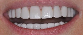
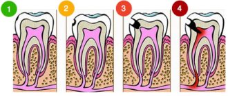
There are 2 main types of gum inflammation – gingivitis and periodontitis. Gingivitis is the initial stage of inflammation, the main symptoms of which are: swelling, redness or bluishness of the gums, pain and bleeding when brushing teeth, bad breath (Fig. 3-4). With periodontitis, these symptoms are accompanied by mobility of teeth, suppuration from periodontal pockets, destruction of bone tissue around the teeth, and in later stages, changes in the position of the teeth (Fig. 5).
Gingivitis and mild periodontitis –
The cause of gum inflammation in both cases is soft microbial plaque, as well as hard supra- and subgingival dental deposits (Fig. 3-5). This is the main cause of inflammation, which is a consequence of insufficient oral hygiene. And the most important part of treatment should not be to reduce symptoms, but to eliminate the real cause of gum inflammation. And for this purpose, dental plaque is removed by a dentist (usually this is done using ultrasound).
Antiseptic rinses act only on the most superficial layer of microbial plaque or tartar. They do not act on the deeper layers, which also consist of pathogenic bacteria, and therefore the use of antiseptics does not lead to complete “disinfection”. Therefore, rinsing with antiseptics and gels for gums (without removing dental plaque) lead only to temporary improvement, and the inflammation in this case will be constant chronic - with periodic exacerbations.
Television advertising very actively promotes all sorts of products for gums, but it does not say that the main treatment is precisely the removal of dental plaque. Remember that a mild form of gingivitis can unnoticeably turn into a severe form of periodontitis if you try to treat the inflammation only with antiseptic rinses and anti-inflammatory gels - without periodic removal of dental plaque by the dentist.
→ How to properly treat gingivitis → Treatment regimens for periodontitis
Toothpastes for gum inflammation -
In complex therapy for the treatment of gum diseases, you can use not only rinses and gel applications, but also special anti-inflammatory toothpastes for gums, which will reduce bleeding and swelling of the gums even faster. Some of these pastes are also suitable for preventing inflammation. We hope that our article on the topic: How to rinse the mouth with gum inflammation was useful to you!
Sources:
1. Dental education of the author of the article, 2. Based on personal experience as a periodontist, 3. PubMed.gov scientific research base, 4. “Optimization of conservative treatment of patients with periodontitis” (Komleva A.S.), 5. Composition of products taken from official websites manufacturers.
Local inflammation of the gums in 1-2 teeth –
In this section we will tell you how to relieve gum inflammation if it occurs in the area of only 1-2 teeth. The reasons for such limited inflammation may be, for example, a localized form of periodontitis or an exacerbation of chronic dental periodontitis. Swelling of the gums with localized periodontitis is usually located in the projection of the interdental space, and closer to the gingival margin (Fig. 9-10). Often, when you gently press on such a swelling, you can see that pus begins to ooze from under the gums.
Swelling of the gums with localized periodontitis: photo
The main causes of periodontitis localized in the area of 1-2 teeth are most often the following factors:
- Traumatic bite (super contact) – so-called “premature biting” may occur in the area of some teeth, i.e. antagonist teeth do not close evenly, and there is premature biting on one of the teeth. The presence of such supercontact causes mechanical overload of the tooth, which leads to destruction of the bone tissue around it and inflammation of the gums. Supercontacts can appear independently, or be the result of poorly made fillings and crowns.
- Overhanging edge of the filling in the interdental space - when treating caries between teeth, the dentist may leave an overhanging edge of the filling, which will injure the gingival papilla in the interdental space. This is a gross mistake by the dentist. In addition to injuring the gums, the overhanging edge of the filling creates conditions for retaining food debris in the interdental space, which also contributes to the development of inflammation.
- The absence of a contact point between the teeth - when the part of the tooth that contacts the lateral teeth in the interdental space is destroyed - it is very important to restore correct contact. This is quite complex and painstaking work that requires skill, and not every dentist knows how to restore the “contact point” between teeth. Lack of good contact will lead to food getting stuck in the interdental space, followed by rotting of food debris and the development of inflammation (especially in patients who do not use dental floss).
- Overhanging edges of crowns –
Poorly made crowns can lead to permanent gum injury. For example, crowns may also have an overhanging edge that can injure the gums. In Fig. 12 you can see an X-ray of the tooth under the crown: in the interdental space, arrows indicate a deep periodontal pocket, which arose due to chronic trauma to the gums by the overhanging edge of the crown.
Inflammation of the gums near the tooth: treatment if the cause is the overhanging edge of the filling or crown, then it is necessary to grind off the overhanging edge of the filling with a bur (if possible, otherwise, completely replace the filling), and make a new crown. If there is no good contact point between the teeth in the interdental space, it is also necessary to replace the poor-quality filling or crown that caused the lack of contact. In the presence of traumatic supercontact, “selective grinding of teeth” is performed.
All of the above is basic therapy aimed at eliminating the causative factor that caused inflammation. Further, depending on the severity of the inflammation and the degree of destruction of the bone tissue around the tooth, curettage of the periodontal pocket can be carried out (with synthetic bone tissue placed in the bone pocket in order to try to restore the bone level), a course of anti-inflammatory therapy, tooth splinting, etc. .
Inflammation of the gums due to periodontitis -
With localized periodontitis, inflammation develops in the interdental space, in which a periodontal pocket is formed due to inflammatory bone resorption.
In turn, swelling of the gums during exacerbation of chronic periodontitis is already associated with the development of purulent inflammation at the apex of the tooth root (Fig. 13). In this case, swelling of the gums or a fistula with purulent discharge will no longer be closer to the gingival margin, but closer to the projection of the apex of the root of the diseased tooth onto the gum. Inflammation of the gums due to periodontitis: photo
The cause of periodontitis (inflammation at the apex of the tooth root) is an infection in the root canals. Periodontitis occurs either as a result of lack of timely treatment of dental caries and pulpitis, or due to poor-quality dental treatment, especially often due to poor-quality root canal filling. Inflammation of the gums during periodontitis is usually limited to 1 causative tooth, but when a large purulent abscess forms, it can spread to several teeth (Fig. 14).
In all cases, inflammation, as a rule, is localized and is located in the projection of the causative tooth. In a periodontitis tooth, a “cyst” (a sac filled with pus) forms at the apex of the root, which causes swelling of the gums. In this case, swelling and swelling of the gums can either constantly increase, or will appear periodically, then disappear, etc.
How to relieve gum inflammation due to periodontitis - first you need to make sure that the inflammation is actually caused by the development of periodontitis. An x-ray will help us with this, which we will compare with the data from a visual examination of the teeth. Externally, the causative tooth will always have either a filling or a carious defect (although if the caries is located in the interdental space, you may not notice it). An x-ray will allow you to see inflammatory changes in the bone tissue in the area of the apex of the tooth root, as well as the quality of root canal filling, if it was previously performed.
Next, dental treatment is carried out. If root canal treatment in a tooth has not been previously carried out, then the nerve is first removed and the root canals are mechanically treated, after which the inflammatory focus at the apex of the tooth root is treated using special pastes based on calcium hydroxide. Next, the root canals are filled with gutta-percha and the tooth crown is restored with a filling or crown. For comprehensive information on the treatment of periodontitis, read the article:
→ Algorithm for the treatment of periodontitis
Types of inflammatory diseases
There are two categories of diseases that provoke the development of such a process. These include:
- Diseases affecting soft tissues. They are caused by injury to the gums. It occurs due to mechanical pressure and chemical burns.
- Diseases of the mucous membrane. Most often they are provoked by infection or fungi. These include diseases such as stomatitis, gingivitis, periostitis, and periodontitis.
Only a doctor can tell you the exact reason. A visual examination of the oral cavity is carried out, and tests for various pathogens will also be required.
Experts' opinion
Numerous clinical studies have proven the therapeutic effect of the Asepta line of drugs.
All focus group patients noted the analgesic effect of the product; after using the rinses, tissue healing was observed in all patients; the hygienic condition of the oral cavity improved in all patients. The products are recommended in the complex treatment of patients, and their availability and ease of use make it possible to recommend them in the clinical practice of dentists[1]. According to the results of clinical studies, after 3 weeks of using ASEPTA® rinse, gum bleeding decreases by 28.3%, inflammation decreases by 32.3% and oral hygiene improves by 33.5%*.
Toothpastes against gum disease
If your gums are bleeding, you should choose the right toothpaste. Aluminum lactate has a good hemostatic effect; chlorhexidine and mineral salts are slightly inferior to it.
As for the other components included in the toothpaste for gums - bisabolol, allantoin, medicinal plant extracts - they do not directly affect bleeding gums. They have an anti-inflammatory effect, as a result of which bleeding is reduced and may disappear. Thus, we can only talk about their indirect effect on bleeding gums, which is why a positive result is observed later than when using a paste with aluminum lactate or chlorhexidine.
At the same time, you need to know that even the best paste only temporarily removes bleeding, but in no case is a remedy. To get rid of the disease, you need to see a dentist.
Precautions for use
Chamomile, sage and other herbs that are used to rinse the mouth are considered completely safe. However, there is always a risk of accidental ingestion of the substance.
Herbal decoctions can be dangerous if the gastric juice is highly acidic. At the same time, an overly concentrated solution can provoke signs of allergies.
It is prohibited to use chamomile decoction internally in the following situations:
- mental disorders, signs of schizophrenia;
- heavy and painful periods;
- damage to the kidneys and urinary tract;
- relapse of gastritis;
- diarrhea.
Doctors advise avoiding the use of chamomile-based alcohol extracts during pregnancy and lactation. In this case, it is necessary to use a decoction or water infusion.
Instructions for use during treatment
Before the procedure, it is necessary to clean the oral cavity from food debris. This will help achieve maximum penetration of the therapeutic composition into the tissue.
While rinsing, you need to keep the infusion in your mouth for quite a long time. In case of severe tissue inflammation, it is worth supplementing the procedure with the application of tampons, moistening them in chamomile oil.
To make the medicine, it is recommended to take 1 cup of flowers and 2 cups of vegetable oil. The composition needs to be infused for 2 weeks in a dark place. After this, you need to strain it and keep it in the refrigerator.
Proper procedure does not cause harm to the human body. The use of chamomile for rinsing the mouth does not have systemic properties.
Rinsing must be carried out according to the rules.
Why salt?
A salt solution was used against tooth pain and gum inflammation back in the days of Stepan Razin. In Rus', they did not know a more effective means of quickly eliminating toothache. Surprisingly, this white powder not only eliminates unpleasant symptoms, but also gives you good health for a long time.
In addition, the salt solution helps reduce tissue inflammation by “pulling” fluid from the inflamed area. Once in the mouth, the salt begins to absorb all the pathogenic bacteria from the gums, the solution penetrates even the narrowest crevices, holes and cracks in the tooth enamel. In addition, the saline solution quickly flushes away food debris, which often causes inflammation.
Back in the 17th century, Anthony van Leeuwenhoek discovered the unique disinfecting characteristics of salt: the scientist measured the number of microbes in a scraping of the oral cavity and realized that rinsing with salt would help prevent many infections and diseases in this area.
- Sodium chloride salt draws liquid from the affected area, and therefore prevents bacteria from multiplying.
- Salt disinfects tissues, washes away harmful microorganisms, disinfecting even the most inaccessible areas.
- The salt solution contains a huge amount of useful microelements.
- Salt does not cause allergies and has no side effects even if the solution is carelessly swallowed.