Many dental clinics offer fillings using several different filling materials. Price lists (including those posted on websites) are also often replete with various commercial names of fillings: Filtek, Spectrum, Aesthetics, Charisma (Karizma), Vines (Venus), Gradia, Estelite, Herculite, Point, Tetrik, Admira, Brilliant, Enamel Plus and others. Patients are asked to make a choice, but this requires dental-level knowledge, which most people, of course, do not possess.
Popular articles on the Internet for the following queries: “which filling is better?”, “which filling material is the best?”, “which filling is better to put?” and so on. – do little to clarify the situation. Most articles (often written by anonymous authors or copywriters) discuss only classes of filling materials: cements, amalgams, glass ionomers, chemical and light-curing composites. After a detailed description of materials that went out of use several decades ago (apparently to give more volume to the text), the conclusion is made that light-curing composites are the best, and if a person has the financial means, then it is advisable to install fillings from them. Either nothing is mentioned about specific brands of heliocomposites, or it is stated that they are all approximately equally good. Or “the latest generation nanocomposites” begin to be advertised.
What fillings do not need to be placed?
The first and most important thing that every patient should understand is that light-curing fillings (photopolymers, heliocomposites) are the only reasonable choice in the 21st century. Even for people on a budget. Other materials are not 10 or 20 percent, but many times worse. Discussing the advantages or price/quality ratio of cement fillings in our time is as stupid as looking for advantages, for example, in reel-to-reel tape recorders. For a long time now, no one has been producing or buying cassette and video recorders, but for some reason some individuals allow themselves to install a 19th-century seal.
Plastic fillings are completely beyond reason. Acrylic oxide and similar anachronisms should have been banned for use a quarter of a century ago, since they are not only unable to stop the further spread of caries, but also provoke its progression. A plastic filling on a tooth is a direct road to pulpitis and periodontitis. You have to hate yourself with a fierce hatred to allow this to happen to you. And clinics that currently use such materials will be deprived of their license.
Amalgam fillings are famous for their durability, but due to toxicity (not for the patient, but for the environment and medical staff), dentists themselves refuse them.
Chemical composites are moderately bad - if a person lives from hand to mouth, then, of course, you can install them (it’s still better than nothing). But for most other people who do not experience urgent need every day, this will not be a justifiable choice. It may happen that such a filling will last for many years, but more often the opposite happens. After 3-5 years (or earlier), caries will develop under the “chemical” filling, and it can either reach pulpitis, or the amount of destruction will become so great that instead of re-installing the filling, the manufacture of an inlay or onlay will be required. As a result, the cost of repeated intervention increases by an order of magnitude due to the savings of thousands of rubles the first time (the approximate difference with the cost of a light-curing composite).
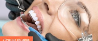
Main stages of filling
Filling the canals is carried out under local anesthesia. Before starting work, sterilization using special means is required. Additionally, preliminary cleaning is carried out. This process prevents bacterial damage and, as a consequence, inflammation.
The root canal filling procedure includes the following steps:
- treatment of caries;
- removal of damaged areas (tissues);
- removal of connective tissue;
- analysis of the anatomical features of the canal;
- treatment of the canal for the subsequent installation of a filling;
- sterilization of the canal using disinfection;
- introduction of material to fill the channels;
- removal of residual filling material;
- hermetically sealed.
Important! Pulp removal is carried out if there is severe tooth decay. This action is aimed at preventing re-infection.
Light-curing fillings are not uniform
Glass ionomer cements with a light-initiated curing mechanism are available. For example, Fuji 2 (Fuji II LC). Compomers (a hybrid material between composites and glass ionomers) are also cured using a lamp. Their most famous representative is Dyract XP. These materials are more reliable than some heliocomposites, but are still significantly inferior to their best representatives. Due to the misconception about the toxicity of composites, some doctors recommend these materials for filling for children. For pediatric dentistry, Fuji and Direct are acceptable options, but composites are still preferable.
Dental composite materials are divided into universal (suitable for filling any cavities), aesthetic for anterior teeth (excellently convey all the nuances of the color of natural teeth) and packable for chewing teeth (have low shrinkage and abrasion, but also low aesthetic characteristics). There is another class of composites – flowable. Previously, they were recommended for filling wedge-shaped defects due to their high elasticity, however, according to modern data, universal composites give better results even with non-carious lesions.1 Therefore, current composites currently have only an auxiliary value: they are used together with other classes of materials, but the main one the volume of the filling does not make up.
Universal heliocomposites are the most widely used type of filling materials in the world. The commercial names of fillings offered to the patient to choose from mostly refer to these materials. They also differ in quality. The percentage of inorganic filler, its chemical composition and the shape of the granules determine the mechanical properties of the filling: polymerization shrinkage, mechanical strength, hardness and wear resistance, polishability and surface smoothness. Some of these parameters antagonize each other. For example, for good polishability, the granules of non-limiting filler (oxides of silicon, zirconium, aluminum, etc.) should be as small as possible, and to increase strength, on the contrary, as large as possible. Therefore, materials advertised as “nanocomposites” cause a wry smile among professionals - the prefix “nano-” attracts illiterate dentists and patients who blindly associate it with high technology. However, for the sake of fairness, quite good material can be presented. For example, Filtek Ultimate.
Teeth filling process
- Initial consultation, taking x-rays.
- Coordination of treatment methods and selection of the type of fillings to be installed.
- Anesthesia before therapeutic intervention (local anesthesia, sedation, in rare cases - general anesthesia).
- Preparation of damaged or caries-affected tooth tissues.
- Grinding and processing the surface of the filling for proper closure.
Filling temporary teeth (baby teeth) follows the same plan, however, in pediatric dentistry, more compatible and safe materials are used.
The best filling materials (as of 2022)
In the dental world, the greatest authority in the assessment of restorative materials currently belongs to the American publication The Dental Advisor. For the last 12 years (from 2010 to 2022), the highest award in the “universal composites” category has been awarded to the Japanese filling material Estelite Sigma Quick. Selection criteria: percentage of inorganic filler, amount of shrinkage, correspondence of shades to the natural color of the tooth, ease of use, radiopacity, polishability. A survey of dentists showed that they switch to Estelite more often than to other filling materials. Clinical success of Estelite is 99%.2
Among aesthetic composites in 2015, 2016, 2022, according to the same publication, the filling material from Germany, Venus Pearl, took the highest position. Its clinical effectiveness is 91%.3 In 2021, it received the title of “preferred product”. It can also be used for chewing teeth.
In 2022 and 2022, first place in the category of aesthetic composites was taken by the Harmonize material of the American manufacturer Kerr Restorative.4
Other good quality filling materials should also be mentioned. These include those who received awards in 2016-2019: Omnichroma, Brilliant EverGlow, Admira Fusion, Filtek Supreme Plus and Filtek Supreme Ultra.
Types of fillings
Dental fillings are distinguished by the time of use and the quality of the material. Temporary fillings are installed for a short period; they close the tooth cavity until the next visit to the dentist. Permanent ones are installed for a long period (1-5 years). They are installed only on sanitized surfaces.
Purpose of permanent fillings:
- prevent harmful microorganisms from entering the deep layers of dentin;
- prevent the development of secondary caries;
- help recreate the shape of the tooth.
When using high-quality materials installed in compliance with the necessary standards, the filled tooth will not differ from other units in the dentition.
Rating of filling materials:
| Filling material | Manufacturer | Rated by The Dental Advisor | Year of last assessment |
| Estelite Sigma Quick | Tokuyama Dental (Japan) | 99% | 2021 |
| Herculite Ultra | Kerr (USA) | 98% | 2016 |
| Tetric EvoCeram | Ivoclar Vivadent (Liechtenstein) | 97% | 2017 |
| Harmonize | Kerr (USA) | 96% | 2019 |
| Filtek Supreme Universal Restorative | 3M (USA) | 96% | 2019 |
| Filtek Supreme Plus | 3M (USA) | 96% | 2018 |
| Filtek Supreme Ultra | 3M (USA) | 96% | 2018 |
| Brilliant EverGlow | Coltene (Switzerland) | 96% | 2018 |
| Beautiful II LS | Shofu (Japan) | 96% | 2018 |
| Grandio SO | VOCO (Germany) | 96% | 2012 |
| Gradia | GC (Japan) | 96% | 2007 |
| Omnichroma | Tokuyama Dental (Japan) | 94% | 2020 |
| Beautiful II | Shofu (Japan) | 94% | 2009 |
| Admira Fusion | VOCO (Germany) | 93% | 2019 |
| Venus Pearl | Kulzer (Germany) | 91% | 2021 |
| Premise | Kerr (USA) | 91% | 2005 |
Where to go
You can make an appointment with a dentist in Ivanteevka at our Sanident clinic. We provide all types of dental care to patients from Shchelkovo and the urban district of Ivanteevka.
In case of significant damage in the oral cavity, our specialists:
- install pins of various types;
- restore the angle of the tooth;
- carry out complete anatomical restoration of the coronal part and other types of work.
High-quality filling and anesthetic materials are used for treatment.
A consultation with a dentist at the Sanident clinic is your first step to a healthy smile!
Material requirements
Dental cements must meet a number of criteria. The main requirements include:
- biological compatibility;
- inertness, resistance to exposure to tissue fluids;
- maintaining plasticity, which is necessary for proper modeling of the filling during installation;
- coincidence of the expansion coefficient with the characteristics of the tooth tissues;
- high adhesion;
- rapid hardening when interacting with saliva;
- color stability, availability of a wide palette;
- no shrinkage of cement after it hardens.
In terms of its parameters, a high-quality filling must match the characteristics of the enamel. It must be resistant to loads, abrasion, and have a long service life.
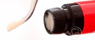
Popular Composite Seals
Composite fillings are commonly used to fill teeth . They consist of active dimethylacrylic polymerizing resins and inactive filler particles. Among the composite fillings the following can be distinguished:
• light-curing fillings, • chemically curing fillings, • thermosetting fillings.
The advantage of their use is high resistance to chewing pressure, high cosmetic qualities, preservation of contrast on X-ray images and excellent adhesion to enamel. They are structurally compatible with tooth tissues.
Restoration in children's practice - to be or not to be?
The particular relevance of the use of composite materials in pediatric dentistry is due to the following factors: in childhood, the percentage of traumatic lesions of the frontal group of teeth is very high, because they are one of the first to erupt and protrude from the occlusal plane of the primary teeth that have not yet been replaced. In addition, some dental pathologies can be observed mainly in children. For example, with destructive forms of hypoplasia or fluorosis, teeth are destroyed so quickly that, due to the desire of doctors to follow the old treatment method (namely, wait until the root tips are closed), the only method of treating such teeth is often orthopedic restoration of the crown part.
For a long time, pediatric dentists were afraid to use composite materials in their practice, citing the following reasons:
* inappropriateness of restoring a tooth that may still be erupting;
* the inability to bring teeth into bite after various types of injuries, because microfractures appear at the root, which, if the load is not applied in a timely manner, can increase and lead to the death of the pulp and resorption of the tooth root.
* unsafe use of composite materials, because they are highly toxic and in teeth with uncovered apices and still wide dentinal tubules can lead to pulp death.
* inappropriateness of using composite materials in the treatment of destructive forms of hypoplasia and fluorosis at an early age, because their abrasion coefficient is lower than that of natural enamel. And in this regard, restorations made from composites require repair or complete replacement after some time.
In addition, often doctors and relatives of patients do not consider aesthetic restoration of teeth at a young age important and limit themselves to temporary structures, forgetting about the psychological aspects. But the trend of today is such that it is fashionable to be healthy and beautiful.
The achievements of modern dentistry dispel fears of using composites in pediatric practice. For example, with regard to toxicity, it is now known that the bonding system has a direct effect on the tooth. The latest generation adhesive systems are not only non-toxic, but may also contain fluorine compounds. The toxic monomer contained in chemically cured composites has practically sunk into oblivion along with the use of chemical composites themselves.
Of course, before starting restoration, it is necessary to carry out all examination methods (x-ray, EDI). However, we must not forget that the protective forces of the child’s body are very great, and in each case we try to individualize the algorithm of actions.
Despite the fact that the evolution of composite materials is advancing by leaps and bounds, pediatric dentistry places increased demands on restorative materials:
— Low toxicity.
— High degree of adhesion of the material to tooth tissues.
— Abrasion coefficient as close as possible to natural tooth tissues.
— Possibility of immediate and final restoration of teeth (both frontal and chewing group).
— Preparation that does not require intervention in healthy tooth tissue.
— Excellent aesthetic characteristics.
From an esthetic point of view, restoring the teeth of young patients is often very difficult. This is due to the fact that the shape and color of children's teeth have a number of features. For example, the macrorelief is characterized by the presence of a scalloped cutting edge that has not yet been subjected to physiological abrasion. The surface layer of enamel in children is formed by the protruding tops of prisms, which gives it the appearance of a “cobblestone street.” In addition, micropores are found in the enamel of children's teeth under a microscope. We must not forget that the Recius lines (enamel growth zones), which form perikemata on the surface, are more pronounced in childhood. All this affects the surface gloss of the enamel and visually makes it brighter. Children are characterized by pronounced mamelons. The most typical for the incisal edge of young patients is the presence of three large mamelons or three mamelons with a split middle one. (Fig. 1)
Fig.1
Tooth color is dictated by the optical characteristics of dentin and enamel. The enamel is responsible for the brightness of the tooth. Enamel is characterized by such a property as opalescence, this is the ability to reflect predominantly short waves (blue) and transmit long ones (orange-red). Dentin is responsible for the richness of tooth color. Dentin of natural teeth has the property of fluorescence. Currently, the identity of the fluorescence of the material and the tooth is becoming an integral requirement for a modern composite. Another optical medium of the tooth is the dentin-enamel junction, which plays a large role in the formation of color.
According to various studies, the majority of teeth belong to shade – A on the Vita scale (Yamomoto 1992, Vanini 1994, Tuati 2000). Due to the fact that the enamel of children is brighter than that of adult patients, the color of their teeth most often corresponds to shades A1, A2 (according to Vita, since the most common lesions in childhood are injuries to the frontal group of teeth, accompanied by a violation of the integrity of the crown angle or the entire cutting edge, pediatric dentists need a material that reproduces all the optical characteristics of the cutting edge of the tooth.
At the moment, the restoration material that best meets all the requirements of pediatric dentistry is Enamel plus.
Enamel plus is a radiopaque, microhybrid, light-curing composite that allows you to achieve excellent results in aesthetic restorations of anterior and posterior teeth, using direct and indirect methods. Еnamel plus is a unique restoration system that makes it possible to reproduce all the optical effects of the cutting edge.
Its creator is Dr. Lorenzo Vanini (Italy). When developing this material, L. Vanini took into account all the components of tooth color. His main task was to create a material, using which it would be possible to obtain a predictable result, which is so important in the daily practice of a dentist. The Enamel plus set includes three basic enamels, seven universal fluorescent dentins, two intensive enamels (for personalizing the enamel on the surface) and opalescent enamels, which can be used to highlight internal incisal opalescences and mamelons. (Fig. 2) In addition, the set includes a Glass Connector . It is a flowable composite that mimics the protein layer of natural teeth and six colors to replicate the characteristics. To determine the color, it is proposed to use the Enamel plus scale, made entirely of composite. (Fig. 3) The set also includes a special color card. This card remains in the medical history, and you can use it in further work. (Fig. 4, 4a)
Rice. 2.
Rice. 3.
Rice. 4.
Rice. 4a.
To obtain maximum results when using the Enamel plus HFO system, it is proposed to use the anatomical layering technique developed by L. Vanini. The anatomical stratification technique involves the construction of lingual enamel, internal dentinal body and vestibular enamel.
Before moving on to consideration of the stratification technique, I would like to note some features of cavity preparation under Enamel Plus. The fact is that preparation for this material is distinguished by the possibility of maximizing the preservation of healthy tooth tissue and does not require modeling a fold on the enamel. It is by increasing the width of the rebate and covering a larger surface of the enamel with a composite material that doctors often try to improve the aesthetics of their restoration (make the transitions of the material to the tooth tissue less noticeable and avoid the appearance of a gray stripe at the border of the filling with the tooth). At the same time, sometimes restorations of large cavities of classes III and IV turn into the production of veneers using the direct method, which is absolutely incorrect in pediatric dentistry, especially in cases where the tooth has not yet fully erupted. When preparing under Enamel plus HFO, a groove is formed on the vestibular enamel and proximal surfaces, along the edge of the cavity being prepared, with a spherical bur, the palatal side is processed at 90 degrees. This preparation technique is very gentle. (Fig. 5, 5a)
Rice. 5.
Rice. 5a.
Rice. Defect in the cutting edge of the 21st tooth.
Rice. Temporary restoration with composite without adhesive preparation.
Rice. Taking a silicone impression.
Rice. Example of a silicone key.
Restoration of dental injuries without opening the pulp.
The most common defect requiring restoration in children is trauma to the frontal group of teeth without opening the pulp. The break line is located parallel or diagonally to the cutting edge. In this case, the medial angle is most often affected.
After completing the color card, preparation and adhesive surface treatment, we begin to restore the lingual enamel. Because enamel in children has a high brightness; most often, we take the shade of enamel GE3. (Fig. 6, 6a)
Rice. 6
Rice. 6a
To simplify the task in case of extensive defects, a silicone block is made, which allows a thin layer to distribute the material and avoid inaccuracies in the formation of the macrorelief. (Fig. 7) When modeling, to create a more regular surface, in addition to conventional trowels, silicone trowels (micerium) are used, which give “finger effect” (Fig. 8).
Rice. 7
Rice. 8
Using a special brush, a thin layer of Glass Connector is applied to the inner surface of the enamel sheet, and it should be located strictly at the enamel-dentin border. (Fig.9)
Rice. 9
After applying the Glass Connector, we begin modeling the dentinal body. To achieve optimal saturation of the restoration, 3 base dentin colors are used. For example, if we want to end up with color A2 (according to Vita), we should start with UD4, then layer UD3 and UD2 - lighter ones.
At the stage of applying the last dentin, mamelons are modeled (Fig. 10, 10a, 11, 11a, 12,12a)
Rice. 10
Rice. 10a
Rice. eleven
Rice. 11a
Rice. 12
Rice. 12a
The finished dentin body is covered with a thin layer of Glass Connector.
To recreate the opalescence of the enamel, opalescent enamel (OBN) is applied between the mamelons and in the incisal area. After this, if necessary, intense white enamels (IM, IW), opalescent enamels (AO, OW) and paints for characterization are applied (Fig. 13, 13a, b)
Rice. 13
Rice. 13a
Rice. 13b
The final stage is the application of the main enamel (GE3) - vestibular enamel sheet. (Fig. 14, 14a, 14b, 15, 15a)
Rice. 14
Rice. 14a
Rice. 14b
Rice. 14c
Rice. 15
Rice. 15a
Thus, with strict adherence to the anatomical stratification technique, the use of Enamel plus material allows one to achieve a natural appearance of teeth in the restoration.
Surface treatment
Includes final modeling of the tooth shape (macro- and microrelief) and surface polishing. To simplify the task, when creating a vestibular convexity, transition lines, and Recius lines, guidelines can be drawn on the surface of the tooth with a slate pencil. Modeling of macro- and microrelief is recommended to be done with diamond burs. After which we begin polishing the surface. To do this, use the polishing system included in the Enamel plus HFO set, which includes three pastes and polishers with a silicone head, goat bristles and a felt disc. (Fig. 16)
Rice. 16
Recommendations for doctors
The requirements for restorations made from Enamel plus are no different from those for any other composite.
We must remember that before starting work, it is necessary to establish individual oral hygiene. After all, good hygiene will prolong the life of any restoration.
The key to the success of your work is high-quality insulation of the working area. From the age of 7-8 years, children can easily tolerate the rubber dam. It is important not to forget that what frightens patients most of all (and, it should be noted, not only children) is the unknown. Therefore, before starting treatment, we show and tell you what it is and why. Let's compare the rubber dam to an umbrella or a cloak for a tooth. Rubber dam is used both for direct restorations and for fixation of indirect restorations.
High-quality finishing and polishing of the surface will not only improve the appearance of your restoration, but will also make it more durable. Despite the fact that we recommend polishing fillings once a year, our foreign colleagues have excellent results 9-10 years ago. Moreover, during this time the patient never came for polishing or simply for a medical examination. It was a completely different tooth that brought him to the clinic. Neither the esthetics nor the marginal fit of the Enamel plus restoration performed for trauma was compromised (Dr. F. Mangani, Italy).
Conclusion
Using Enamel plus HFO, the pediatric dentist will receive the final result of the restoration immediately after a dental injury, detection of caries or any other destructive process.
Due to the high adhesion force of the material (30 mPa), excellent strength characteristics, and the degree of abrasion as close as possible to natural enamel (Fig. 17 diagram). Enamel Plus HFO makes it possible to restore teeth even with partial eruption. Moreover, after complete eruption, after a year, five or even ten years, the restored tooth will look natural.
Rice. 17 diagram
Author:
Timoshenko O.A., dentist, Department of Pediatric Dentistry, Sechenov Moscow Medical Academy