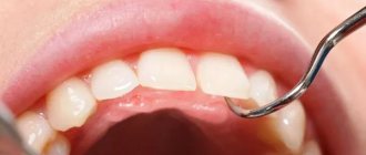
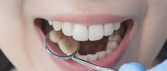
Are you suffering from severe, lingering toothache? Does it come on in attacks, worsen at night, and radiate to the ear or temple? Most likely, this is a three-channel pulpitis. The disease is quite dangerous, especially if you do not seek medical help in a timely manner. Dentistry "MasterDent" offers high-quality services in this area at affordable prices. European materials that have undergone numerous studies are used in treatment. They have proven their high performance in practice. When treating root canals, the clinic’s specialists use modern methods.
Now 1 out of 5 people who come to the dental office suffer from pulpitis. What is meant by this name? Pulpitis is a disease in which the “nerve” of the tooth, the pulp, becomes inflamed. Our molars, which dentists call molars, usually have 3 canals. Each of them contains a bundle of nerves and blood vessels. If the inside of the tooth, the pulp, becomes inflamed, the process is very painful. This occurs due to swelling and compression of the pulp. Therefore, as a result, a person experiences extremely unpleasant sensations.
Even if the patient goes to the dentist in a timely manner, treatment may take several visits. The fact is that three-channel pulpitis is treated in stages. Each channel is processed separately and the entire procedure can take a long time. For example, working with a multi-channel tooth takes a specialist up to one and a half hours. If a doctor uses a microscope for treatment, it takes about 2 hours.
Features of the structure of teeth, their roots and canals
There are no two identical root dental systems, which is explained by the purely individual structure of a person’s teeth. In addition, the root system of incisors, canines and molars is arranged in accordance with their purpose:
- Ones and twos (incisors) are needed for biting food.
- Fours and fives (premolars) perform the initial chewing function.
- Sixes and sevens completely grind food.
Based on this, it becomes clear that the seventh tooth requires more nutrients than the fifth. It must be strong and hardy, therefore it has a more developed channel system. Despite the fact that the 6th tooth in the lower jaw performs the same functions as the seventh, it usually has fewer canals. This is due to the fact that there is less chewing load on it.
For a detailed study of the structure of the dentofacial apparatus of a particular patient, radiographic examination is used.
Tooth structure
Each dental unit consists of:
- crowns - the area above the gum;
- neck - the area between the crown and the root;
- root - the area under the gum.
Inside the crown is the pulp, which passes into the root canals. At the end of the root there is a small apical opening through which blood vessels and nerve endings pass, starting from the main neurovascular bundle and ending in the pulp.
When a person’s pulp becomes inflamed, not only it, but also all root canals need to be cleaned of infected tissues, since they are “communicating vessels.” If even one canal is left uncleaned, pathogenic microorganisms will continue to develop inside the dental unit, which will lead to its removal. That is why the doctor must know the exact number of canals in the tooth.
How many nerves are there in a human tooth?
Thanks to the nerve, the tooth can respond to external stimuli. After removing the pulp and filling the canals, the dental unit loses sensitivity, as it is deprived of a nerve. But due to the removal of blood vessels, problems begin with its blood supply and mineralization. The crown becomes less durable and more prone to various chips and breaks. The enamel quickly darkens, and it cannot be properly bleached even with strong chemicals.
Before removing the pulp, the patient is sent for an x-ray to find out how many canals are in the operated tooth: a person has only one dental nerve in a tooth, but there may be several canals
. This preparation allows for depulpation to be carried out competently and quickly.
Types of Root Canals
There are several options for the structure of dental canals:
- in the root there is one canal passage, which corresponds to one apical foramen;
- in the root there are several canal branches that connect in the area of a single apical foramen;
- two different branched passages have one mouth and two apical openings;
- canal cavities in one root merge and diverge several times;
- three root canal passages emerge from the same orifice, but have 3 different apical openings.
There can be as many channels as there are roots, but often their number differs. Several types of canals may be present in one molar and premolar.
How many canals in a person’s teeth - table
According to statistics, the number of channels depends on the depth of the tooth.
: The deeper it is located in the jaw, the more canals it has. This is due to the increased load on the molars located at the base of the dentition.
Typically, teeth in the upper jaw have more canals. But this pattern is not observed in all patients.
The table below presents average statistical data on how many canals are in a person’s teeth above and below.
| Dental unit | Number of channel passages | |||
| Fangs | Upper | 1 | ||
| Lower | 2 | |||
| Incisors | Upper | 1 | ||
| Lower | Central | in most cases 1, less often 2 | ||
| Lateral | 1 or 2 (about the same probability) | |||
| Premolars | Upper | First | most often 2, but sometimes first premolars with 1 or 3 canals are found | |
| Second | in most cases 2, sometimes 1 or 3 | |||
| Lower | First | 1 or 2 | ||
| Second | 1 | |||
| Molars | Upper | First | 3 or 4 with equal probability | |
| Second | in most cases 3, sometimes 4 | |||
| Third | around 5 | |||
| Lower | First | most often 3, sometimes 2 or 4 | ||
| Second | usually 3, but there are roots with 4 channels | |||
| Third | no more than 3 | |||
Number of canals in teeth in the lower jaw
The teeth on the lower and upper jaws are significantly different from each other. This is partly due to the uneven load and different functions. Typically, teeth in the lower jaw have fewer canals. But each specific case requires detailed study. Therefore, the dentist first sends the patient for an x-ray, and only then proceeds to open the crown and treat pulpitis.
It is impossible to start treating caries and pulpitis based only on encyclopedic information, because:
- The 6th tooth of the lower jaw can have any number of canals - from 2 to 4;
- in the 5th tooth below there is usually only 1 canal, but in approximately 10% of patients there are quints with 2 canals;
- In the 4th tooth there is usually only 1 canal, but in about a third of cases there are 2.
The eighth tooth on the lower jaw is the most “unpredictable”. Exactly how many canals are in the wisdom tooth located below can only be determined using x-rays. Officially, there are no more than 3 of them, but during the treatment of caries, additional cavities usually open. It is precisely because of its incomprehensible structure and inconvenient location that the figure eight is most often removed.
It is impossible to treat a dental unit without studying the structure of its root and canal system. This can only aggravate the pathology and lead to complications.
Number of canals in teeth in the upper jaw
The root system of the teeth of the upper jaw is more complex and branched. This explains the longer treatment of upper molars and the frequency of repeat visits due to incompletely sanitized dental cavities.
Features in the structure of the canal system of teeth in the upper jaw:
- The 6th tooth of the upper jaw is most often three-channel. But sometimes there are also four-channel first molars.
- The fourth and fifth teeth from above are most often two-canal, but sometimes single-canal and three-canal premolars are found.
- The 4th upper tooth usually has 2 canals, but sometimes there are premolars with 1 or 3 canals.
The “wise” eight on the upper jaw is a four-channel tooth. Third molars with 5 canals are extremely rare. However, in dentistry, even cases of the presence of eight-channel wisdom teeth located at the top have been recorded.
Diagnostics
Before performing dental procedures, it is extremely important to identify the features of the canal-root system in order to clean it completely. The problem is that the dentist is not always able to determine the number of strokes visually and by palpation (using a probe). They often narrow at the mouth, and the probe does not pass through the narrowing, overgrow against the background of a long-term inflammatory process, or have an atypical structure.
That is why, in most cases, the doctor sends the patient for dental x-rays before starting treatment. The photographs show the number of roots, the structure of the cavities, and indirect signs of the inflammatory process. In complex diagnostic cases, they resort to computed tomography, which helps to more clearly visualize all tooth structures.
In addition to topical diagnostics, it is also important to measure the length of the canal in order to understand at what depth the filling material should be inserted.
Channels in baby teeth
There are as many nerves in baby teeth as there are in molars—one. In addition, temporary units are similar to permanent ones in the structure of the root system. That is, a milk tooth such as the upper six or second molar has a canal system similar to its molar brother, the second premolar.
Nerve endings perform standard functions:
- signal about developing caries;
- responsible for the growth and development of teeth;
- control the flow of water and nutrients to dentin and enamel.
The root canals of baby teeth are also treated and filled, but the tactics of their treatment depend on how long ago they erupted. Under the temporary units, permanent ones are formed, so treatment should be aimed at preserving them. Milk teeth can only be removed if the permanent teeth are ready to emerge.
The roots of permanent incisors, canines and molars do not form immediately, but over the course of about 3 years. Treatment of permanent teeth with unformed roots also differs from the standard one. The canals in the teeth of patients four, five, six years old (depending on the rate of formation of the dentoalveolar apparatus) are filled with a special paste with calcium and fluoride, which helps close the roots.
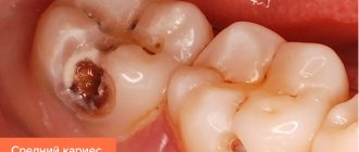
What diseases cause inflammation of dental canals?
Root canals can become inflamed due to the development of the following diseases:
- caries;
- pulpitis;
- periodontitis.
An accurate diagnosis of inflammation of the pulp and canals of the tooth can only be determined by a dentist after X-ray diagnostics and a visual examination of the oral cavity.
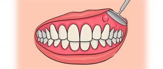
Root canal treatment
The treatment plan for dental canals consists of several stages:
- First, access to the problem area is freed: using a special dental instrument, the filling or the area of the crown damaged by caries is removed.
- Then the contents of the pulp are removed, and the canals are cleaned mechanically using antiseptic drugs.
- After this, the root is prepared for filling. At this stage, the dentist can form the correct conical shape of the canal passage.
- Then the canals are carefully sealed. If baby teeth are treated, the dentist uses a special filling paste, which gradually dissolves as the root dissolves.
- After this, a filling is placed on the crown.
This treatment regimen is standard and does not depend on exactly how many canals there are in the diseased tooth. The main thing is that all dental canals are cleaned, treated with an antiseptic and carefully closed. If treated incorrectly, it may be necessary to remove the tooth and visit an oral surgeon.
Teeth can be single-channel, two-channel, three-channel and even eight-channel. If one of the ducts becomes inflamed, it is necessary to clean and seal not only it, but also all other canals, since the infection could penetrate into them.
Prevention
In order not to have to endure unpleasant moments during dental treatment, it is better to promptly identify and, as far as possible, prevent caries and pulpitis. To do this, you need to follow the recommendations of dentists:
- Brush your teeth regularly.
- Use dental floss that gets into hard-to-reach places.
- Rinse your mouth after eating.
- Give up bad habits (smoking, drinking alcohol).
- Drink coffee and tea less often.
- Don't eat foods that are too cold or hot.
- Visit your dentist once every six months, even if nothing bothers you.
- Follow a diet rich in vitamins, phosphorus, fluoride, calcium.