Bone exostosis or osteochondroma is the most common skeletal tumor. It accounts for about 20% of all cases of the formation of bone tumors and almost 40% of the development of all benign bone tumors. Most often it is detected before the age of 20, although it is usually asymptomatic. Only in isolated cases does pain and other disorders occur. Nevertheless, the formation of a growth on the bone necessarily requires consultation with a doctor, since malignancy or malignancy of osteochondroma is rare, but still possible.
What is bone exostosis
Osteochondral exostosis is a benign osteochondral formation on the outer surface of the bone. Most often, it is detected at a young age before the end of skeletal growth, including in children.
The formation of osteochondroma provokes a disruption of the natural processes of resorption (resorption) and the formation of new bone tissue (remodeling).
Exostosis is a growth formed by spongy bone and having a cortical layer (a strong, hard “shell”). It can grow on a stalk or be attached to a bone with a wide base. On the outside, the bone growth is covered with cartilage tissue, which has much in common with articular cartilage. Its thickness does not exceed 1 cm. The top layer of exostosis is also called a cartilaginous cap.
Osteochondroma is a consequence of impaired bone growth in the area of the epiphyseal plates, i.e., their displacement from the anatomically correct position with the “throwout” of cartilage tissue to the side. Subsequently, just like the metaphysis, it ossifies in the direction from the base to the apex, which leads to the formation of bone exostosis with a cartilaginous cap. At the same time, it continues to increase in size until the growth zones close, and can appear in absolutely any bone. The bony part of osteochondroma is formed from unevenly distributed bone beams. Between them there is adipose tissue with islets of hematopoiesis. Bone septa are formed due to enchondral ossification of cartilage. This raises the possibility of the presence of calcifications in the cancellous bone.
The metaphyseal areas of long tubular bones are most often affected:
- femoral (30%);
- shoulder (10-20%);
- tibial (15-20%).
Metaphysis (sometimes neck) is a small part of a tubular bone that is located between its epiphysis (head) and diaphysis (body). At 18-25 years of age, it stops growing and ossifies, which indicates the completion of bone formation.
Thus, articular exostosis is often observed - the formation of a growth on the upper (proximal) part of the tibia or lower (distal) part of the femur, i.e. the tumor is localized just below or just above the knee. Damage to flat bones, i.e. exostosis of the rib, scapula, pelvic bones, are less common. Osteochondromas of the bones of the fingers, toes, and clavicle are diagnosed even less frequently. The most rare occurrence is the formation of bone growths in the spine.
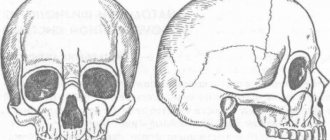
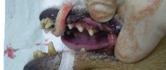
Also, bone exostosis can form on the lingual or buccal surface of the jaw. At the same time, it can take the form of a ridge, protrusion, mound, or take on bizarre shapes. In such cases, patients are advised to consult a dentist.
Features of the postoperative period
In order for the wound to heal as quickly and easily as possible, it is important to follow the doctor’s recommendations during the rehabilitation period.
- Do not damage the seam
- Carry out hygiene procedures carefully (but under no circumstances abandon them)
- Take prescribed medications, including antibiotics, if the dentist considers it necessary to prescribe them
- Refuse hot or spicy foods, follow a semi-liquid diet, consume food and drinks only at room temperature.
Exostosis in a child
It is in children that osteochondroma is most often first diagnosed, which is due to its formation from cells of the epiphyseal plate that is present only until the end of the growth period, which is adjacent to the metaphysis. It is also called the bone growth zone, since it is a hyaline cartilage whose cells are in constant miotic division. As a result, new chondrocytes (cartilaginous tissue cells) are formed, forming the epiphyseal plate, and the old ones are shifted to the metaphysis and subsequently replaced by osteoblasts (bone tissue cells).
In infants, violation of the rules for the prevention of rickets, in particular excessive use of vitamin D preparations, increases the likelihood of the formation of exostoses.
After puberty, the growth plates gradually close and are replaced by bone tissue, transforming into a thin epiphyseal line. If hormonal imbalances occur during this period, it is possible that the growth zones may remain open, which creates the preconditions for the formation of osteochondromas.
Usually, until the age of 7-8 years, bone exostosis does not manifest itself in any way in a child and makes itself felt only during the period of intensive growth, i.e., at 8-16 years, since it also begins to grow actively. In young children, such growths are present in the metaphysis area immediately near the epiphyseal plate, but subsequently move away from it and approach the diaphysis. Therefore, by how far the bone exostosis in children is from the epiphysis, the time of its formation is determined.
The growth of the neoplasm continues until the end of the growth period.
Types and stages of development
Exostosis can be present only on one bone, i.e., be solitary, or affect all metaphyses of the bones of the skeleton. In the second case, they talk about a generalized form of the disease, which is more common in men and is mainly hereditary. It is also called multiple exostotic chondrodysplasia. In this case, several bones or the vast majority of them are affected, but the formations have different shapes and sizes.
Depending on the shape and direction of growth, there are:
- hilly;
- linear;
- spherical exostoses.
Single exostoses have a narrow or wide base, while multiple ones are usually spherical or oval growths measuring 2-12 cm, sometimes more.
In its development, any osteochondroma goes through 3 successive stages:
- the formation of cartilaginous exostosis, which is not determined by palpation (palpation);
- ossification and active growth of growth;
- stopping the growth of the bone part of the neoplasm while maintaining the possibility of increasing the size of the cartilaginous cap.
Bone exostoses can already be felt during examination and can lead to discomfort, pain during physical activity and other symptoms.
Based on the nature of development and clinical picture, osteochondromas are divided into the following types:
- With normal growth rate. The osteochondral formation grows slowly, and there is a correspondence between the growth rate of the cartilage and the affected bone. Such osteochondromas have the most favorable prognosis, since after the end of the growth period they do not tend to continue to grow and almost never become malignant.
- With a high growth rate. An increase in the size of the growth occurs due to the proliferation of cartilage tissue and can also continue after the completion of the formation of the skeleton due to the preservation of the growth zone. In such cases, removal of exostosis is indicated, since there is a fairly high risk of malignancy.
- Malignant exostosis has the most unfavorable prognosis. Most often, osteochondral neoplasms of the ribs, pelvic bones, scapula and spine degenerate into chondrosarcoma or osteogenic sarcoma.
Classification
The classification is based on several characteristics. Based on quantitative characteristics, solitary (single) and multiple (generalized) forms are distinguished.
According to the stages of osteochondroma formation:
- The neoplasm is formed from cartilaginous tissue and cannot be detected by palpation.
- Ossification of the growth and its increase. The bone tissue is covered with cartilage, and active growth continues.
- The growth of the bone part of the tumor stops, but the growth of the cartilaginous “cap” of the tumor may continue. Determined by palpation examination. During physical activity, it can manifest as pain and impaired limb function.
Based on the shape and direction of tumor growth, there are hill-shaped, linear and spherical exostoses.
Causes of exostosis
There is still no consensus on the reasons for the appearance of exostoses. A number of authors believe that they are of a tumor nature. But most agree that they are a consequence of disorders of enchondral ossification processes that arise as a result of dysembryogenesis (disorders of embryonic development). Therefore, today the main theory of the formation of bone exostoses is the displacement of the epiphyseal plate during intrauterine development of the fetus. It is a bone growth zone located directly below the epiphysis and is responsible for its lengthening. Therefore, the disease is considered congenital, and its development continues throughout the entire period of growth.
The following can increase the risk of osteochondroma:
- ionizing radiation (in 10% of cases the disease develops in patients who underwent radiation therapy in childhood);
- disturbances in the functioning of the endocrine system, taking hormonal drugs;
- smoking and alcohol abuse of parents.
The disease can also be hereditary and transmitted to the child from one of the parents. Especially often, multiple bone lesions due to exostoses are genetically determined.
The acquired nature of the disease cannot be ruled out. The following can provoke the appearance of neoplasms:
- bone injuries;
- microtraumas caused by excessive physical activity, which is typical for professional sports;
- infectious diseases and chronic inflammatory processes of any localization (for example, a heel spur often develops against the background of plantar or plantar fasciitis);
- pathologies of the periosteum, degenerative-dystrophic diseases of cartilage, ankylosing spondylitis;
- microcirculation disorders in soft tissues;
- muscle dystrophy;
- obesity;
- severe forms of allergic diseases;
- compression of the limbs, including an incorrectly applied plaster cast or tourniquet.
Wearing improperly selected clothing and shoes, in particular frequent hypothermia of exposed areas of the body, increases the risk of developing bone tumors.
Of particular importance in the development of osteochondroma is hormonal imbalance in children during puberty. Often the formation of bone exostoses is observed against the background of increased synthesis of sex hormones in adolescents. This can provoke the growth zones to remain open, which can subsequently cause gigantism. Also, an increase in the size of osteochondral exostoses can continue after the closure of growth plates in women as a result of hormonal changes.
Reasons for appearance
It is not precisely determined why osteophytes appear on the gums. But factors contributing to the disease include:
- Hereditary predisposition;
- frequent inflammation, purulent processes leading to atrophy, deformation of the jaw bone and nearby tissues;
- injuries of the dental system, especially accompanied by fractures of the facial part of the skull with improper reposition of bone fragments;
- complex tooth extraction;
- advanced periodontitis, periodontal disease;
- bite pathologies;
- congenital abnormalities of the jaw.
Often the pathology appears in childhood or adolescence. Also, the appearance of jaw growths may be associated with dysfunction of the endocrine system.
Symptoms
The clinical picture depends on the form of the disease, location, size, shape of exostoses, and the degree of their influence on surrounding tissues. Single neoplasms are usually immobile and their growth is slow. Therefore, in most cases they are asymptomatic and there is no pain. The condition of the skin with solitary exostoses is usually not changed.
But in some cases, there is active growth and acquisition of large osteochondromas. They can mechanically compress blood vessels, nerves, and joints passing near them and provoke the development of reactive bursitis and myositis. This is accompanied by the appearance of pain of varying severity, swelling, redness of the tissues, and sometimes a feeling of numbness. If joint exostosis is present, the range of motion may be limited.
What symptoms exostosis will have depends largely on its location. With multiple exostoses, often the first signs of the disease are stunted growth, valgus deformity of the knee joints and clubhandedness. Fractures of the osteochondroma pedicle may also occur.
Exostosis of the knee joint
It may be caused by the formation of a growth at the end of the tibia and or femur. Exostosis of the tibia provokes severe deformation of the knee joint and is easily detected with the naked eye. Upon palpation, it is possible to detect a dense but painless formation under the skin, which can be either smooth or rough. Reaching large sizes, it provokes pain in the knee when walking (especially often in women who tend to wear high-heeled shoes). In this case, exostosis of the joint tends to injure the adjacent soft tissues, which provokes the development of tendonitis (inflammation of the tendons) and bursitis (inflammation of the synovial bursa).
Damage to the femur in the early stages of development is asymptomatic. But when the tumor reaches a large size, pain in the thigh and dysfunction of the affected limb may occur. Often there are many areas of deformation up to the covering of its entire surface. Since the femur is located deep in the soft tissues, it is difficult to detect exostoses during palpation.
Exostosis of the calcaneus (calcaneal spur)
This is a well-known type of osteochondroma. The growth can take on different shapes, which determines the characteristics of the symptoms that arise. With a spherical or mushroom-shaped neoplasm, after it reaches a size of 3-4 cm, pain appears when walking and the inability to fully step on the foot. Since the growth is located in an area that is constantly subject to friction from shoes, the skin in its projection gradually becomes coarser and thickens. A characteristic symptom is an increase in pain in the morning and a gradual decrease in its severity during the day. In the evening, swelling of the lower extremities is often observed.
Spinal exostosis
The lesion can occur at any level, but the thoracic region is most often affected. The neoplasm is located in the area of the arches or processes of the vertebrae. It can grow towards the vertebral or spinal canal or in an anterior direction. The first option is more dangerous, since it is capable of mechanically compressing the spinal cord and the nerve roots extending from it. This provokes the development of the so-called radicular syndrome with sharp, severe pain, radiating in accordance with the level of damage to the body part, impaired sensitivity and mobility.
Exostoses of the ribs
The formation of growths on the ribs is more often observed with curvatures of the spine, which also lead to deformation of the chest. Their growth causes fixation of the pathological position of the chest and limitation of the vital volume of the lungs. As a result, respiratory function disorders may occur, which provokes the appearance of signs of hypoxia (oxygen starvation). To a greater extent, the lack of oxygen affects the brain, which can be accompanied by dizziness, headaches and other disorders. The heart, liver, spleen, and other internal organs are also sensitive to changes in the gas composition of the blood.
Why should jaw osteophytes be removed?
A bone growth on the gum is not dangerous until it begins to grow. Increasing in volume, the osteophyte puts pressure on the dentition and bone structures. This leads to tooth displacement, malocclusion, and jaw deformation. Large growths impede the movements of the tongue, complicate diction, and interfere with normal chewing of food. Large growths prevent prosthetics and implantation. Osteophyte of the jaw will not disappear on its own. The only effective method of treatment is surgical removal of the pathological formation.
Exostosis of the humerus
The humerus is one of the most common sites of osteochondroma. During its formation, the following may be observed:
- dull pain in the shoulder area that occurs when moving the arm with a large amplitude;
- a feeling of stiffness in movements, present mainly in the morning, until you manage to warm up;
- limited mobility of the upper limb in the shoulder;
- dystrophy of the muscles of the shoulder and forearm, which in difficult cases can be seen with the naked eye.
After operation
For speedy healing after surgery, the patient should follow the following doctor’s recommendations:
- Don't overheat . Until the wound heals, avoid playing sports, visiting bathhouses, sunbathing on the beach and taking hot baths. An increase in temperature promotes bleeding from the wound.
- Gentle nutrition . During the healing period, avoid rough, dry, spicy and sour foods. You should also not eat food that is too hot or too cold. The consistency of the food should be liquid or puree.
- Avoid smoking . Nicotine and combustion products of tobacco impair wound healing.
Continue brushing your teeth, but avoid the surgical area with your toothbrush. After each meal, rinse your mouth with antiseptic solutions prescribed by your doctor. To relieve pain in the first days after surgery, take painkillers.
Complications
Cartilaginous exostosis in children has a dangerous negative impact on the condition of the growth plates, which can result in shortening of the lower leg, thigh, forearm, shoulder, etc. This can also cause curvature of the arms and legs, increase the risk of fractures and cause disability.
Patients with severe forms of the disease feel inferior, which negatively affects their mental and emotional state.
The most dangerous complication of osteochondroma is its malignancy, i.e. transformation into a malignant tumor - chondrosarcoma. The highest risk of malignancy is with multiple exostoses. In this case, growths localized in the pelvic bones are more likely to undergo malignancy. Less commonly, exostoses of the ribs, scapula, and spine turn into malignant neoplasms. Single osteochondromas transform into a malignant tumor in no more than 1% of cases.
Atypical growth can be observed in any part of the exostosis: the cartilaginous cap, at the base or in the middle part.
List of sources
- Zatsepin, S.T. Bone pathology of adults / S.T. Zatsepin. – M.: Medicine, 2001. – 640 p.
- Neustadt, E.L. Tumors and tumor-like bone diseases / E.L. Neustadt, A.B. Markochev. – St. Petersburg: Foliant, 2007. – 344 p.
- Epidemiology of skeletal tumors: educational manual / Chernyakova Yu.M., Ivanov S.A., Yadchenko V.N. – 2012.
- Volkov, M.V. Bone diseases in children / M.V. Volkov. – M: Medicine, 1985. – 511 p.
- Lanzman Yu.I. Benign tumors / Bone tumors. – Tomsk: Tomsk University Publishing House, 1990. – P. 88158.
Diagnostics
Diagnosis of bone exostoses is not difficult. If these symptoms occur, you should consult an orthopedist. Based on the patient’s complaints, medical history and examination results, the doctor can already assume the presence of a neoplasm.
To clarify its size and other features, radiography is prescribed. It is carried out in 2 projections. Osteochondroma on x-rays is visualized as a shadow with smooth, clear contours and a pedicle or wide base connected to the bone. If there is no calcification in the cartilaginous layer, it will not be visible on x-ray.
To obtain additional data, the following may be assigned:
- CT scan, which makes it possible to detect the presence of bone marrow contents in exostosis and trace its connection with the medullary canal of the affected bone;
- MRI, which provides accurate data on the thickness of the cartilage cap and thereby clarifies the nature of the neoplasm (malignant tumors are characterized by a thickness of the cartilage tissue covering them of more than 2 cm).
If there is a suspicion of the development of oncology, patients are advised to undergo a biopsy - taking a fragment of exostosis for histological examination. In this way, benign osteochondroma is differentiated from malignant neoplasms.
If exostoses are detected in childhood and adolescence, it is imperative to conduct a full endocrine examination and determine the content of all significant hormones in the blood.
Treatment of exostoses
When diagnosing exostosis, treatment is prescribed only when symptoms appear. Otherwise, dynamic monitoring of the tumor is sufficient. If the tumor bothers the patient, depending on its type, the nature of the manifestation of osteochondroma, conservative treatment is selected or surgical intervention is prescribed. But the main method of treating exostosis is surgery.
Conservative, i.e., non-surgical, therapy is indicated mainly for exostosis of the calcaneus and rib, but only in the early stages of development. First of all, the use of special orthopedic insoles, made individually, and the replacement of regular shoes with models with offset edges are prescribed. This helps eliminate mechanical pressure on the tumor, reducing the load on the Achilles tendon and calcaneal tubercle.
If severe pain and inflammation occur, drug therapy is prescribed, including:
- NSAIDs in the form of topical agents (Nimid, Dolaren, Ketoprofen, Voltaren);
- drug blockades with the introduction of a mixture of anesthetics and corticosteroids (performed for severe pain that cannot be eliminated by other means).
Courses of physiotherapeutic procedures are also indicated. Shock wave therapy (SWT) is the most effective. It involves the impact of infrasonic acoustic waves on the affected area. The mechanism of action of the method is based on the cavitation effect that occurs at the interface between media. The acoustic resistance of soft tissue is less than that of bone. Therefore, sound waves penetrate through them and affect bones and cartilage. This provokes an improvement in blood supply, restoration of normal cartilage tissue and bones, and a reduction in the size of tumors.
Additionally, we may recommend:
- electrophoresis with the introduction of anesthetics;
- ultrasound therapy;
- laser therapy;
- Ural Federal District;
- magnetotherapy.
Surgery to remove exostosis
Surgical intervention is indicated for:
- large size of osteochondroma and the presence of persistent pain;
- development of complications (tendinitis, bursitis, vascular, neurological disorders, etc.);
- bone deformations as a result of the growth of exostosis;
- fracture of the base leg;
- malignancy of the tumor.
For children with bone exostosis, surgery is prescribed only in extreme cases. As a rule, it is carried out only if the testimony remains after reaching adulthood.
But if there are contraindications, surgery is not performed. This:
- purulent-inflammatory processes in the area of the upcoming intervention;
- exacerbation of chronic diseases;
- decompensated form of diabetes mellitus;
- acute infectious diseases.
Treatment of exostosis with surgery involves its complete excision with capture of the cartilaginous cap. There are several methods for removing osteochondral growth. A specific one is selected based on the location of the osteochondroma and its size. Usually preference is given to marginal resection of the growth, i.e., its removal within healthy tissue. Only in some cases is it necessary to resort to corrective osteotomy with concomitant resection of osteochondroma, but the cost of removing exostosis in this way is higher, since the operation also corrects bone deformation.
After surgery, complete recovery is observed in 98% of cases. Therefore, almost everyone who has once removed an exostosis forgets about it forever.
Marginal resection is a relatively simple type of surgery. Its essence is to expose the affected bone and remove the entire neoplasm in the front of healthy tissue along with the surrounding capsule using a sharp chisel, drill, oscillating saw or bur. After removal of the pathological formation, curettage of the maternal bone is performed using cutters and bur. In some cases, the extent of resection can be large and create the need for bone grafting using autografts or special implants. It is important to remove the entire cartilaginous cap and neoplasm, otherwise a relapse may develop.
Carrying out a corrective osteotomy involves cutting the mother's bone using an osteotome and removing the osteochondroma. After this, the bone fragments are fixed in the desired position using special plates, screws or knitting needles.
Treatment and prevention of exostosis of the jaw
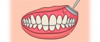
Surgical excision of the bone growth is the only effective treatment for exostosis. The intervention is performed under local anesthesia, so the patient does not experience discomfort. It is worth saying that there are two techniques for performing the operation. The choice of a specific technique depends on the location of the osteophyte:
- Removal of the palatine torus. In this case, the doctor makes a small linear incision, as well as two releasing incisions - in front and behind. After this, the dentist peels off the mucous membrane and removes the osteophyte. Extraction can be carried out either as a single block or in fragments. Next, the bone tissue is smoothed, and then interrupted sutures are applied.
- Removal of alveolar osteophytes. The procedure for performing the manipulations is no different from the previously discussed technique. The main difference is the configuration of the cut - in this case it has a trapezoidal shape. Otherwise, surgical treatment of maxillary and mandibular osteophytes is the same as removal of the palatine torus.
As for prevention, there are no specific measures to prevent this disease. The only thing that can be recommended is to visit the dentist regularly, at least twice a year, for examinations.
Still have questions? Sign up for a consultation at our clinic or call:
Rehabilitation
After removal of the exostosis, the patient remains in the hospital for 3 days, the sutures are removed on days 7-10. Performing marginal resection does not require any serious restrictions even in the early postoperative period. The patient can completely return to his usual lifestyle after the stitches are removed, and forget about exostosis immediately after the operation.
When performing a corrective osteotomy, complete bone fusion occurs after 6 weeks, but full recovery may require up to 3 months. At this time, it is important to strictly follow all the doctor’s recommendations and not to overload the operated part of the body. All patients who have undergone osteotomy are prescribed drug therapy, exercise therapy, and often physical therapy. The duration of rehabilitation in such cases depends on the type of surgical intervention performed and the individual characteristics of the patient.
Thus, osteochondroma is a very common type of tumor. At the same time, statistics on the frequency of its occurrence are not entirely objective, since exostoses often do not appear throughout a person’s life, and therefore are not diagnosed. However, if the formed formation bothers the patient, causes him pain, or reduces the quality of life, you should not hesitate to contact an orthopedist. A specialist will be able to assess the degree of danger of the tumor and, if necessary, remove it using the most gentle method possible.
Indications
- Rapid growth of osteophyte;
- exostosis after wisdom tooth removal;
- discomfort, pain;
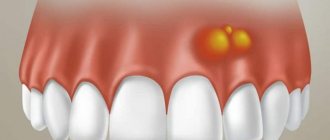
- the appearance of cosmetic defects on the lower or upper jaw (they look like white balls on the jaw, noticeable when smiling or talking);
- the need for implantation, removable or fixed prosthetics;
- the risk of tumor transformation from benign to malignant.
If you need to install prostheses or implants, exostoses will become an obstacle to the procedure. Dentures will injure the bone growth on the gum, and implants will not be able to take root normally in the bone due to the pressure of osteophytes.