An abnormality such as bone spurs is often ignored until it actively progresses. Patients often come to the clinic late, which entails complex treatment, including surgery. Exostosis is a pathology in which hardened areas of cartilage tissue protrude above the jaw are formed. The causes of this phenomenon are mechanical damage, trauma, tooth extraction and other diseases. Chewing functions are preserved, and soft tissues are also not damaged. But as the protrusions grow, thinning of the mucous membrane and ruptures occur, causing injury to surrounding areas.
Causes
Reasons for the formation of growths:
- inflammatory, infectious processes, purulent accumulations;
- mechanical impacts, injuries causing bone destruction, jaw displacement and other damage;
- deviations of row segments, formation of nodular hardenings;
- disorders of the endocrine system, hormonal levels.
One of the reasons for the development of pathology is complex tooth extraction. Often, when a wound heals, the tissue around it becomes overgrown with moving areas. If done incorrectly, compactions may appear and exostosis will gradually develop.
Signs
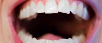
The main symptom is the presence of a dense tubercle near the place where the tooth was removed. At first it does not manifest itself in any way, except that it disturbs a little in the mouth, but again this depends on the location of the compaction. The lump is growing very quickly. When the maximum size is reached, the following signs are present:
- Aching pain that intensifies when eating.
- The cone takes on a bright scarlet hue.
- At maximum sizes, it interferes with the normal position of the tongue.
Symptoms
The initial stage of the pathology practically does not manifest itself. Symptoms generally have a vague picture, which is why patients rarely see a doctor in a timely manner. But as the condition worsens, the following symptoms appear:
- growths with a pronounced convex shape appear on the tissues; the structure can be embossed or smooth;
- there is a feeling of a foreign object in the mouth;
- pain appears, sometimes quite severe;
- partial jaw dysfunction is observed;
- the shade of the mucous membrane changes to bright;
- capillary obstruction appears in the area of the growth.
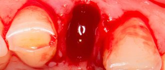
Something sharp sticks out of gum after tooth extraction
“I had a tooth removed, and now there is a sharp piece of root sticking out of my gum. What should I do?"
This is a fairly common complaint from patients after tooth extraction. Most patients are sure that a piece of a poorly removed tooth root is sticking out of the gum. In fact, this also happens sometimes, of course, but extremely rarely. But almost always the matter is completely different.
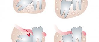
The tooth is located in the bone in its own socket, which follows the shape of its roots. The tooth socket smoothly transitions into the tooth, thinning along the edge. When a tooth is removed, the socket rises on its own, without the tooth inside. Naturally, the edge of the socket becomes the highest point of the bone in a given place, and since it is very thin, it becomes sharp to the touch. This sharp edge is called exostosis.
The first time after removal, the patient does not really notice such things, because... first anesthesia, and then swelling hide this sharp edge. And then the swelling goes away, and the bone begins to stick out through the thin mucosa. And this is already becoming noticeable both on the tongue and on the finger, with which patients, despite the ban, still regularly touch the extraction site.
Plus, the chewing process on such a sharp bone becomes very painful. The patient immediately blames the doctor for everything and does not even want to go to him, hoping that the “tooth fragment” will soon come out on its own. But, you understand, this is not a fragment and it will never come out on its own.
Therefore, there are only two ways to solve the problem.
The first and most correct
Contact the doctor who removed the tooth. The doctor will administer a small anesthesia and, under this anesthesia, gently smooth down the sharp edge. Sometimes with the help of a bur, and sometimes simply by squeezing the edges of the hole.
In any case, there is no need to remove the bone too much, because... it is very valuable for future implantation (if it is planned, of course). It is enough to simply smooth the edge of the bone slightly so that it does not scratch.
If implantation is not planned, then there is no need to remove a lot of bone either, because Even for a simple removable denture, bone is a very valuable resource. The more bone, the better the denture will fit on the jaw, the more stable it will be when chewing, the longer it will last, and the less bone will settle under the denture over the years.
The second method is a little dubious
If the protrusion of the bone is not so sharp, does not cause severe pain, and you cannot get to the dentist, then you can try to exercise the gum a little over this exostosis.
Old surgeons advised patients to smooth out such bone protrusions several times a day with an ordinary teaspoon or tablespoon. On the one hand, they still become smaller over time as a result of tissue resorption and sedimentation, on the other hand, such a massage causes thickening of the mucous membrane in a given place and the bone through such a “callus” is felt much less.
It is not so easy to smooth the bone by hand and cause the mucous membrane to grow, but they say that sometimes it works.
Indications and contraindications
The growth is treated surgically. Indications for using this option for removing pathology are:
- the bone growth grows quickly;
- pronounced cosmetic changes, progression of pathology;
- physical discomfort, the growth becomes too large;
- before prosthetics.
There are a number of the following contraindications to surgical intervention:
- pathologies of the endocrine system;
- adrenal dysfunction;
- diabetes of any form;
- bleeding disorders.
Treatment
Before treatment, the doctor conducts an examination and prescribes a number of diagnostic measures. It is necessary to perform fluoroscopy, determine the level of blood clotting, and determine whether there are contraindications to surgery. If there are no contraindications, the doctor removes the growth:
- anesthesia is performed (for uncomplicated pathology, the operation is performed under local anesthesia);
- the oral cavity is treated with antiseptic drugs;
- an incision is made on the gum, then the mucosal tissue is dissected to open access to the bone;
- the growth is removed using a laser beam or chisel (depending on the state of the pathology);
- the bone tissue is smoothed to an even texture, at which time cooled water is supplied to the surface;
- The tissues are sewn together and a bandage is applied.
The duration of the procedure depends on the complexity of the treatment. For mild cases, the operation takes about forty minutes; for complex pathologies, removal may take an hour and a half.
During the recovery period, the patient must follow the doctor's recommendations. For the first day, the intake of food and solid foods is limited, smoking and drinking alcohol are excluded. It is necessary to avoid physical activity, which can provoke bleeding and cause swelling.
Why remove exostosis?
Exostoses, as a rule, do not hurt or cause discomfort. Most often they form in children under the age of 20 during the formation of the skeleton and bone tissue, however, pathology also occurs in adults.
It is necessary to remove bone growths for several reasons:
- violation of aesthetics: exostoses look like white balls on the gums, which are noticeable when smiling,
- growths often increase in size,
- possible transition to a malignant form,
- if it is necessary to install implants or prostheses, exostoses will become an obstacle to the procedure, since the prostheses will injure the growths, and the implants simply cannot be fixed due to a violation of the quality of the bone tissue.
Diagnosis of the disease
Expert opinion
Vasiliev Alexander Alexandrovich Implant surgeon Work experience 9+
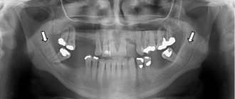
“Exostoses do not always have external manifestations - often they can only be detected on photographs when diagnosing other diseases. However, the examination uses an x-ray - a targeted image of one tooth. Or, more effectively, an orthopantomogram. It allows us to examine the condition of the jaw bone and tooth roots along the entire row, because growths are often formed in multiple quantities.”
GENERAL VIEW
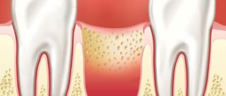
Exostosis of the oral cavity is the proliferation of bone and cartilage tissue in a certain area of the jaw row. Visually, it looks like a lump or nodule that tends to grow rapidly.
The sharp edges of the hardened cartilage tissue that forms exostosis can damage the mucous membrane covering it, leading to injury and severe pain, making it difficult to open the mouth and eat.
On the upper jaw, exostotic formations are most often localized in the area of masticatory elements on the outer or inner side of the row. When the lower jaw is affected, growths form at the base of the incisors, canines and premolars.
As a rule, cartilaginous growths have a benign structure, but there are known cases of their transformation into malignant neoplasms.
Diagnostics
The defect rarely manifests itself with clinical symptoms. Most often, growths are discovered during the diagnosis of another disease. The doctor makes a diagnosis based on the patient’s complaints, the results of a clinical examination, and an x-ray. On an orthopantomogram or x-ray, the spines look like a formation not fused with the mucous membranes with clear boundaries.
Stages and methods of treatment
Exostosis of the jaw bone is treated surgically. There are two methods of manipulation. The choice of technique is determined by the location of the defect.
Removal of the palatine torus:
- small linear incision;
- detachment of the mucoperiosteal flap;
- excision of the growth with a block or sawing to remove pieces;
- revision of the wound, return of the gum flap and suturing of the wound.
The procedure for excision of alveolar osteophytes involves the surgeon performing the above actions. The difference lies in the technique of making the cut. The doctor makes an incision in the gum in the shape of a trapezoid.
The surgical procedure is performed using a bur, laser, or piezo knife. During the process of excision of spines, the doctor removes the surface layer of the adjacent part of the periosteum. This is necessary to reduce the risk of re-formation of pathology. If the patient has a deficiency of jaw bone tissue, the surgeon creates a cavity during the intervention and places osteoplastic material into it. The wound is then stitched.
Complications
Failure to follow the recommendations of the treating dentist can lead to postoperative complications. There is a risk of sutures coming apart due to damage to the wound by hard food and the development of an inflammatory process due to ignoring hygiene rules. If negative phenomena occur, you must see your doctor.
Prevention
To prevent exostosis of the lower jaw, following simple rules:
- daily oral hygiene;
- timely clinical examinations by a dentist;
- avoiding jaw injuries;
- treatment of diseases of teeth and gums at the initial stage.
Dear patients, systematic self-diagnosis has been proven to be effective. It is necessary to independently examine the oral cavity for the presence of pathological formations, palpating the palate and the surface of the jaws.
Price
The price of surgical treatment is determined by the complexity of the clinical situation, the size of the exostosis of the upper jaw and the surgical technique. In most dentists, the procedure is performed on a turnkey basis and costs about 3,000 rubles. The price includes: anesthesia, surgeon's work, consumables and doctor's examinations.
What to do if a growth appears
The best solution is to visit a dental surgeon. After all, without proper diagnosis, it is impossible to understand what needs to be done with a growth on the gum. In general, when the lump is small and painless, people are in no hurry to seek medical help, hoping that everything will go away on its own. But there are a number of defining signs, the presence of which is necessary to visit a doctor. These include symptoms such as:
- rapid increase in the size of the growth,
- the osteophyte is large in size,
- pain: in adjacent teeth, during brushing, when chewing food,
- decreased smile aesthetics if the lump has grown on the vestibular side,
- potential problems with teething, with the formation of a child’s bite,
- if you plan to install a prosthesis or orthodontic device (braces, plates, aligners, trainers),
- if a person wears a mouthguard: an athlete, a patient with bruxism, etc.
Features of the appearance of bumps on the gums
The formation of a lump on the gum occurs as a result of the development of a pathological process, the cause of which may be a non-infectious or infectious process. In most cases, the formation is a “bag” of pus, but compaction from periodontal tissue cannot be ruled out.
A lump on the gum can be caused by pathogenic bacteria (infectious origin) or tissue trauma (non-infectious). The pathological process is also distinguished by the nature of its course: chronic and acute.
Mostly, an acute form of pathology initially develops with the manifestation of pain and signs of inflammation. If left untreated, the formation can lead to complications or become chronic with periodic relapses. Treatment for a chronic gum lump may require surgery.
Progress of the operation
Before you begin the operation, you first need to carry out some kind of preparation. The patient must undergo blood tests to exclude the presence of contraindications. These include diabetes mellitus, HIV, and poor blood clotting. In order to confirm the diagnosis, an x-ray is required. The operation itself is performed as follows:
- Thorough treatment of the oral cavity with antiseptic drugs.
- Introduction of an anesthetic drug.
- Removal of exostosis. This can be done with a standard set of cutting tools or using a laser. The latter method is practiced only in prestigious clinics where the appropriate equipment is available.
- Finally, stitches are placed to stop the bleeding. After a couple of days they are removed; some dentists use absorbable surgical thread.
REHABILITATION
Removal of exostosis is not a simple operation, so the recovery period most often does not exceed a week.
This time is enough for the gum tissue to recover after the cut, and for the bone to take on its natural shape.
To ensure an easy recovery period and to avoid postoperative complications, the patient should follow these rules:
- regularly take antibiotics and other medications prescribed by a specialist;
- avoid eating foods at too high or low temperatures;
- limit physical activity;
- stop smoking and drinking alcohol;
- exercise caution during daily hygiene procedures.
Do bumps on your gums hurt?
Painful sensations due to gum disease do not always occur. Often such pathologies are asymptomatic, which leads to delays in visiting the dentist and the occurrence of complications.
In non-infectious processes, swelling and formation on the gums are predominantly not accompanied by localized pain. The sensations may be general. So, if the jaw is injured, the entire area of impact may hurt. A cancerous tumor generally manifests itself as pain only after the beginning of the 2nd and subsequent stages. Teething can be painful, but when it comes to baby teeth, permanent teeth cause only minor discomfort in most cases.
Severe pain is characteristic of periostitis, which occurs as a complication of inflammatory processes affecting the periosteum and jawbone. With gingivitis and periodontitis, moderate pain may occur with mechanical and thermal effects, as well as with pressure. Pain with exostosis and epulis is felt with mechanical damage to the seal or the concomitant development of inflammation.
Despite the fact that periodontitis is an infectious process in which suppuration of tissue occurs, pain is not felt, especially in the presence of a fistula through which pus comes out.
As a rule, patients are driven to visit the dentist by pain. If they are absent, even if a lump is detected on the gum, going to the dentist is postponed. Such negligence often becomes a fatal mistake, since the period in which the tooth could be saved is missed. In addition to tooth extraction, a complication may be the spread of inflammation and abscess to healthy gum tissue and teeth.