The cervical spine is the most mobile part of the spine. In addition, the neck is a complex anatomical area where blood vessels, trachea, and esophagus pass. Therefore, disc protrusion in the cervical spine is often the cause of both neurological symptoms and vascular disorders associated with effects on the vertebral arteries. As a rule, disc protrusion is associated with degenerative-dystrophic changes in the spine (osteochondrosis). Degenerative processes in the spine tend to progress. Therefore, timely and adequate treatment of protrusion of the cervical spine allows not only to relieve symptoms, but also to prevent the transformation of the protrusion into a disc herniation and avoid the need for surgical treatment.
But there may be other reasons for the development of disc protrusion. Disc inflammation, damage, or degeneration can cause a range of symptoms that vary depending on the severity of the condition. A bulging disc can lead to neck pain that radiates to the arm, numbness, tingling, motor disturbances, muscle spasms, or some combination of these symptoms. The most common degenerative changes and disc protrusion occur at the C5-C6 and C6-C7 levels. Medical statistics indicate that disc protrusion in the cervical spine occurs in 60% of people over 40 years of age. It is worth noting the fact that representatives of the stronger sex get sick more often.
Causes
- Degeneration or wear that leads to disc failure.
- Repetitive flexion (extension) and twisting movements, especially in combination. These movements have the greatest impact on the outer annulus fibrosus of the disc.
- Discs are most susceptible to injury in a sitting or bending position because pressure on the disc is shifted anteriorly and the vector of force forces the contents of the disc posterolaterally toward the thinner, weaker portion of the annulus fibrosus.
- Fall or injury such as a car accident.
- Repetitive stress injuries from overhead work or weightlifting.
- Genetic factors may, to a certain extent, contribute to the early development of degenerative changes in the discs (osteochondrosis).
- Smokers are more likely to develop early disc degeneration
- Infection (discitis)
Symptoms
Disc bulges in the cervical spine are quite common, and most are not symptomatic. Symptoms of disc protrusion depend on the location of the bulge and the soft tissue structures that are affected. The process may involve: the back of the head, neck, arms, shoulder girdle and hands. Symptoms can range from minor discomfort to severe and persistent.
Most often, the following symptoms occur:
- Pain in the neck, head, shoulder and arm, worsens with minimal strain on the arm, coughing, sneezing;
- Crunching and pain in the neck when turning or tilting the head;
- Muscle spasm and postural changes;
- Numbness, tingling in the arms or legs, burning between the shoulder blades;
- Headache, which usually begins in the neck and spreads to the back of the head and temples;
- Dizziness and even fainting when suddenly turning the head;
- Decreased range of motion in the neck, difficulty moving the head and neck;
- Sitting for long periods of time can cause neck and arm pain;
- Pain in the neck or radicular pain radiating to the arm if the disc compresses a nerve root.
- Weakness, muscle atrophy, or decreased strength in the arms may occur if motor neuron compression occurs.
- Feeling weak and tired;
Other symptoms of cervical disc protrusion include tinnitus, decreased visual acuity and hearing, and sometimes pain in the heart area.
The action of the secretion of the salivary gland of leeches
Getting into the inflamed area of the oral cavity and into the blood, the substances contained in the saliva of leeches:
- slow down clotting;
- reduce the likelihood of blood clots;
- have anti-inflammatory, bactericidal effects;
- stimulate the immune system;
- improve drainage in lymphatic channels;
- ensure the extraction of blood along with fluids of the affected tissues;
- remove venous congestion;
- increase blood supply to the injection area.
Red flags for neck pain
A serious underlying cause is more likely in people who:
- The onset of symptoms appeared before the age of 20 years or after 55 years.
- Weakness involving more than one myotome or sensory disturbance involving more than one dermatome.
- Intense or increasing pain.
Red flags suggesting possible malignancy, infection or inflammation:
- Fever.
- Unexplained weight loss.
- History of inflammatory arthritis.
- A history of malignancy, drug abuse, tuberculosis, AIDS, or other infection.
- Immunosuppression.
- Pain that gets worse, is very intense and/or disturbs sleep.
- Lymphadenopathy.
- Local point tenderness over the vertebral body.
Red flags suggesting myelopathy (spinal cord compression):
- Progression of symptoms.
- Gait disturbance; weakness or impaired motor function in the hands; loss of bladder/bowel control.
- Lhermitte's sign (bending the neck causes an electric shock sensation that travels down the spine and into the extremities).
- Pathological symptoms in the lower extremities (Babinski reflex, hyperreflexia, clonus, spasticity) and in the upper extremities (atrophy, hyporeflexia).
- Various sensory changes (loss of proprioception more noticeable in the arms than in the legs).
Red flags indicating the possibility of severe skeletal injury:
- Trauma history.
- Previous neck surgery.
- Osteoporosis or risk factors for osteoporosis.
- Increase and persistence of pain.
Red flags indicating vascular insufficiency:
- Dizziness and darkening before the eyes (compression of the vertebral artery) when moving, especially when straightening the neck.
- Dizziness, syncope episodes.
Indications for the use of HIRUDOTHERAPY:
Diseases of the cardiovascular system:
- IHD, angina pectoris, post-infarction cardiosclerosis
- Hypertension stage I-III.
- Atherosclerosis of various localizations
Respiratory diseases:
- Bronchitis acute, chronic
- Pneumonia (recovery period)
- Bronchial asthma, COPD
- Chronic gastritis (except for erosive forms in the acute stage)
- Peptic ulcer of the stomach and duodenum (not exacerbation)
- Chronic pancreatitis
- Chronic colitis, enterocolitis
- Transient cerebrovascular accidents: transient ischemic attacks, hypertensive cerebral crises
- Ischemic stroke (recovery period)
- TBI, concussion, concussion
- Migraine
- Headache
- Trigeminal neuralgia
- Facial nerve neuropathy
- Tunnel syndromes
- Dorsopathies
- Polyneuropathy
- Ankylosing spondylitis
- Various diseases and injuries (sprains, bruises, dislocations, hematomas, contractures)
- Limb fractures
- Arthritis of various etiologies and localizations
- Arthrosis
- Tendon diseases (tendonitis, tendovaginitis)
- Bursitis, periarthritis
- Inflammatory processes (abscesses, boils, carbuncles, phlegmons, felons, at the stage of infiltration or after surgery; osteomyelitis)
- Prevention and treatment of postoperative scars, infiltrates
- Condition after reconstructive operations
- Diseases of the veins of the lower extremities (varicose veins, thrombophlebitis, trophic ulcers)
- Haemorrhoids
- Mastitis
- Obliterating endarteritis
Differential diagnosis of neck pain
As a rule, most back pain is associated directly with changes in the discs (protrusion, disc herniation), however, for an accurate diagnosis it is necessary to use medical imaging methods.
Differential diagnoses include:
- Cervical spondylosis.
- Traumatic prolapse of an intervertebral disc.
- Neck pain: acute neck strain, postural neck pain, or pain associated with whiplash injury
- Headache.
- Referred pain - for example, from the shoulder.
- Malignancies: primary tumors, metastases or myeloma.
- Infections: discitis, osteomyelitis or tuberculosis.
- Fibromyalgia.
- Vascular insufficiency.
- Psychogenic neck pain.
- Inflammatory diseases (for example, rheumatoid arthritis).
- Metabolic diseases: Paget's disease, osteoporosis.
What is resection
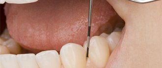
Removal of a dental cyst is most often carried out by excision of the root and removal of the affected fragments. Then, to avoid the spread of infection to adjacent areas of hard and soft tissue, the canal is sealed. The complex of these surgical procedures is called root apex resection. In fact, this is the simplest way to remove a dental cyst, often used when identifying the disease in the initial stages. It is important that the operation is tooth-saving, that is, it makes it possible not to part with the molar. If the element can be preserved completely, then it can serve a person for a lifetime. If a fragment of the unit remains, it is used as a basis for further prosthetics. Tooth-preserving methods are valued by doctors because they allow not to remove a unit and maintain the integrity of the row. Therefore, resection of the root apex is used not only to remove a dental cyst, but also in some other cases.
- When an element covered by a crown becomes inflamed. It would seem that the obvious solution is to remove the prosthesis, remove the abscess, and install a new structure. However, this manipulation is more expensive and time-consuming than root resection.
- If the base under the fixed implant pin begins to tear. It is impossible to remove it, so the only way out in such situations is excision of the root and elimination of the lesion.
- Root canal abscess resulting from poor-quality filling or the use of low-grade materials. Such cases are especially common in systems with curved roots.
- When osteomyelitis is detected. There is a need to cut out the affected areas of bone tissue bordering the root trunks. This helps prevent infection of deep structures.
- In case of detection of foreign objects in the channels, for example, boron particles. This happens when using cheap soft metal equipment. Our clinic uses modern instruments based on titanium alloys.
REMEMBER! Properly performed resection not only allows for prompt removal of the dental cyst, but also strengthens the bone tissue, which over time completely restores function. However, even such an effective operation has contraindications. Resection is not done if the roots of diseased and healthy elements are located too close; when there are extensive crown chips; with abnormally high mobility of units; in the stage of acute periodontitis. It is not recommended to perform manipulation for people with diabetes, mental and nervous disorders, chronic heart and vascular diseases, pathologies of the lungs and thyroid gland.
Diagnosis of disc protrusion
Neck pain from radiculopathy and the absence of red flag symptoms usually do not require imaging or other special tests. However, for verification purposes, the doctor may prescribe the following research methods:
Blood tests
may be prescribed to exclude infectious or inflammatory diseases (a decrease in hemoglobin may be a sign of anemia; an increase in ROE may be a sign of both infection and inflammation; the level of rheumatoid factor and HLA-B27 determination may indicate systemic connective tissue diseases).
Medical imaging tests are very important, but they must be interpreted in light of the clinical picture, since positive results are quite common in people without any complaints:
• Radiography
. Plain x-rays of the cervical spine can be used to evaluate chronic degenerative changes, metastatic tumors, infection, spinal deformity, and motion segment stability. Interpretation of study results is often difficult because degenerative signs are almost universal in people over 35 years of age.
• MRI.
This imaging method may be prescribed if radiography does not allow the genesis of symptoms to be determined. MRI should be performed to diagnose disc protrusion (herniation) and the presence of compression of nerve structures.
• CT myelography
can be used if there are any contraindications to MRI.
•EMG (ENMG
) - these methods can be informative in the presence of radiculopathy or the need to determine the level of damage and differentiate cervical radiculopathy from nervous compression syndromes.
Contraindications to the procedure
All diagnoses that require the application of worms as part of hirudotherapy have a common criterion. Procedures cannot be performed with a critically low hemoglobin indicator, anemia, or hemophilia. It is prohibited to use such techniques on cancer patients. You should not apply leeches to pregnant women, women during lactation, or small children.
Age requirements are partly added to the standard requirements. After all, periodontitis and periodontal disease are largely diseases of mature and elderly people. Over the years, blood clotting may worsen, and the number of allergic reactions may increase. Therefore, treatment of dental problems with leeches should be carried out only after a particularly thorough examination by the treating periodontist.
Treatment
As a rule, treatment of protrusion of the cervical spine uses conservative methods, and surgical treatment is rarely required. Conservative treatment relieves symptoms and stops the progression of the disease.
The doctor prescribes a course of treatment for the patient, taking into account the clinical picture, age, gender and characteristics of the body. Treatment of protrusion of the cervical spine can take up to one and a half months. The patient undergoes procedures 2-3 times a week. Neck pain can usually be relieved after 1-2 weeks of treatment. The following methods can be used in the course of treatment:
Why is a cyst on the root of a tooth dangerous?
A dental cyst is an inflammatory neoplasm at the apex of the root in the form of a capsule with purulent contents. As it grows, it melts the bone tissue around the tooth and fills the resulting space. The insidiousness of the disease is that it can develop over years, grow to enormous sizes, without causing any discomfort. Most often it is discovered incidentally during X-ray examinations for other reasons.
Odontogenic cyst on the root of the lower tooth in the picture
The asymptomatic course is explained by the body’s defense systems when the immune system copes with the pathology. But it is dangerous because the cyst reaches significant sizes without treatment and can cause unpleasant consequences:
- On the upper jaw - spread into the maxillary sinuses, the occurrence of odontogenic sinusitis.
- In the lower jaw there is damage to the mandibular nerve and anemia of half of the lower lip; even jaw fractures are possible due to melting and thinning of the bone tissue.
Annual preventative examinations at the dentist allow you to identify possible inflammatory processes at an early stage and save the tooth. The larger the cyst, the less likely it is that the tooth can be saved.
Against the background of acute respiratory infections or other conditions when the immune system is weakened, the tooth may become uneasy (pain when biting, swelling and redness of the gums, the feeling of a “growing” tooth). This happens due to an increase in the content of infected fluid and pressure inside the capsule. It is possible for the contents to escape through the fistulous tract. And this is the best outcome, since the patient in this case, as a rule, goes to the clinic.
Surgery
The most common disc-related surgery is a discectomy, in which the disc is removed through an incision. In the cervical spine, discectomy can be performed either anteriorly or posteriorly. In some cases, vertebral fusion surgery may be recommended. Most patients who have a discectomy recover fairly quickly from surgery with the help of physical therapy. Discectomy is usually performed when a herniated disc is present, as most disc bulges can be successfully treated conservatively.
Are there any complications?
Like any surgical procedure accompanied by the use of anesthesia, removal of a dental cyst can sometimes lead to undesirable consequences. They arise, as a rule, in cases where the doctor does not fully follow the chosen method, if not all necessary diagnostic tools were used at the preparatory stage, or if inappropriate equipment was used. In our clinic, there are no cases of complications after removal of a dental cyst due to the skill of the doctors, the availability of modern equipment, and the use of advanced techniques. But every patient should know about the possible consequences of the operation. Symptoms such as increased body temperature, pain in the treated area, swelling, hematomas are inevitable, but taking antibiotics, regular ice compresses, and lotions will eliminate the discomfort very quickly. However, there are more serious complications after removal of a dental cyst.
- Vascular ruptures occur when a road is laid to provide access to the root. Therefore, a blood clotting test is required.
- Injuries to nerve bundles that cause pain after surgery cannot be ruled out. Taking analgesics will help cope with this.
- Sometimes, if hygiene measures are not followed, secondary suppurations occur. To avoid this, a course of antibiotics is prescribed.
- Incomplete removal of the affected area may cause re-infection. The physician must be extremely careful when cleaning the cavity and examining it before suturing.
- Careless actions of an inexperienced physician can lead to perforation of the walls of the maxillary sinuses. Today such cases practically never occur.
The listed complications after removal of a dental cyst at the VivaDent clinic are impossible. We employ real professionals in their field, we have installed the most modern equipment that allows us to solve the most complex problems. We are confident of a successful result even when other medical institutions do not undertake treatment for dental cysts. Much depends on the patient himself. It is important to follow your doctor's recommendations in the first days after surgery. At this time, you need to limit the consumption of hot, cold and hard foods, do not use hard brushes or aggressive rinses, do not overcool or experience high physical activity, take vitamins and anti-inflammatory drugs. Taking good care of your own body will reduce the rehabilitation period after removal of a dental cyst to a minimum. The staff of the VivaDent clinic will make every effort to rid the patient of oral problems forever.