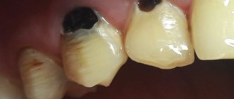
The doctor makes the diagnosis of “post-extraction alveolitis” based on a visual examination. To confirm the diagnosis, X-ray diagnostics is prescribed. An x-ray can show changes in tissue, identify tooth fragments, a cyst or granuloma, which could cause inflammation.
The final result of treatment of alveolitis should be:
- Elimination of inflammation;
- Cleaning the hole from foreign bodies;
- Complete healing of the hole;
- Exclusion of the development of complications.
Treatment at the dentist
The doctor begins treatment by washing the socket and treating it with antiseptic solutions, and then cleans the socket from the remains of the blood clot using a surgical curette. Then the wound is dried with a gauze swab and treated with an anesthetic and antibacterial agents. The wound is covered with a bandage, which is designed to protect the open surface of the wound from possible biological, chemical or mechanical irritants. To check the possibility of detecting pieces of the extracted tooth in the socket, an x-ray can first be taken.
The procedure for cleaning the hole as a whole is carried out according to the following scheme:
- Local anesthesia is performed;
- The well is washed with an antiseptic solution (hydrogen peroxide solution 3%, chlorhexidine 0.05% or furatsilin 0.02%), and food debris and necrotic masses are removed;
- If there are foreign bodies in the hole (root fragments, cysts and granulomas), they are removed using special instruments;
- After cleaning the hole, it is treated with antiseptic solutions, dried with a sterile gauze swab, and turunda with an antiseptic and an anesthetic is injected into it. With a mild degree of inflammation, a turunda may not be necessary; thorough cleansing of the hole and subsequent care for it is sufficient;
- In case of a pronounced necrotic process in the hole, rinsing and application with trypsin is carried out - this is an enzyme preparation that accelerates the breakdown of dead tissue. Trypsin has anti-inflammatory and anti-edematous effects, helps clean the socket from necrotic masses and pus;
- After completing all the manipulations, the patient goes home, the doctor appoints appointment days when he needs to come for antiseptic treatment of the hole and change the turunda, if one has been installed. If necessary, a course of antibiotics is prescribed.
For alveolitis, antiseptic baths (not rinsing) with solutions of chlorhexidine 0.05% or miramistin 0.01%, which can be purchased ready-made at the pharmacy, are effective. When performing baths, the solution is taken into the mouth and held for several minutes, then carefully spat out. To relieve pain and reduce the degree of inflammation, nimesulide or ibuprofen are prescribed.
To prevent the turunda from falling out of the socket, during treatment you need to eat soft, pureed food and do not chew on the sore side. If the turunda does fall out, you need to rinse your mouth with chlorhexidine solution and immediately consult a doctor.
If this is not possible, you should rinse the hole yourself from food debris: to do this, bite off the sharp tip of the needle from a 5 ml syringe, bend it a little, and disinfect it with pure alcohol. A chlorhexidine solution is drawn into the syringe, the needle is inserted into the hole (not too deep) and the piston is pressed intensely to create a liquid pressure that can remove food debris.
All cases of alveolitis treatment are individual, so it may be necessary to visit the dentist up to several times.
With a favorable course of the healing process, the pain goes away, and the inflammatory process gradually subsides and disappears after a few days.
If the process, despite the procedures performed, progresses, then after antiseptics, gauze tampons soaked in propolis tincture or camvorophenol solution (10%) are inserted into the hole. A tetracycline-prednisolone cone inserted into the well has a good antibacterial effect.
Procedures for continuing treatment at home
Treatment of alveolitis at home is carried out when acute symptoms have been eliminated, antiseptic turundas are no longer needed and the goal is to speed up the healing of the hole. For this use:
- Mouth rinses with infusions of chamomile, calendula, and sage. These medicinal plants have anti-inflammatory properties and accelerate healing processes. Rinsing is carried out several times during the day, always after meals.
- Applications with dental adhesive paste Solcoseryl. The drug belongs to the group of tissue regeneration stimulators: it accelerates healing, improves the supply of cells with oxygen and nutrients. The paste has a pleasant mint taste and contains the anesthetic component polidocanol 600. To properly apply the paste, you need to dry the hole with a gauze swab and fill it with Solcoseryl, lightly moisten the paste with water on top. The procedure is repeated several times a day as necessary; the paste is able to protect the socket throughout the day from mechanical and chemical influences.
- Taking vitamin preparations. You can purchase any vitamin and mineral complex at the pharmacy (Vitrum, Duovit, Complivit); Vitamins are taken to increase the body's defenses and shorten the recovery period.
Additional physiotherapy treatment (click to expand)
Physiotherapeutic treatment is an important addition to drug therapy; with the help of physiotherapy, the intensity of inflammation can be significantly reduced and healing time can be accelerated. For alveolitis, the following techniques are used:
- UV therapy - the hole is irradiated with short-wave ultraviolet light, which kills pathogenic microorganisms and reduces the level of inflammation.
- SMV therapy is a method of treatment with an electromagnetic field, based on the effect of centimeter waves on the area of inflammation. The procedure helps improve blood circulation and metabolism, due to which toxic substances are removed from tissues faster and regeneration processes are accelerated. SMV therapy also has an analgesic effect.
- UHF therapy – the body is exposed to a high-frequency electromagnetic field. For alveolitis, UHF therapy is used if the patient's regional lymph nodes are enlarged.
- Electrophoresis – medications are injected into inflamed tissue using electrical impulses. For post-extraction alveolitis, electrophoresis is used to reduce pain. For this purpose, solutions of novocaine, lidocaine, trimecaine are used.
- Fluctuarization is a treatment technique with pulsed currents of a sinusoidal shape with a low frequency. As a result of the procedure, blood circulation and lymph flow improves, swelling resolves, and the level of inflammation decreases.
- Laser therapy - the hole is exposed to infrared laser radiation, which has an anti-inflammatory effect, reduces swelling and redness of soft tissues, and accelerates healing.
Complications of alveolitis
If alveolitis is not treated in a timely manner, the following complications may develop:
- Odontogenic sinusitis is an inflammation of the maxillary sinus caused by the spread of infection from the inflamed sockets after the removal of premolars or molars of the upper jaw;
- Phlegmon - purulent inflammation spreads to the surrounding soft tissues;
- Acute periostitis - pus accumulates in the periosteum area;
- Odontogenic osteomyelitis is a purulent-necrotic lesion of the jaw bone;
- Sepsis - an infection enters the bloodstream, causing it to become infected.
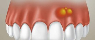
Alveolitis itself is not so dangerous, but its complications are life-threatening. Therefore, if you notice symptoms of alveolitis, contact your doctor immediately.
Alternatives to wisdom tooth removal?
Is it possible not to remove a wisdom tooth if it rarely bothers you or doesn’t bother you at all? Can! But it is necessary to understand the consequences of the decision made. If the tooth is only partially in the oral cavity and the gums above it periodically become inflamed, this can cause a lot of inconvenience. Near the location of the wisdom tooth there is a masticatory muscle, which can be affected by the inflammatory process in case of gum inflammation. As a result, it becomes painful to open your mouth and eat.
An unerupted figure eight located deep in the bone can push the teeth apart, which can lead to pain in the area of the second molar (seventh tooth) or even damage to it (see x-ray). Pressure on neighboring teeth can cause a shift in the dentition and, as a result, malocclusion and the formation of crowded teeth.
The patient's role in the prevention of alveolitis
According to statistics, in most cases, the cause of the development of post-extraction alveolitis is the patient’s incorrect actions after tooth extraction, ignoring the recommendations of the attending physician. After tooth extraction it is prohibited:
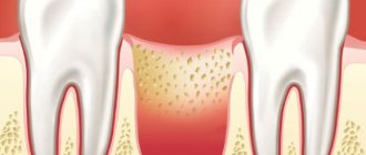
- Remove the blood clot from the socket. The blood clot that forms in the hole after tooth extraction prevents microbes from entering the wound, and the healing process under it proceeds quickly and without complications. Patients can remove the clot by touching it with the tongue, with fingers, by vigorously rinsing the mouth immediately after tooth extraction, and also while eating solid foods.
- Do hard physical work, sports, take hot baths, go to the sauna. Intense loads and elevated temperatures provoke the opening of the wound, and bleeding resumes; pathogenic microorganisms can enter the wound.
- Smoking, drinking alcohol. Bad habits lead to excessive irritation of the mucous membranes, healing of the hole is much slower, and inflammation may occur.
To speed up healing and prevent the development of inflammation in the socket, you need to:
- In the first few days after tooth extraction, review your menu, excluding spicy, too salty, sour, and hot dishes. All food should be soft, pre-chopped;
- Maintain careful oral hygiene. The presence of chronic inflammation of the gums and carious teeth in the mouth can cause infection of the socket, so after tooth extraction you need to carry out antiseptic baths (they should be prescribed by a doctor);
- After each meal, rinse your mouth with clean water to remove any remaining food; this must be done very carefully so as not to remove the blood clot covering the hole;
- Brush your teeth carefully, trying not to touch the socket with the brush.
Preparing for wisdom tooth removal
Your surgeon will explain to you what needs to be done and how to prepare before tooth extraction. If you smoke, it is advisable to stop smoking one day before surgery, as this significantly reduces the likelihood of such complications after surgery as infection of the extracted tooth socket, which significantly lengthens the healing time of the postoperative wound. The result of wound infection is the development of inflammation and pain in the postoperative area.
If you plan to remove all your wisdom teeth under general anesthesia (anesthesia), the anesthesiologist will give recommendations on how to prepare for anesthesia. It is usually recommended not to eat or drink for about 8 hours before going into anesthesia.
If there is an inflammatory process in the area of the teeth that are supposed to be removed, the operation is usually postponed for several days until the inflammatory phenomena subside, antibiotics are prescribed and the area of inflammation is “washed” (under the hood) with antiseptic solutions
The role of the doctor in excluding the possibility of alveolitis
Actions of the dental surgeon to exclude alveolitis:
- Thoroughly clean the socket so as not to leave a tooth fragment or cyst in it. After extracting a tooth, especially a damaged one, a control x-ray of the socket is often taken;
- Be careful and avoid injury. The tooth extraction was accompanied by bone injury due to the inept actions of the doctor. Usually the bone is injured during complex extraction, when tooth extraction cannot be carried out using conventional instruments. Difficult procedures include the removal of unerupted and incorrectly positioned wisdom teeth, multi-rooted teeth, and teeth with curved or destroyed roots. An inexperienced doctor may not understand the situation and damage the bone;
- Control during anesthesia. An overdose of anesthetic leads to a sharp decrease in the amount of blood released after tooth extraction and insufficient formation of a blood clot;
- Prescribing appropriate antibacterial drugs, especially if the tooth extraction was difficult or was carried out against the background of purulent inflammation.
Therefore, it is very important to seek help from competent and experienced specialists who are fully familiar with all the intricacies of the tooth extraction procedure.
Sometimes alveolitis can develop despite preventive measures taken due to certain reasons. These reasons include:
- Reduced immunity of the patient, inability of tissues to regenerate;
- Tendency to bleed, resulting in a blood clot not forming;
- An increase in the amount of the hormone estrogen in women when taking hormonal contraceptives or certain diseases, which leads to the destruction of a blood clot.
A timely visit to the dentist if alveolitis develops is the key to a quick cure. Patients of our clinic who have undergone tooth extraction surgery receive detailed recommendations on oral care; if they are strictly followed, the likelihood of developing alveolitis is excluded.
Wisdom tooth.
The third molar, also known as the wisdom tooth, usually does not begin to emerge until age 17. If it has not appeared before the age of 25, then most likely it either does not exist at all, or it cannot squeeze into the dentition due to lack of space in it. The wisdom tooth becomes impacted.
Very often, a wisdom tooth cannot erupt because it is located perpendicular to the neighboring teeth, rests against them and pushes them. If a tooth cannot erupt for a long time, it is called impacted. Impacted wisdom teeth are often the cause of:
- Inflammation of the gums and the development of pericoronitis (inflammation of the gums overhanging the wisdom tooth);
- Caries of neighboring teeth, because the gap between an unerupted wisdom tooth and a healthy second molar is almost never cleaned when brushing your teeth and a large amount of bacterial plaque and food debris accumulate there;
- Development of an abscess in the retromolar region (the area behind the wisdom tooth)