Pulpitis accounts for approximately a quarter of all dental diseases. Pulpitis is an inflammatory process of the internal tissues of the tooth (pulp). The pulp is a unique tissue in the human body. It is a complex of connective tissue, blood vessels and nerves. The pulp has a complex structure and ensures the normal functioning of teeth. Inflammation of the pulp significantly affects not only the health and aesthetics of teeth, but also the patient’s quality of life. Moreover, without comprehensive treatment, pulpitis is prone to complications, which makes treatment difficult. A timely visit to a doctor will help get rid of the disease and prevent its reappearance.
What is pulpitis and how does the disease progress?
Pulpitis develops when carious destruction of the integrity of the tooth occurs when food and microorganisms penetrate the tooth cavity (pulp). Another way of developing the disease is infection from neighboring affected teeth through blood vessels, which occurs much less frequently and is called retrograde pulpitis. There are other, rare pulpitis:
- Traumatic, develops as a result of a crack, break, or chip of a tooth.
- Concrete occurs when there is excessive deposition of mineral formations that begin to replace the internal tissues of the tooth.
- Inflammation develops due to incorrect treatment, the use of low-quality materials, and accidental opening of the pulp.
Regardless of the type of infection, inflammation requires immediate treatment. Among the factors that increase the likelihood of developing pulpitis are: insufficient hygiene, osteoporosis and diabetes.
There are three stages of pulpitis: acute, chronic and aggravated chronic pulpitis. According to the ICD-10 classification used when making a diagnosis, the disease is divided into several categories.
Pulpitis acute
- K04.00 - initial inflammation of the pulp.
- K04.01 - acute focal pulpitis.
- K04.02 - purulent abscess.
The diagnosis is made when the pulp first comes into contact with infection. This includes the serous form of the disease, focal and purulent abscess.
First, the tooth becomes sensitive to temperature: hot or cold. Unlike caries, pain does not subside when the irritant is removed. Then comes the diffuse phase - the pain intensifies at night, is periodic, and often radiates to the temple or other parts of the face. With the transition to the chronic phase, the pain disappears, or is characterized by the patient simply as an unpleasant sensation.
Chronic pulpitis
- K04.03 - fibrous.
- K04.04 - gangrenous.
- K04.05 - pulp polyp.
Chronic pulpitis occurs after the acute phase of the disease. It is characterized by (irreversible) destructive changes in tissues, divided into three types according to the type of changes:
- Fibrous
- the most common sluggish form of chronic pulpitis. It is characterized by the growth of connective, fibrous-granulation tissue in the pulp.
- Gangrenous
- This is a putrefactive inflammation, which is marked by a characteristic odor from the mouth. The gums often swell, and fistulas may form through which exudate flows out.
- Hypertrophic
- the most rare pulpitis, characterized by the growth and exit of the pulp into the carious cavity.
The chronic form is not painful, but severe pain may occur periodically.
Note! In the acute stage, the infected tooth mainly reacts to cold; in the chronic form, the source of pain is exposure to high temperature.
Complicated forms
- K04.1 - pulp necrosis.
- K04.2 - pulp degeneration.
By complicated course of the disease we mean both inflammation of the entire pulp and simultaneous damage to several canals. When the pulp degenerates, denticles are formed - dense mineral formations (stones). They cause pain when there is a sudden change in body position, as this causes them to shift. For example, such patients experience pain when flying, going up in an elevator, or playing sports.
Necrosis refers to the death of cells in the neurovascular bundle of the dental root. In fact, this is the final stage of the disease. Necrosis can occur due to pulpitis, damage to the blood supply to the tooth, or under the influence of toxic components of the filling material.
Symptoms
Acute pulpitis:
- Severe throbbing pain radiating to neighboring teeth, ear, throat, temple.
- Reaction to hot and cold, including cold weather.
- Painful attacks occur after certain periods of time and intensify in a supine position.
Fibrous pulpitis:
- The pain is aching in nature, worsening in the evening and at night.
- A feeling of “bursting” and heaviness in the affected tooth.
- A sharp temperature reaction that continues after removal of the irritant.
Gangrenous pulpitis with an open lesion cavity:
- Discomfort and heaviness in the area of the affected tooth.
- Pain when biting.
- Reaction to hot drinks and food with prolonged persistence of aching pain.
- The enamel is gray, the gums are red and swollen, there is a fistula.
- Putrid odor from the mouth.
Gangrenous pulpitis with a closed lesion cavity:
- Intense pain attacks.
- Vivid reaction to hot drinks and food with pain persisting for a long period of time.
These symptoms help you pre-diagnose the presence of pulpitis and consult a dentist as soon as possible.
Causes
Treatment of the disease primarily depends on the causes of its occurrence. Factors that often lead to inflammation:
- Untreated caries in a timely manner. In a mild form, the disease does not affect the pulp in any way, since the carious lesion extends only to the superficial tissues. As the crown is destroyed, caries affects all cellular structures and reaches the roots.
- Chronic periodontitis (except for mild forms). Deep periodontal pockets that form with this pathology reach the root plexuses. Pathogenic microorganisms that multiply in these cavities spread throughout the tooth and reach the pulp.
- Injury. Impact, bruise and other external influences contribute to the disruption of the established blood supply process and lead to a pathological process.
- Poor quality dental treatment. If the dentist did not clean the tooth cavity well before placing the filling, leaving carious particles, they will spread into deeper layers and reach the neurovascular bundle. Also, due to the fault of the doctor, the patient may receive a thermal burn of the pulp if the specialist neglected the rules while drilling the crown and did not cool it enough with water. The disease is also provoked by overdrying of the dentinal tubules by air flow.
- Exposure to acids, alkalis, medications, toxic filling compounds and other chemicals.
- Blood infection. Infection can penetrate into the pulp not only through carious holes, but also during sepsis.
- The patient's individual predisposition to tooth wear and the formation of mineral deposits in the pulp chamber.
Types of pathology
There are 2 main forms of the disease: acute and chronic. In the first case, a person suddenly experiences a sharp, paroxysmal pain (most often at night), which does not depend on external factors and does not go away when the irritants are eliminated. At first, a dark hole appears on the enamel, which grows over time and affects deep tissues, including reaching the canals.
If the inflammation does not go away after 3 weeks, the disease becomes chronic. Dull aching pains appear with a certain frequency and are disturbing not only at night.
Types of acute pulpitis:
- Focal. It is observed only for a few days. The pain lasts up to 20 minutes, with the intervals between attacks being about 2-3 hours. There is swelling of the gums.
- Diffuse. It extends to the coronal part, nerve endings and root. There is a disruption in the blood supply to tissues. The pain becomes throbbing and lasts much longer than with the focal form.
- Purulent. The cavity of the affected unit fills with pus. A painful pulsation is felt. The patient's condition is constantly deteriorating.
- Serous. Most often, this type is observed in children (pulpitis of temporary teeth). This is a pathology of infectious etiology, which is accompanied by short attacks of pain.
Classification of chronic dental pulpitis:
- Fibrous. Is a consequence of acute. It can proceed latently for several months with periodic periods of exacerbation. The gums are not swollen, the pain is aching and occurs infrequently.
- Gangrenous. This is a complication of acute fibrotic disease, in which the death of pulp tissue and destruction of the coronal part occurs.
- Hypertrophic. In the carious cavity, tissues of a bright red hue are clearly visible, which constantly bleed, especially when pressed.
Symptoms
Despite the fact that each type of disease has its own distinctive features, there is a list of symptoms characteristic of all forms of pathology. At the initial stage of the disease, the tooth begins to react painfully to cold and hot, as well as to other irritants. Then a sharp throbbing pain appears, which intensifies at night and when lying down. Soreness occurs spontaneously or under the influence of irritating factors.
External manifestations of the disease, regardless of its type:
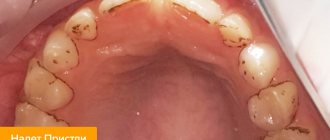
- darkening of the enamel;
- tooth mobility;
- bleeding;
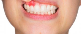
- redness of the gums;
- swelling of the tissues around the diseased tooth;
The chronic form of pulpitis can be asymptomatic. It is characterized by a putrid odor from the mouth and aching pain.
Important! Very often the patient cannot understand which tooth hurts. The pain may radiate to the neck or ears. To determine the location of inflammation, a doctor’s examination and additional diagnostic tests are necessary.
Diagnostic methods
To make an accurate diagnosis, the dentist conducts and prescribes the following studies:
- inspection of the damaged cavity using a mirror and probe;
- checking the tooth’s reaction to temperature fluctuations (thermometry);
- exposure of the crown to a weak electric current (electroodontodiagnosis), with the help of which one can distinguish the disease from deep caries and determine in what form it occurs;
- X-ray.
Features of treatment
The therapeutic regimen directly depends on the stage of the disease and the location of inflammation. Treatment methods for pulpitis used in dentistry:
- Biological. Only the source of infection is eliminated, the progression of the disease is stopped, and the inflammation gradually goes away. Medicines are introduced into the cavity, which stop the growth and reproduction of pathogenic microflora and help eliminate inflammation.
- Conservative (vitalization). The doctor preserves the living pulp, but removes its coronal part. The tooth is completely preserved, while its functioning is restored.
- Devitalization. A special paste is placed into the drilled hole, which promotes the complete death of the pulp. The neurovascular bundle is removed, then the specialist places a temporary filling. When tissue inflammation completely disappears, the filling material is replaced with a permanent one. Devitalizing paste is used by pediatric dentists for tooth pulpitis in a child.
- Surgical. Surgical removal is divided into amputation (partial removal of pulp tissue) and extirpation (the pulp is completely removed).
Attention! The sooner the patient contacts the dentist, the higher the likelihood that the tooth can be saved. A qualified doctor will be able not only to cope with swelling and inflammation, but also to restore the integrity of the crown and restore the lost whiteness of the enamel.
Is it possible to cure a disease with folk remedies?
Pulpitis requires qualified medical care and cannot be treated on its own. Home methods for relieving pain and reducing inflammation can only temporarily alleviate the patient’s condition. Without timely conservative or surgical treatment, severe complications are possible.
Possible complications
If you do not receive treatment on time or do it incorrectly, the risk of complications increases, such as:
- periodontitis (damage to hard tissues around the tooth root with pulpitis);
- periostitis (inflammation of the periosteum, or flux);
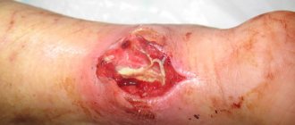
- abscess (purulent inflammation accompanied by intoxication of the body);
- sepsis (blood poisoning);
- chronic pathologies of internal organs and systems.
Preventive measures
In order to avoid long-term treatment and surgical intervention, you need to take care of the prevention of pulpitis by adhering to the following rules:
- brush your teeth 2 times a day, rinse your mouth after eating, remove plaque and tartar in a timely manner;
- visit the dentist for preventive examinations at least once every six months;
- do not use toothpicks or other traumatic devices;
- eat a balanced diet, ensure that the body receives a sufficient amount of vitamins and minerals;
- give up bad habits (smoking, drinking alcohol), large amounts of sugar and junk food;
- treat caries and other dental pathologies in a timely manner, without bringing them to severe stages.
Sharp pain radiating to the neck, temples or ears is a reason to urgently visit a dental clinic. If treatment is not started in time, serious complications are possible, including complete tooth loss and a septic process.
By following preventive measures, you can protect yourself from the occurrence of pulpitis and other oral diseases.
Diagnosis of the disease
During the appointment, the doctor interviews the patient. Determines the nature of pain, the conditions for the manifestation of pain, intensity and duration. Then, he conducts an external inspection and tapping using hand tools. This allows us to identify the presence of a carious cavity, the density or, conversely, softness of the pulp, and assess the condition of the gums. After this, an x-ray is prescribed. It shows the condition of internal tissues and channels, the localization and extent of inflammation.
In some cases, electroodontodiagnosis is prescribed—this is an analysis of the tooth’s reaction to electricity. The study provides insight into the integrity and functionality of the nervous system. Normally, the pulp responds with a slight pain signal to the electricity passing through it. As caries progresses, degenerative processes occur that reduce the sensitivity of nerve receptors.
Why does purulent pulpitis occur?
Precursor of purulent pulpitis in children
and adults is acute pulpitis. The formation of pus occurs from the serous exudate that is present in the nerve. Due to an increase in its volume, metabolic processes occurring in the nerve suffer, which stimulates the accumulation of lactic acid and, as a result of this process, a decrease in the protective function of cells. It is accompanied by tissue breakdown and abscess. The infection that provokes the development of purulent pulpitis penetrates the pulp in different ways:
- from a carious cavity in the absence of treatment;
- during the treatment of periodontitis with dissection of the gums;
- through blood and lymph in infectious diseases.
Treatment of pulpitis
Pulpitis requires repeated visits to the dentist. Treatment of the initial stage of damage to incisors and canines takes 1–2 visits, more complex cases require 3–4 visits to the doctor. With serous pulpitis, complete recovery is possible with preservation of tissue viability. The first stage of therapy is opening the pulp chamber and removing carious areas. Further steps depend on the degree of tissue damage.
1. Conservative treatment
Drug therapy is recommended at the onset of the disease, if it is not aggravated by the inflammatory process. Antibiotics may be prescribed to stop the infection. At the next appointment, using an x-ray, they check whether the inflammation has been eliminated. If the dynamics are positive, a permanent filling is installed. Otherwise, more drastic measures are required: removal of the entire pulp. If you keep the pulp in the canals, the tooth will continue to be nourished and strengthened by dentin.
Devital extirpation
During the first appointment, the carious cavity is opened so that the doctor has access to the pulp. A devitalizing paste is placed there. Namely, today it continues to be briefly called “arsenic”. However, in fact it is a paste based on formaldehyde, resorcinol and paraformaldehyde. They are safe compared to arsenic compounds and are non-toxic. You can walk with a devitalizing paste, which is fixed with a temporary filling, for one week. Usually within 7 days it completely kills the pulp tissue. After the specified period, the dentist removes the paste; it cannot be left in the tooth cavity longer, otherwise it will poison the adjacent tissues, and the tooth will soon crumble.
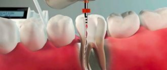
At the next appointment, the doctor removes the pulp. Large, curved canals are carefully cleaned with special instruments. This procedure is called nerve ablation. Next, the empty canals are disinfected and filled with filling material. It is necessary to fill them completely, otherwise there is a risk of re-inflammation; for this, the doctor measures the length of the canal with apex locator electrodes. They are lowered into each channel, since their length can be different. The obtained data is compared with an x-ray.
The canal is treated, disinfected, and dried. Afterwards, the doctor fills it with special material. Our clinic uses the method of three-dimensional obturation of root canals with thermally modified gutta-percha. Gutta-percha pins are placed in the canals so that the sealer does not reach the upper hole by a few millimeters. Part of the pin is heated and removed. The dentist can monitor the process using x-rays.
When the canals are filled, the dentist again places a temporary filling. It is necessary for the gutta-percha to completely dry out and remove all moisture from it. This takes 4-5 days, after which you can install a permanent filling and restore the tooth.
The main disadvantage of devital extirpation is the duration of treatment, consisting of several visits to the doctor’s office:
- The first step is to open the pulp, remove carious lesions and apply a devitalizing paste.
- The second visit is to remove the nerve, clean the canals and fill them with gutta-percha.
- The final stage is filling and restoration of the tooth.
Prevention of pulpitis in children
Treatment of pulpitis in permanent teeth in children, as well as their “predecessors” - baby teeth - is a rather complex procedure that may require several visits to the doctor. Preventing a disease is easier than treating it - all you need to do is pay attention to hygiene procedures and teach your child how to brush their teeth correctly from a very early age.
It is also important to monitor the child’s diet - strengthen the enamel with solid foods (carrots, apples, etc.), provide a balanced diet to saturate bone tissue with minerals. It is better to limit sweets and give only water at night. Prevention of caries is the basis for preventing its complications, including pulpitis.
What can happen if pulpitis is not treated?
Since in some cases the disease occurs without pain, many people put off going to the dentist until the last minute. Do not forget that while you are inactive, the disease progresses. Refusal to treat pulpitis can cause serious complications:
- Flux
- periostitis, a pathological inflammatory process developing from the periosteum.
- Periodontitis
- inflammation of the tissue around the root of the tooth with the possible formation of purulent pockets and destruction of the periapical bone tissue.
- Pulp gangrene
(necrosis) is the death of cells in the internal tissue of the tooth.
- Sepsis
- blood poisoning that develops when microorganisms enter the general bloodstream (usually occurs with reduced immunity).
To avoid such dangerous complications, you need to visit the dentist once every six months. Patients at risk should be examined more frequently (once every three months). The risk group includes patients with diabetes, oncology and other diseases that reduce immunity. If you notice symptoms of caries, go to the doctor immediately.
Complications
In the absence of adequate treatment, pulp inflammation spreads beyond the boundaries of the root canal, to the periodontium and periosteum. In addition to the formation of a purulent focus (abscess) that opens onto the mucous membranes of the oral cavity next to the affected tooth, there is a possibility of developing more serious complications:
- flux or inflammation of the periosteum, which leads to the loss of a diseased tooth, and sometimes healthy teeth located nearby;
- osteomyelitis or purulent inflammation of the bone, which requires complex, expensive therapy;
- phlegmon - lesions of the soft tissues of the face, their “melting” with purulent exudate.
Each of these complications can, with a certain degree of probability, lead to life-threatening conditions, for example, sepsis.
What might you encounter after treatment?
Some patients report discomfort after filling. It is important to distinguish whether this pain is correct (post-filling) or one that requires immediate consultation with a doctor.
Mild pain is almost normal after pulpitis treatment. This is due to the fact that a serious intervention was performed, especially if inflammatory and pathological processes had previously occurred. Temporary pain occurs during the first couple of days and occurs when chewing. There is no need to go to the dentist, wait a while, the pain will go away.
The pain is sharp, intensifying and occurring throughout the week may indicate the continuation of the inflammatory process. Swelling of the gums indicates the same thing. In this case, you need to visit your dentist.
Where to contact?
The “Good Hands” dental clinic has everything necessary for successful treatment of purulent pulpitis. We employ highly qualified specialists who constantly improve their skills. They have modern equipment and effective drugs at their disposal. Treatment is carried out using gentle techniques and high-quality anesthetics, which makes it as comfortable as possible for our patients. You can schedule a free consultation with us and find out all the necessary information right now by filling out an application on our website or calling us by phone.