Problem: parents with a 12-year-old teenager contacted the Dial-Dent orthodontic department. The boy's fangs stick out strongly, and his other teeth grow crooked. The parents wanted advice on how to correct protruding fangs, what the treatment consisted of, and how long it would last.
Solution: orthodontist O.A. Baranova performed treatment with Damon braces, aimed at normalizing the position of the fangs, straightening the teeth and correcting the bite.
Content
- Features of fangs
- What to choose?
- Which implantation to choose?
- How is implantation performed?
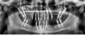
- Perform triple implantation
A canine is a single-rooted tooth used for grasping, holding and tearing food. Humans have a pair of fangs on the upper and lower jaws. They are called "triples" because they are considered third after the two incisors. These teeth are tall and cone-shaped.
PROMOTION
Osstem dental implantation turnkey
18,000 rub.
Is it possible to put braces on fangs?
Dystopia can and should be corrected for both a five-year-old child and an adult. The question of how this can be done will be decided based on the complexity of the anomaly formed, the degree of its neglect and, of course, the age of the patient.
If the reason for the lack of space is an erupted wisdom tooth, you will have to get rid of it to get the necessary free space in the jaw. You can install metal or sapphire braces, which will move the canine to the place of the least valuable, previously removed first premolar. Finally, surgery may be performed to reposition the displaced eye tooth.
Even the best braces in Moscow will not make the correction process quick. The easiest way to return the fangs to the correct position is for 14-15 year olds. How long do adults need to wear braces ? At least 1.5–3 years - it all depends on the type of orthodontic structure, the material of the clasps and the patient’s dental characteristics.
What to choose?
If in the case of other teeth in the dentition, you can always discuss prosthetics or implantation, then when it comes to restoring the troika, the answer is almost clear - only implantation! Why is that? Due to the shape, size and functionality of the canine, it undergoes the greatest stress during the process of biting and chewing food. Therefore, it is important to ensure reliable and sustainable results. In this case, bridge prosthetics is not suitable due to the fact that the canine is located at the intersection of two zones - the frontal and chewing. Here the jaw arch bends, which means that even the strongest bridge will fail very quickly. In addition, the use of a bridge in this area of the jaw poses serious risks to the supporting teeth.
Unilateral canine agenesis: clinical case and literature review
Dental agenesis or hypodontia is one of the most common anomalies of the human dental system, which is characterized by the initial absence of one or more teeth. This condition can occur in association with a certain genetic syndrome or separately (non-syndromic). The absence of one or more permanent teeth without systemic pathology is the most common phenotype of hypodontia.
Both external conditions and genetic factors can cause disruption of the development of the tooth germ, although hypodontia mainly occurs on the basis of the genetic aspect.
Recent reports have shown that in the Caucasian population, the incidence of hypodontia in permanent teeth (excluding third molars) is in the region of 4.5-7.4%.
In syndromic oligodontia, permanent canines are often missing. Congenital absence of permanent canines in non-syndromic patients is described sporadically in severe hypodontia and oligodontia, while isolated cases of absence of permanent upper canines are quite rare. Previous studies have revealed the approximate frequency of such pathology – 0.07 and 0.13%.
This clinical case demonstrates a unilateral absence of a permanent upper canine tooth. A review of the literature on the frequency, etiology and diagnosis of pathology was also carried out.
Clinical case
In October 2013, an 18-year-old woman came to the Hacettepe University Clinic for a regular dental examination. The general health condition was satisfactory, no systemic pathologies were identified. Intraoral examination revealed a class I relationship between the molars, a correctly positioned central line and a slightly increased vertical overlap. Between 22 and 23 there was a 2 mm trema. Clinical examination of the oral cavity revealed a small canine with signs of wear on the right side, the tooth was immobile (Photo 1). There is a small carious lesion on the distal surface of the tooth. To detect the permanent canine, palpation of the transitional fold and palatal surface of the alveolar process was performed. Palpation is ineffective.
Figure 1: Intraoral view of the upper dental arch showing a small canine with signs of wear.
An OPTG was performed to identify the location of the permanent canine and other possible anomalies. The x-ray showed that the permanent canine bud was absent, and that in the dental arch there was a primary canine with a characteristic short root, a small crown and a thin enamel structure. The primary canine had grade 1 root resorption, and all third molar buds were also absent (Photo 2). According to the patient, no teeth were removed and there was no history of trauma. The family history is also without any peculiarities or similar pathologies. No other dental anomalies were identified. However, in the area of the left third molar of the mandible, the radiograph revealed an irregularly shaped area of radiopacity (Photo 2). The lesion was diagnosed as osteosclerosis on a targeted photograph (Figure 3).
Figure 2: OPTG showing the absence of the right permanent canine and all third molars. A primary canine with signs of root resorption is located in the dental arch. Pay attention to the radiopaque lesion in the area of the left third molar of the mandible
Figure 3: Spot X-ray of the left postmolar area showing the area of osteosclerosis
The patient was sent to the Department of Therapeutic Dentistry for treatment of a carious tooth.
Discussion
Many studies have examined developmental disorders and explained these conditions using anatomical and evolutionary models. According to Bolk's theory of terminal reduction, due to the phylogenetic evolution of the human species, reduction of distal elements of the dental arch is much more common than mesially positioned teeth: thus, the most commonly missing second premolars, upper lateral incisors and third molars. In adapting Burler's concept to human occlusion, Dahlberg defined the canine as a key tooth that sits on its own in a specific location and has great stability and is therefore rarely congenitally absent.
The causes of congenital absence of teeth are very different. Severe hypodontia is usually associated with genetic disorders such as Witkop syndrome, ectodermal dysplasia, and Rieger syndrome. Mild and moderate hypodontia can occur due to improper formation of the bud, various injuries, Down syndrome and syndromes associated with cleft lip and palate.
The etiology of dental agenesis is subject to active debate. Graber states that congenital absence of teeth is an inherited phenomenon and is possibly transmitted to other generations as an autosomal dominant trait with incomplete penetration and variable expression. Brook proposed a multifactorial theory of hypodontia with a combination of polygenic and external influences. The role of genetics has been confirmed, but there is controversy on the subject of environmental influences due to a study on monozygotic twins: Markovic studied cases of hypodontia in twins and identified one pair in which unilateral canine absence had the appearance of a mirror image, which spoke primarily of a genetic basis for the phenomenon hypodontia. However, Kindlan demonstrated that genetic coding is not the only controlling factor in dental agenesis in twins, as it revealed differences in facial features and the extent of hypodontia. It has recently been suggested that isolated hypodontia of permanent canines is associated with mutations in the WNT10A gene.
The absence of a permanent canine rudiment is more common in women and mainly affects the upper jaw on the left. Statistics on agenesis of the upper canines vary from study to study. Harris and Clark found only two cases of congenital absence of the upper canines among 600 black American citizens (0.4%), while they could not find a single case among 1,100 whites. Cho described the absence of permanent canine rudiments in 32 cases out of 69,852 Chinese children. Fakuta Found only 42 cases with a frequency of 0.13% among 35,927 studied. Davis also reported five similar cases among 1093 Chinese children (0.46%). In European studies, Fekonja reported one case of agenesis of the upper permanent canine among 212 patients who received orthodontic treatment (2.1%), while Rozsa found an incidence of 0.27% in his study. Sisman showed a rate of 0.37% in an analysis of Turkish orthodontic patients.
It has already been said that the absence of one canine is more common than multiple canine agenesis, and basically this anomaly is combined with congenital agenesis of other teeth, microdontia, delayed tooth development and eruption, supernumerary teeth, odontomoma, taurodontia and additional cusps of teeth. In our clinical case, no additional anomalies of the dentofacial system were identified.
If clinically permanent canine teeth are not found in the oral cavity, several other conditions should be considered. For example, if palpation on the buccal side is ineffective in an 11-year-old child, it is possible to assume ectopic eruption or impaction of the tooth. Bone diseases, cysts, tumors can cause ectopic eruption and delay this process. In a rare situation, transposition with first premolars or lateral incisors is possible. Migration of the upper canine past the midline is known as transmigration, an uncommon anomaly that may occur when the canine is absent from the mouth on examination. Early diagnosis of permanent canine agenesis helps to carry out timely rehabilitation, reduce treatment time, complexity and avoid complications. Thus, if a canine tooth is absent in the oral cavity during a clinical examination, it is necessary to conduct an X-ray examination to clarify the condition and identify associated anomalies.
Congenital absence of upper permanent canines requires a specific treatment plan. Many factors must be taken into account: the condition of the primary teeth, the patient's occlusion (crowding or tremors, midline shift), type of facial growth and patient preference. Treatment may include removal of primary teeth to facilitate spontaneous or orthodontic closure of the site, or retention of primary teeth until growth is complete to preserve alveolar bone and create a favorable situation for implant placement without bone grafting.
In this case report, the main goal was to preserve the temporary canine. It was decided to observe the patient, since tooth root resorption had already begun. In the near future, the girl will need orthopedic rehabilitation.
Authors: Nagihan Koc, L. Berna CaLJrankaya, Nursel Akkaya
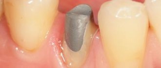
How is canine implantation performed?
- Diagnostic stage – the condition of the bone tissue, the general condition of the body is assessed, and the absence of contraindications is checked.
- Implant installation is performed under local anesthesia. The doctor opens the area of mucous membrane above the fang, removes the bone flap, forms a bed and inserts a titanium implant into it.
- The wound is sutured.
- After a week, the stitches are removed.
- Within 3-5 months, the implant takes root without load (a temporary prosthesis is not installed).
- After making sure that the implant has fused well with the bone, the doctor opens the mucosa above the top of the implant (local anesthesia is used).
- An individual abutment is made or a universal adapter is used.
- A gum former is installed, since the canine is located in a visually open area of the row.
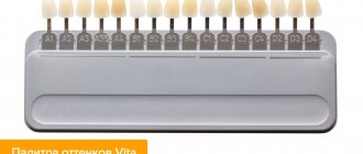
- Impressions are taken from the abutment, a crown is made, which will exactly repeat the canine in shape and match the color of the neighboring teeth.
Braces and wisdom teeth removal
On the Internet you can find a lot of information that before orthodontic treatment it is imperative to eliminate the eighth teeth. However, this statement is not entirely correct: “eights” are not always disposed of, but only in cases of extreme necessity. As a preventative measure, removing third molars is incorrect and even dangerous. Correctly erupted “eights” are the same part of the dentofacial apparatus as other teeth, provided, of course, that they have antagonists. Moreover, extracting the “eight” is not such an easy process.
Interesting fact!
In cases where there is clearly not enough space in the bone for the correct placement of teeth, specialists can remove the “eights” without waiting for them to erupt.
Nevertheless, when a patient is indicated for removal, experts pay attention to the wisdom teeth first - and here's why.
- They erupt later than others, between 16 and 27 years, which often negates the results of treatment carried out in adolescence.
- The process of their eruption is often associated with various complications (in about 75% of cases) - and this also affects the location of other teeth.
- “Wise” teeth sometimes remain impacted and do not participate in the bite, which makes them an unnecessary vestigial organ and a prime candidate for removal. However, there are situations when the patient simply does not have third molars or their extraction is not enough to correct the anomaly - in this case, it is necessary to resort to the removal of other teeth.
The second stage of transposition treatment is moving the teeth into place.
Next, it was necessary to move the crown of the first premolar 1.4 from the palatal position and the reverse overlap to its rightful place. This required the separation of the bite with onlays. And, of course, the long and labor-intensive stage of normalizing the position of the tooth roots began. It is at this stage of treatment that the canine root is partially located outside the alveolar process of the upper jaw and, accordingly, the risk of gum recession is high. After a year and a half (!) of treatment, we managed to move the crowns and roots of the teeth to the correct position. And the third stage of treatment began
Tooth extraction before installing braces and treatment time
In most cases, contrary to popular belief, extraction does not increase the treatment time, but, on the contrary, shortens it. For example, if the patient has severe crowding, then extraction will speed up the process of moving teeth by at least a couple of months, if not more. However, if the situation allows you to do without removal, then the fate of the tooth depends on the patient’s decision. In addition, you should not be afraid that after extraction there will be gaps between the teeth - they will close during orthodontic treatment. Usually it takes from three months to six months to eliminate them, depending on which tooth was removed. The gaps close at a rate of approximately 1 mm per month, so the smaller the tooth removed, the faster they will heal. As mentioned earlier, the smallest holes remain after removing the “fours”. Extraction is usually performed a week before braces are installed.
Important!
Removing an inflamed tooth during orthodontic treatment can negatively affect the timing of correction, therefore, before installing braces, the oral cavity must be completely sanitized.
Eye teeth in teenagers
The permanent lower eye teeth are formed at the age of 9-10 years. The upper fangs appear after 1-2 years.
In a healthy child, the formation of permanent eye teeth is painless. If this process is accompanied by pain, then this indicates a serious problem. This usually happens if diseases of the oral cavity occur, such as gumboil, pulpitis, periodontitis or other inflammatory processes. In such a situation, you should not try to get rid of the problem yourself. The most you can do is take a painkiller and immediately go to the dentist.