At the end of the last century, malignant tumors of the oral cavity were considered a concomitant disease of wastrels due to the abuse of tobacco and alcohol. Today, experts also include the human papilloma virus among the causes of cancer in this localization, so the frequency of all head and neck tumors has increased significantly.
- What precedes cancer
- When should a malignant process be suspected?
- Stages of lip cancer
- How to treat
Head and neck cancer ranks sixth in the world among all malignant tumors and will soon be in the TOP-5, but, unlike its “brothers” in localization, the detection rate of lip cancer is not increasing, but is steadily decreasing.
The incidence of lip cancer has decreased by more than 16% over the past 5 years. In 2022, carcinoma was detected in 2,235 Russians, with men getting sick 2.7 times more often.
- Out of 100 thousand people, 29 Russians develop the disease, and at a more mature age than a decade ago.
- The average age of affected women is 75.5 years; in them, the tumor is often localized on the upper lip.
- In men, cancer is diagnosed on average 8 years earlier, in most cases on the lower lip.
- In 87.6%, the disease is detected at stages 1-2, stage 3 with metastases to the lymph nodes is diagnosed in 8%, and stage 4 in 4.6%.
Etiology of neoplasms
External factors remain the main reasons for the development of lip cancer in patients of all age groups. The pathological process develops against the background of regularly repeated mechanical, chemical, temperature and meteorological influences.
The most common mechanical injuries are damage to the red border of the lips during shaving, systematic removal of keratinized epithelium (biting with teeth or tearing off with fingers). Often the cause of damage to the lips is cuts that appear due to low-quality dentures or sharp edges of damaged teeth.
Oncologists include smoking and drinking excessively hot food and drinks as dangerous thermal effects. Regular interactions with carcinogens during the patient’s professional activities are included in the group of chemical factors. Dangerous weather conditions include excessive amounts of ultraviolet radiation, high humidity and low air temperatures. In 10–15% of cases, cancer of the lower or upper lips develops against the background of human infection with the herpes simplex virus type 1.
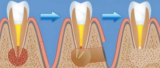
Venous lake removal
Removal of a venous lake can be carried out in several ways:
- Removing a venous lake with a scalpel threatens bleeding during the procedure;
- Cryosurgery and excision of the venous lake with an electrocoagulator are highly traumatic and have a high probability of destruction of adjacent healthy tissue;
- Laser removal of venous lake should be considered the most effective.
Rice. 3. Stages of laser removal of venous lake on the lip
Classification of pathology
The type of malignant neoplasms under consideration is represented by two types. The classification of lip cancer involves the separation of keratinizing and non-keratinizing tumors. The first type of neoplasm accounts for up to 95% of clinically registered cases of pathology. Cancer of this type has a favorable course - slow growth, moderate invasion of adjacent tissues, and rare metastasis.
Non-keratinizing lesions of the red border of the lips are more aggressive. The tumor grows rapidly, affects adjacent tissues, and forms numerous foci of metastasis. Lymphogenic metastases develop in the submandibular and mental lymph nodes, hematogenous - in the lungs. Secondary tumors of the first type are detected in 8% of patients, of the second - in 2%.
Classification
To classify the disease, the international TNM system is used, where:
- T (tumor) stands for tumor;
- N (nodulus) are lymph nodes that are affected by a tumor;
- M (metastasis) – metastases outside the lymph nodes.
T symbol gradation:
- Tx – there is no data to evaluate the primary neoplasm;
- Тis – preinvasive carcinoma (invasion of the lamina propria or intraepithelial invasion);
- T1 – tumor size up to 2 cm in greatest dimension;
- T2 – tumor size in greatest dimension is 2-4 cm;
- T3 – neoplasm in greatest dimension – more than 4 cm;
- T4a – the neoplasm spreads to neighboring tissues and organs (inferior alveolar nerve, skin of the nose or chin, floor of the mouth, etc.);
- T4b – the neoplasm affects the pterygoid processes, the masticatory zone, the base of the skull, and the carotid artery.
Gradation of the N symbol, which indicates metastases (or lack thereof) in regional lymph nodes (L/N):
- NХ – there is no data for assessing regional l / y;
- N0 – regional lymph nodes are not affected;
- N1 – in one lymph node on the affected side there are metastases up to 3 cm in size in the greatest dimension;
- N2 – in one lymph node on the affected side there are metastases 3-6 cm in greatest dimension; in several lymph nodes on the affected side in the greatest dimension there are metastases up to 6 cm; contralateral or bilateral involvement of lymph nodes with metastases up to 6 cm in greatest dimension;
- N2a – in one lymph node on the affected side there are metastases 3-6 cm in greatest dimension;
- N2b – on the affected side there are metastases in several lymph nodes up to 6 cm in the greatest dimension;
- N2c – contralateral or bilateral involvement of lymph node metastases up to 6 cm in greatest dimension;
- N3 – there are metastases in the lymph nodes more than 6 cm in greatest dimension.
M symbol gradation (distant metastases):
- M0 – no distant metastasis;
- M1 – distant metastases are present.
Stage according to TNM classification
| Stage | T | N | M |
| I | T1 | N0 | M0 |
| II | T2 | N0 | M0 |
| III | T3 | N0 | M0 |
| T1-T3 | N1 | M0 | |
| IV | T4 | N0 | M0 |
| Any T | N2,3 | M0 | |
| Any T | Any N | M1 |
Symptoms of pathology
The primary signs of lip cancer are lumps or ulcers on the surface of the anatomical area in question. Some patients complain of moderate itching, which intensifies at night or during meals. In the absence of treatment, the size of the primary lesion increases, and the soft tissues lose their original shape. Acute pain syndrome does not allow the patient to eat solid food. The upper and lower lips do not close together, and drooling appears. Cosmetic defects intensify, the tumor process affects the mucous membrane of the oral cavity, gums, cheeks and tissues of the lower jaw.
An external examination of the affected area allows the oncologist to see a dense formation with a grayish-brown surface covered with ulcers and cracks. In the later stages of lip cancer, signs of weeping appear and the tumor begins to bleed. Several seals can be combined into one large unit. Invasion of the primary lesion into bone tissue often leads to destruction of the lower jaw.
How to treat
Recommendations for the treatment of lip cancer are based not on clinical studies, as is customary for the vast majority of malignant tumors, but on decades of practical experience. It so happens that, due to the rarity of the disease, no randomized clinical trials have been conducted anywhere in the world. The choice of treatment is determined by the size of the primary tumor and, of course, the expected cosmetic defect. Even a small lip tumor changes a person’s quality of life much more than all other cancers. It is too noticeable, as is the scar that remains after its removal.
When treating cancer, it is important that there are no malignant cells in the surgical wound, so the tumor must be retreated in all directions. The lip itself is small, so even with a tumor measuring 5 mm, a postoperative scar of several centimeters will remain. Only after treatment of very small and superficial tumors do minor defects remain. Unfortunately, it is impossible to treat the patient in such a way that there are no defects left at all, therefore, when choosing a treatment method, they are guided by the least functional deformation and the minimum undesirable aesthetic result.
In this situation, the determining factor will be the decision of the patient who chooses the treatment option that will result in the least severe psychological consequences for him. Surgical treatment is preferable based on results, but radiation therapy, if possible, will leave fewer “traces.” With a large and superficial tumor of the lower lip, for example, a good result is likely after radiation therapy, which cannot be achieved if the cancer grows in the jaw bone. In this situation, the treatment option would be surgery.
Squamous cell skin cancer is very responsive to chemotherapy, but how lip cancer will react to cytostatics in each specific case can only be assumed, since serious and reliable studies of the effectiveness of drug therapy in this localization have not been conducted. However, for large inoperable tumors and relapses after excision, combined chemoradiation treatment has a good effect. If there are metastases to the nearest lymph nodes, the question of radical removal of regional lymphatic collectors with subcutaneous fat is raised.
Treatment of cancer in this location is purely individual, because the reconstructive possibilities of restoring lost lip volume and microsurgical leveling of the postoperative defect tend to zero. However, in most cases the patient has every chance of recovery. In case of lip cancer, it is very important to get to a good oncologist surgeon in time.
Other materials:
Alternative medicine in cancer treatment
Cancer recurrence
New perspectives for personalized cancer therapy
Book a consultation 24 hours a day
+7+7+78
Diagnostic procedures
When the first signs of lip cancer are detected, the patient should contact an oncologist. The doctor will examine the affected area and palpate the lips, gums, cheeks, and regional lymph nodes. The condition of the outer covers of the red border of the lips will allow the oncologist to make a preliminary diagnosis. If signs of a malignant process are detected in the neoplasm, the patient receives a referral for an ultrasound examination of the lips, radiography of the lower jaw, and panoramic tomography. The oncologist will collect biomaterials (smear-imprint) from the tumor for laboratory tests. Histological analysis is performed after a biopsy of the primary cancer site.
If the patient has symptoms of lymphatic metastasis, the doctor takes a biopsy sample from the lymph nodes. The search for hematogenous metastases is performed using chest x-ray or ultrasound.
When should a malignant process be suspected?
A long-term pathological process on the skin of the lip without a tendency to cure with active therapeutic measures should always raise suspicion that it is of poor quality. If the “spot” does not respond to local treatment and increases in size, you should immediately consult an oncologist.
The appearance of ulcerations and bleeding, or any thickening where there used to be soft skin, should be alarming. These symptoms can be either single or in combination, but even with a single symptom, the lack of treatment result within 3 weeks can be regarded as a bad sign, which can only be “clarified” by a biopsy of the skin defect. And the sooner it is done, the better.
Therapeutic measures for a confirmed diagnosis
The treatment strategy for lip cancer is determined by the oncologist, taking into account the stage of development of the malignant neoplasm. A significant factor is the patient's chronic diseases and the presence or absence of a secondary infection.
Stage one lip cancer allows for surgery, during which doctors excise the affected areas. Sometimes oncologists refer patients for radiotherapy. The second stage of the pathological process forces doctors to pre-irradiate the tumor. After this, radical surgery is performed. Stage three cancer will require long-term radiotherapy. The affected areas are the red border of the lips and the lymph nodes affected by metastases. The remains of the tumor and lymph nodes are excised during surgery.
Stage four cancer requires more complex treatment. The patient is undergoing radiotherapy and chemotherapy. After this, surgeons perform a wide excision of the affected tissue. At the terminal stage of the pathological process, people suffering from a malignant tumor of the lips receive palliative treatment (courses of chemotherapy and radiotherapy).
What precedes cancer
The lips are muscles covered with tissue and skin, which is called the “red border”. The inner part, covered with the mucous membrane, anatomically belongs to the vestibule of the oral cavity, and tumors arising there are no longer considered labial. A malignant tumor of the lip can arise out of nowhere - practically out of nowhere, and since this disease is considered to belong to a not very socially prosperous population, cancer in this localization is often preceded by skin diseases of the red border of the lips. These pathological conditions are classified as precancerous, although not all of them become fertile ground for the development of a malignant process.
Precancerous processes are similar in appearance, but differ in cellular structure. Precancerous lips are characterized by hyperplasia - excessive cell growth, frequent cell division, however, within the strictly prescribed framework of nature, and not endlessly, as in a malignant process. Cells of irregular shape appear, prone to rapid keratinization, which is manifested by hyperkeratosis - scaly dry skin. All lip diseases are seriously treated surgically or with close-focus radiotherapy.
- Previously, Bowen's disease was classified as an obligate precancer - a condition that, if persisted for a long time, was highly likely to develop cancer. In reality, cancer develops at the site of this “sore” in approximately every sixth patient. Today the disease is already considered cancer in situ - stage 0 cancer. With Bowen's disease, a spot with small nodules and papillae, velvety or smooth, sometimes with superficial ulcers - erosions, lives and grows on the lip for a long time.
- Erythroplasia Keira, a bright red lump with clear contours that rises above the skin, is no less likely to become a cause for the development of lip cancer. Over time, the lump ulcerates and is also considered stage 0 cancer.
- Manganotti's abrasive cheilitis is a pathology in which most often polished erosions with raised edges appear in the center, become covered with crusts and even heal on their own, but certainly recur.
- In young men, a bulging area with white scales surrounded by inflamed tissue may appear on the lower lip - this is limited hyperkeratosis of the red border of the lips.
- The lip is also affected by a variety of leukoplakia, often with small warts, plaques and erosions, which threatens every fourth patient with lip cancer.
- Keratoacanthoma is a semicircular fat cyst covered with scales, with a depression in the center similar to a volcanic crater. The pathology affects the lower lip, as a rule, of rural men, and in the elderly it is single, and in the young it consists of several nodules.