Summary
Breakage of nickel-titanium instruments is a major problem in endodontic treatment. A study examining the failure of nickel-titanium files due to torsional loading found that the majority of such failures occurred at the tip of the tool within the last millimeter and in instruments that had a small taper and/or size. Consequently, the tip of small instruments is at higher risk of breakage due to torsional loading, and to prevent this, the use of torque-reducing motors is necessary to reduce apical pressure and prevent the instrument tip from becoming stuck in the root dentin. In contrast, flexural fracture occurs as a result of repeated subthreshold loading resulting in metal fatigue. Numerous studies examining the causes of overload and breakage of machine-assisted nickel-titanium files have confirmed that by pre-expanding the root canal using hand instruments and creating a glide path before using machine instruments, a significant reduction in the rate of breakage can be achieved. This fact highlights the importance of manually pre-expanding the root canal and creating a “glide path” to reduce the incidence of mechanical instrument failure. To pre-expand the root canal and create a “carpet path,” hand-held steel instruments are most often used. Unfortunately, they have a number of disadvantages due to their relative rigidity and the presence of an aggressive tip, which can lead to the formation of ledges or changes in the course of the root canal in curved and/or calcified canals. For this reason, a new PathFile™ (Dentsply Maillefer) set for glide path creation and root canal pre-expansion was recently introduced, consisting of three machine-made nickel-titanium instruments.
PathFile™ are the first instruments specifically designed and intended for the mechanical creation of carpet and pre-enlargement of the root canal. The combination of a low 2% taper, a square cross-section and 4 cutting edges makes these instruments highly flexible, durable and effective, enabling fast and safe treatment of even severely curved and/or calcified root canals. Preliminary scientific research and clinical trials have confirmed that PathFile™ instruments are highly effective in treating severely curved root canals, allowing the creation of a perfect “glide path” without altering the course of the root canal, even if the working length is incorrectly determined.
Reciprocating tip
In 1964, the Giromatic system was introduced for mechanical treatment of root canals. The system was developed to save time on canal processing and is currently represented by a contra-angle handpiece with special files resembling pulp extractors (Ripsi file) or having a screw shape (Hele file). The constant rotation in the tip is transformed into another - a quarter turn movement. In some tips, reciprocating movements in the vertical direction of 0.8 - 1 mm (Endo - Cursor) are carried out every quarter turn.
The poor clinical outcome of these techniques may have been due to the inadequate design of the endodontic instruments used. In the 1980s, these problems were overcome with the development of "Dynatrat" files, S-shaped files with a smooth tip without grooves (guide tip only). They were used to process the coronal 2/3 of the canal, and then the apical third was processed manually.
It should be noted that many works have appeared comparing manual and machine processing of canals. It was noted that during machine processing, especially in curved canals, the canal very often acquired an “hourglass” shape. In addition, the problem arose of clearing the channel from sawdust, and the constant threat of blocking the channel with it. Gaining time in preparation turned into losing time in cleaning the canal. The time required was comparable to or even greater than manual preparation. Losing the “channel sense” led to complications.
Introduction
The use of machine-made nickel-titanium instruments has radically changed the technique of mechanical root canal preparation and the prognosis in difficult clinical situations. Numerous “invitro”1−13 and “invivo”14−17 studies have shown that machine-made nickel-titanium instruments are superior to steel hand files in the quality of root canal formation and the possibility of their use in canals with pronounced curvature, reducing the risk of creating ledges or straightening the canal curvature . Schäfer16 conducted an in vivo study in which 110 root canals were treated with machine nickel-titanium instruments and 84 canals with hand instruments. All canals were processed by 8 experienced doctors. Before the start of treatment and after obturation of the canals, x-rays of each tooth were taken.
Straightening the curvature. Nickel-titanium machine tools.
Work time. Manual steel files.
Rice. 1. Probability of root canal straightening and working time after in vivo root canal debridement using nickel titanium machine tools and hand steel files (adapted from Schäfer et al.16).
Straightening of the root canal curvature was assessed using a computer image analysis program. Root canal preparation using machine-assisted nickel-titanium instruments significantly reduced working time and resulted in less straightening of the canal curvature compared to the use of hand instruments16 (Fig. 1). In another in vivo study, Sonntag et al17 compared the risk of complications in root canal preparation with hand-held steel instruments and machine-operated nickel-titanium instruments. All clinical cases were performed by students. The results demonstrated that even in the absence of sufficient experience on the part of the practitioner, the quality of root canal preparation using nickel-titanium instruments was higher than that using manual steel files, while the number of shoulders was significantly reduced and the integrity of the apical foramen was maintained.17 Unfortunately, the use machine-made nickel-titanium instruments have serious limitations as they are associated with an increased likelihood of file breakage within the canal compared with steel instruments.17,18 Suter et al. When assessing in vivo the possibility of successfully removing a broken instrument from a root canal, success rates are reported to be 87% of cases. Studies of removed instrument fragments confirm that machine-made nickel-titanium instruments break more frequently than hand-operated steel instruments.18 Sonntag et al.16 report that breakage of nickel-titanium instruments is a major problem in endodontic treatment. Studies evaluating the influence of various factors on the breakage of machine endodontic nickel-titanium instruments have demonstrated that file breakage occurs primarily under the influence of torsional load19−25 and instrument fatigue.21,23,26−28 Torsional load depends on the contact area of the instrument with root canal dentin, the taper and diameter of the instrument, the area of the instrument subjected to stress, the strength (cross-sectional shape) of the instrument, the design of the working part, and the torsional load applied to the instrument.27,28 A study on the failure of nickel-titanium files due to torsional load has shown that the majority of such failures occur at the tip of the tool within the last millimeter and for instruments that have a small taper and/or size.24,25,27,28 Consequently, the tip of small-sized instruments is at higher risk of failure due to torsional loading, and to prevent this, the use of torque-reducing motors is necessary to reduce apical pressure and prevent the instrument tip from becoming stuck in the root dentin.27,28 In contrast, flexural fracture occurs as a result of repeated subthreshold loading, resulting in metal fatigue.
The bending load depends on the radius of curvature and size of the root canal, the rotational speed and flexibility of the instrument, the characteristics of the nickel-titanium alloy, the presence of intracanal obstructions, and sudden changes in the course of the root canal (for example, in the case of fusion of canals or the presence of additional canals).27,28 Numerous Studies examining the causes of overloading and failure of machine-assisted nickel-titanium instruments have confirmed that a significant reduction in the incidence of machine instrumentation failure can be achieved by pre-enlarging the root canal using hand files and creating a “glide path” before using machine instruments. In a study of the effect of pre-enlargement of the root canal on the failure rate of 4% taper nickel-titanium machine instruments, Roland et al.29 concluded that “pre-enlargement of the root canal with hand files followed by the use of machine instruments allows the instruments to be used more times before failure than with the isolated use of the crown down technique recommended by the manufacturer.” Peters et al.,30 examining the physical performance of machined nickel-titanium ProTaper instruments in the treatment of curved maxillary molar canals “in vitro”, showed that “even when significant load was applied in some clinical cases, no ProTaper instrument failed in the presence of adequate "carpet path" Blum et al.31, after analyzing the mechanical preparation of extracted teeth using ProTaper machine instruments, determined that “particular attention in a precise root canal protocol should be given to the use of small, flexible hand-held steel files to ensure that each part of the root canal has sufficient space for unobstructed machine access.” tools in the process of further machining...". Berutti et al32 evaluated the effect of manual root canal pre-expansion and torque on failure rates using ProTaper machine instruments. In this study, the authors used 400 plastic training blocks divided into 2 groups. All blocks were processed with ProTaper instruments, but one group had the canal pre-dilated manually using hand instruments to ISO #20 before using machine files. The results of the study showed that after manually pre-expanding the root canal, ProTaper instruments were able to process significantly more plastic blocks before failure occurred32 (Figure 2).
Rice. 2.
Number of plastic training blocks processed to failure by ProTaper S1, without pre-expanding the canal to a #20 ISO file and with pre-expansion. Adapted from Berutti et al.32 No pre-expansion. Pre-extension to ISO file #20.
Finally, Varela et al33 examined the effect of manual canal pre-expansion on the failure rate of three different nickel-titanium machine instruments (ProFile, ProTaper and K3) when used in canals of extracted teeth with curvature greater than 30º. The authors demonstrated a significant reduction in the incidence of file breakage when performing preliminary manual root canal expansion before using machine instruments. In this study, there were no significant differences between the three types of instruments used.33 All of the above studies suggest that the beneficial effect of pre-manual reaming is to reduce the likelihood of the tip of the weakest instruments becoming stuck in the root canal.28−33 In addition, As an explanation for the reduction in machine instrument failure in curved root canals, not only the presence of a smooth “carpet path” to prevent dangerous deformation of the instrument tip must be taken into account, but also the reduction in bending load.28,30,32
Preliminary enlargement of the root canal and creation of a “carpet path” is most often carried out using manual steel files. Unfortunately, these instruments have a number of disadvantages due to their relative rigidity and the presence of an aggressive tip, which in curved and/or calcified canals can lead to the formation of a shoulder or change in the course of the root canal.34 For this reason, the new PathFile™ set was recently introduced (Dentsply Maillefer) for creating a “carpet path” and preliminary expansion of the root canal, consisting of three machine-made nickel-titanium instruments.
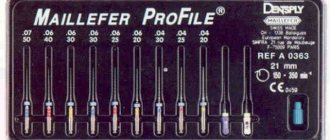
Rice. 3.
PathFile™ No. 1−3 (Dentsply Maillefer). PathFile™ No. 1 (0.13 mm). PathFile™ No. 2 (0.16 mm). PathFile™ No. 3 (0.19 mm).
LIST OF REFERENCES USED
- 1. Leontyev V.K., Pakhomov G.N. Prevention of dental diseases. – M., 2006. – 78 p. Leont'ev VK, Pahomov GN Profilaktika stomatologicheskih zabolevanij. – M., 2006. – 78 s.
- 2. Yanushevich O. O., Kuzmina E. M., Kuzmina I. N. Dental morbidity in the population of Russia. – M., 2009. – 78 p. Yanushevich OO, Kuz'mina EM, Kuz'mina IN Stomatologicheskaya zabolevaemost' naseleniya Rossii. – M., 2009. – 78 p.
- 3. Maksimovsky Yu. M., Mitronin A. V. Therapeutic dentistry. Cariesology and diseases of hard dental tissues. Endodontics: a guide to practical exercises: a textbook for students of institutions of higher professional education studying in the specialty 05/31/03 “Dentistry” / pod. total ed. Yu. M. Maksimovsky. – M.: GEOTAR-Media, 2016. Maksimovskij Yu. M., Mitronin AV Terapevticheskaya stomatologiya. Kariesologiya i zabolevaniya tverdyh tkanej zubov. Endodontiya: rukovodstvo k prakticheskim zanyatiyam: uchebnoe posobie dlya studentov uchrezhdenij vysshego professional'nogo obrazovaniya, obuchayushchihsya po special'nosti 05.31.03 “Stomatologiya” / pod. obshch. red. Yu. M. Maksimovskogo. – M.: GEOTAR-Media, 2016.
- 4. Qaed N., Mourshed B., Al-Shamiri H., Alaizari N., Alhamdah S. The Effect of surface topographical changes of two different surface treatments rotary instrument // Journal of Clinical and Experimental Dentistry. 2022. 0–0. – doi: 10.4317/jced.54472. BROKEN LINK
- 5. Arantes WB, Silva da CM, Lage-Marques JL, Habitante S, Rosa da LC, Medeiros de JM SEM analysis of defects and wear on NiTi rotary instruments // Scanning. 2014. No. 36. R. 411-418.
- 6. Pedulla E., Plotino G., Grande NM et al. Shaping ability of two nickel-titanium instruments activated by continuous rotation or adaptive motion: a micro-computed tomography study // Clin Oral Investig. 2016. No. 20. R. 2227-2233.
- 7. FKG Dentaire SA The XP-endo Finisher file Brochure. https://www. fkg.ch/sites/default/files/fkg_xp_endo_brochure_en_vb.pdf.
- 8. Debelian G., Trope M. Cleaning the third dimension // Endodontic Practice. 2015. August. P. 18-21.
- 9. Mitronin A.V., Korchagina M.A., Dzaurova M.A., Galieva D.T., Mitronin V.A. Evaluation of the effectiveness of using a rotary instrument with zero taper when removing the smear layer // Endodontics today. 2022. No. 4. pp. 8-12. Mitronin AV, Korchagina MA, Dzaurova MA, Galieva DT, Mitronin VA Estimate effektivnosti ispol'zovaniya rotacionnogo instrumenta s nulevoj konusnost'yu pri udalenii smazannogo sloya // Endodontiya today. 2017. No. 4. S. 8-12.
- 10. Rzhanov E. A., Kop’ev D. A. Method for assessing the probability of failure of a nickel-titanium tool depending on the duration of its operation in conditions of a curved channel. Experimental research. 2011. No. 2. pp. 66-72. Rzhanov EA, Kop'ev DA Metod ocenki veroyatnosti polomki nikel'- titanovogo instrumenta v zavisimosti ot prodolzhitel'nosti ego raboty v usloviyah iskrivlennogo kanala // Eksperimental'noe issledovanie. 2011. No. 2. pp. 66-72.
- 11. Manak T.N., Devyatnikova V.G. Assessment of physical and mechanical properties of ni-ti endodontic instruments // Dentist. Minsk. 2012. No. 3 (6). pp. 45-48. Manak TN, Devyatnikova VG Ocenka fiziko-mekhanicheskih svojstv ni-ti ehndodonticheskih instrumentov // Stomatolog. Minsk. 2012. No. 3 (6). S. 45-48.
- 12. Sattapan B., Nervo GJ, Palamara JE, Messer HH Defects in rotary nickel-titanium files after clinical use // Journal of Endodontics 2000. No. 26. R. 161-165.
- 13. Tripi TR, Bonaccorso A., Tripi V., Condorelli GG, Rapisarda E. Defects in GT rotary instruments after use: an SEM study // Journal of Endodontics. 2001. No. 27. R. 782-785.
- 14. Dmitrieva L. A., Mitronin A. V., Sobkina N. A., Pomeshchikova N. I. Efficiency of using self-adapting SAF files according to the results of laboratory studies // Endodontics today. 2013. No. 3. pp. 39-42. Dmitrieva LA, Mitronin AV, Sobkina NA, Pomeshchikova NI Effektivnost' ispol'zovaniya samoadaptiruyushchihsya fajlov SAF po rezul'tatam laboratornyh issledovanij // Endodontiya today. 2013. No. 3. S. 39-42.
- 15. Mitronin A.V., Gerasimova M.M. Endodontic treatment of pulp and periodontal diseases (Part 1). Aspects of the use of antibacterial drugs // Endodontics today 2012. No. 1. pp. 9-14. Mitronin AV, Gerasimova MM Endodonticheskoe lechenie boleznej pul'py i periodonta (ch1). Aspekty primeneniya antibakterial'nyh preparatov // Endodontiya today. 2012. No. 1. pp. 9-14.
- 16. Luzi A., Forner L., Almenar A., Llena C. Microstructure alterations of rotary files after multiple simulated operative procedures // Medicina Oral, Pathología Oral y Cirugía Bucal. 2010. No. 4. R. 658-662.
- 17. Ned Tijdschr Tandheelkd. Treatment of a fractured endodontic instrument in the root canal // EDITION????? 2015. Dec. No. 122 (12). 663-5.
- 18. Ramos Brito AC, Verner FS, Junqueira RB, Yamasaki MC, Eritas DQ Detection of Fractured Endodontic Instruments in Root Canals: Comparison between Different Digital Radiography Systems and Cone-beam Computed Tomography. J Endod. 2022. Apr. No. 43 (4). R. 544-549.
- 19. Hülsmann M., Rümmelin C., Schäfers F. Root canal cleanliness after preparation with different endodontic handpieces and hand instruments: a comparative SEM investigation. J Endod. 1997. No. 23 (5). R. 301-306.
- 20. Kim HC, Yum J., Hur B., Shun Pan GC Cyclic fatigue and fracture characteristics of ground and Twisted Nickel Titanium rotary files // Journal of Endodontics. 2010. No. 36. R. 147-152.
- 21. Larsen CM, Watanabe I., Glickman GN, He J. Cyclic fatigue analysis of a new generation of nickel titanium rotary instruments // Journal of Endodontics. 2009. No. 35. R. 401-403.
- 22. Lopes HP, Elias CN, Vieira MV et al. Fatigue Life of Reciproc and Mtwo instruments subjected to static and dynamic tests // Journal of Endodontics. 2013. No. 39. R. 693-696.
The article was published in the magazine “Endodontics today” Volume 17, 02/19
PathFile™ Tools Sequence
The sequence of using PathFile™ tools is very simple (Fig. 4):
1 – Primary navigation and examination of the root canal with a K-file No. 10, which should freely enter the canal to the working length. To speed up this stage, if necessary, use endolubricants containing EDTA.
2 – Determination of working length using an electronic apex locator and (or) radiography.
3 – Pass PathFile™ No. 1 (0.13 mm) to working length.
4 – Pass PathFile™ No. 2 (0.16 mm) to working length.
5 – Pass PathFile™ No. 3 (0.19 mm) to working length.
6 – You can then begin using the nickel titanium files using standard techniques (if using the ProTaper system, use file S1).
PathFile™ are used in a gentle reciprocating motion at 300 rpm, motor torque of approximately 5 Ncm until full working length is reached. Significant apical loading on instruments should be avoided. The use of relatively high motor torque is not harmful given the square cross-section of the tool and the results of a study by Berutti et al.38 which demonstrates that the use of high torque allows nickel-titanium machine tools to machine significantly more channels before failure.38 The time required to work with three PathFile™ files per working length does not exceed 3 – 5 seconds for each tool; increasing the working time is useless, but not dangerous, since PathFile™, thanks to their high flexibility, do not change the course of the canal even in the event of errors in determining the working length. Copious irrigation is recommended after each instrument, although PathFile™ coils do not become clogged with dentinal filings and these instruments do not cause filing blockage of the apical foramen.
Rice. 4.
Sequence of using PathFile™ tools. X-ray before treatment (4A). Determination of the working length of the palatal and medial buccal 1 canals (4D). Radiograph showing the master pin in the medial buccal 1 and medial buccal 2 (4E), and in the distal and palatal canals (4F). X-ray immediately after treatment (4G) and 1 year later (4H).