Types of chancre
Chancre is an ulceration that occurs 3-90 (on average 21) days from the moment of infection with syphilis, more often in the area of penetration of the pathogen into the patient’s body.
First, a pink spot (inflammation) forms on the skin, then a dense nodule appears in this place, after 7-10 days it becomes necrotic and turns into an ulcer or erosion. If the resulting chancre is not accompanied by itching or pain, then it is called syphiloma. Syphiloma has a regular round or oval shape and clear boundaries that protrude above the skin. The edges are dense, roll-shaped. The bottom of the syphiloma is red, less often – meat-colored, maybe red-brown, and the chancre itself looks varnished, due to the characteristics of the wound.
The size of syphiloma varies from 5 to 30 mm, but there are ulcers of larger or smaller diameter. The main symptom of chancre is a cartilaginous compaction at the base of the ulcer, which can be felt upon palpation. When pressed, the chancre is painless. The skin around the ulcer is clean, without signs of inflammation.
Chancre is formed due to the penetration of Treponema pallidum (spirochete) into the body. For reproduction, the temperature of the human body is suitable for it and after entering the body it actively forms syphilomas. The chancre themselves can be of different types, shapes and sizes.
Classification
By quantity
Single - chancre in the form of a separate neoplasm, or multiple, which looks like several ulcerations.
By origin
Twin chancres occur during simultaneous infection - when the pathogen enters the patient’s body not in one, but in several “places”. That is, instead of one chancre, two or more can be found.
Chancres formed as a result of infection at different times. They appear one after another, in approximately the same place.
The so-called “kissing” chancre, which occurs on contacting surfaces.
Concomitant diseases can contribute to the formation of multiple syphilomas:
- scabies
- acne
- skin injuries
By depth of penetration into tissue
Syphilomas have different depths of tissue damage.
Ulcerative lesions affect the deep layers of the skin. They can pass through the dermis - right down to the subcutaneous tissue. I have a rough bottom covered with purulent plaque. Erosion is located closer to the surface. The bottom is smooth, shiny, and shaped like a saucer.
To size
In addition to standard sizes from 5 to 10 mm, there are giant and dwarf chancres.
Giant chancres are extremely rare and can reach up to 20 cm in diameter. They are localized mainly in areas of accumulation of fatty tissue: on the abdomen, thighs, and pubis. Syphilomas are called dwarf and are the size of a poppy seed - 1-2 mm; they can only be seen through a magnifying glass. They are rare.
By location
Chancre can be located on different parts of the body:
- genital syphilomas are located on the genitals
- extragenital - on any other parts of the body outside the genitals (Fig. 1)
- bipolar syphilomas occur simultaneously both on the genitals and outside the perineum.
Figure 1. Chancre on the index finger of the left hand and in the area of the inner corner of the left eye. Source: CC0 Public Domain
By shape
In addition to the classic round shape, chancre can have a different appearance.
Slit-shaped syphilomas look like cracks. They can form in the corners of the mouth, on the tongue, near the anus. They are rare. Located in the corners of the mouth, they can be perceived by the owner as non-dangerous “jams”.
Cortical chancre is not like ordinary syphilomas; it is not concave in shape and is covered with a crust. It usually forms on ulcers that are located in places where their contents dry out easily: the surface of the nose, face, corners of the mouth.
Diphtheritic chancre is covered with an ash-gray film, similar to diphtheria. Happens quite often. It can be localized in any area.
The burn chancre quickly increases in diameter, while its borders lose their regular shape and clear outline, and the bottom changes from smooth to red-grained (Fig. 2). Typically, primary syphilomas do not tend to grow. This is an exception.
Figure 2. Burn syphiloma. Source: CC0 Public DomainCaption
Erosive chancre includes many erosions; ulcers can merge. Formed exclusively on the mucous membranes of the genitals.
Chancroid herpetiformis is named for its resemblance to genital herpes. This is an erosive formation, in the field of which there are many small ulcers with clearly defined edges. Similar to Folman's balanitis, but in this case the ulcers do not merge.
Signs and symptoms of syphilis in women during the primary period of the disease
Chancroid in women has its own characteristics, which depend on their location:
- erosive chancres are more often located in the area of the labia majora, indurative edema is less often recorded;
- chancre located in the area of the urethra always has a dense base;
- in chancre located in the area of the vulvovaginal fold, the compaction at the base is not pronounced;
- primary syphilomas, located on the cervix, are erosions of round shapes, with a flat bottom, clearly defined boundaries, intense red color, scanty serous discharge;
- when lymphatic capillaries are damaged and lymph outflow in the area of the clitoris and labia is impaired, unilateral indurative edema occurs, characterized by a significant increase in the organ, which becomes dense, dark red in color, often with a bluish tint. After pressing, there is no hole left. There is no pain. The swelling subsides slowly even with treatment.
- Primary vaginal syphiloma is extremely rare;
- in the area of the nipple in women, chancre is recorded in the form of erosions, usually single, with pronounced compaction at the base, covered with a crust;
- The crescent-shaped crack has a primary defect localized at the base of the nipple.
Rice. 12. Primary syphilis in women on the labia majora (photo on the left) and cervix (photo on the right).
Rice. 13. The first signs of syphilis in women are primary ulcers on the genitals.
Rice. 14. The photo shows an atypical form of chancre in a woman - indurative edema (photo on the left) and a primary defect in the nipple area (photo on the right).
The appearance of an erosive ulcer on the genitals or inducing swelling of the labia majora is the first sign of syphilis in women.
Localization
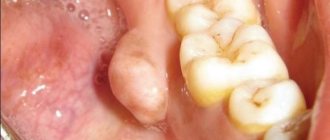
Since syphilis is transmitted primarily through sexual contact, chancroid is most often localized on the genitals. However, clinical practice shows that syphilomas are almost as often found in the mouth and anus.
This means that chancre can appear anywhere, the place of its localization can be:
- penis and scrotum
- labia and clitoris
- posterior commissure and anal area
- pubis
- oral cavity: lips, gums, tongue and throat
- inner thighs
- chest and stomach
- face
- rarely - eyelids, conjunctiva of the eyes
Chancre can be located inside the genital organs, for example, on the walls of the vagina or cervix, then syphiloma is difficult to detect.
Signs and symptoms of syphilis in men during the primary period of the disease
Hard chancre in men has its own characteristics, which depend on their location:
- small erosive chancres are often located on the head of the penis;
- ulcerative chancres are located in the head groove, they are large in size, and have a powerful infiltrate at the base;
- primary syphilomas, located on the frenulum of the penis, have an elongated shape, are easily injured and bleed during erection;
- in primary syphilomas located on the sides of the frenulum, there is no compaction at the base;
- in the region of the edge of the foreskin there are linear chancres;
- the seal at the base of the chancre, located in the area of the inner layer of the foreskin, looks like a visor;
- the hard chancre located on the crown of the glans penis resembles a swallow's nest;
- when located in the urethra, chancres have a dense consistency, are painful on palpation, always bleed, urination is also painful, scanty serous-bloody discharge is noted, in some cases, when cured, a cicatricial narrowing of the organ occurs;
- when the lymphatic capillaries are damaged and the outflow of lymph is impaired in the area of the scrotum and penis, indurative edema occurs, characterized by a sharp increase in the organ, which becomes dense; after pressing, no hole remains. There is no pain. The swelling subsides slowly even with treatment.
Rice. 10. Primary syphilomas in men - localization on the head of the penis (photo on the left) and in the area of the inner layer of the foreskin (photo on the right).
Rice. 11. Ulcerative chancre, located in the head groove, is large in size, has a powerful infiltrate at the base and is linear in direction.
Stages of development
Incubation period
The incubation period - from the moment of infection to the appearance of symptoms of the disease - lasts on average from 2 weeks to 2 months, although observations of longer duration are known. It all depends on the state of immunity at the time of infection. Long asymptomatic periods usually occur in people who were taking antibacterial drugs to treat other diseases at the time of infection.
During the incubation period, Treponema pallidum multiplies in the lymphatic system until it reaches a maximum concentration. They then enter the bloodstream and spread throughout the body. At this time, the disease does not manifest itself in any way and is not detected in blood tests, but the person is already infected. Without realizing it, he or she is putting the sexual partner at risk if they have unprotected sex.
Important! Treponema pallidum can reproduce in a small temperature range, around 37 °C. Therefore, to treat syphilis, the method of pyrotherapy is practiced - increasing body temperature. The patient is given drugs that increase body temperature, as a result of which treponema is deprived of the opportunity to reproduce. This method is considered the most effective for nonspecific treatment of syphilis.
Chancre formation
Figure 3. Development of syphiloma.
Source: CC0 Public Domain Once in soft tissue, treponema begins to actively multiply. During this period, a slight increase in temperature and an increase in nearby lymph nodes, which can be palpated, are possible. Inflammation develops at the site of infection - this is how immune cells try to destroy the enemy.
From the outside, the inflammation site looks like a bright pink patch of skin. Then a characteristic dense nodule forms on the skin - the rudiment of the future chancre (Fig. 3). It enlarges, thickens and after a week and a half ulcerates.
If the ulcer is covered with a crust, then the chancre is also called cortical. If you press on it, a yellowish liquid will come out, in which there is a high concentration of treponemes. This type of syphiloma is called “crying chancroid.”
Discharge from chancre is contagious, and contact with it can transmit infection. This danger is especially high when chancre is located in the mouth - there is a high risk of infecting a partner with syphilis even through a kiss.
The formation of chancre indicates the end of the incubation period and the onset of the primary stage of syphilis. Chancre does not bother the wearer with either itching or pain, which is bad - a person may simply not pay attention to it and waste time.
The primary stage is the most favorable for the destruction of the pathogen. During this period, a complete and rapid recovery is possible with timely administration of antibacterial therapy. In the future, coping with the disease will become more and more difficult.
Healing chancre
An ulcerated chancre lasts 6-7 weeks, then healing begins. Erosion can pass without a trace, but sometimes it leaves a dark pigment spot. When the ulcer heals, a scar remains. A few days before the chancre disappears, profuse itchy rashes may appear on the body.
At this stage, syphilis enters the secondary stage.
Important! Healing of chancre is often mistaken for recovery. This is wrong. In fact, the disease continues to develop, spreading throughout the body.
What is the difference between soft chancre and hard chancre?
There is a chancroid similar to hard - soft chancre (chancroid). It, like syphilis, is sexually transmitted, but occurs due to infection not by Treponema pallidum, but by bacteria of the genus Haemophilus influenzae.
Chancroid differs from hard chancroid in the following ways:
- there is no hard cartilaginous base (that’s why chancroid is called “soft”)
- the edges of the ulcer are not hard, but soft, spreading
- characterized by copious discharge of pus
- painful
- color hot pink, red
Complications of primary syphilis
When a secondary infection occurs, the clinical manifestations of chancroid may change. Inflammation of the glans penis (balanitis) and the inner layer of the foreskin (posthitis) may develop. Balanoposthitis causes the development of complications such as narrowing of the foreskin (phimosis) and pinching of the head of the penis by the ring of the foreskin (paraphimosis). In weakened individuals, gangrenization and phagedenism develop. Decreased immunity and poor hygiene contribute to the development of complications.
Complications in men develop when hard chancre is localized in the coronary sulcus or on the inner sheet of the foreskin.
- When a trichomonas or bacterial infection is attached, acute inflammation develops around primary syphilomas, which is complicated by narrowing of the foreskin, which makes it impossible to open the head of the penis. The penis enlarges, turns red and becomes painful. Forcible opening of the glans penis can lead to paraphimosis. Failure to provide medical care in a timely manner can lead to necrosis of the foreskin and tissues of the glans penis.
- When infected with B. fusiformis, gangrenization of chancre develops. A black scab forms on the surface of a hard ulcer, which, when sloughed off, reveals a deep ulcer. There is severe swelling and hyperemia of the surrounding tissues. The patient's general condition deteriorates sharply.
- The surrounding tissues are swollen and hyperemic. It's getting worse.
- As the disease progresses, the process spreads beyond the chancre. Bleeding occurs. As a result of necrosis, the head of the penis may be torn away, causing perforation and destruction of the urethra (phagedenism).
Rice. 19. In the photo, urethral chancre complicated by balanoposthitis.
Rice. 20. Complications of chancroid in men - phimosis.
Rice. 21. Complications of chancroid in men - paraphimosis.
Rice. 22. Complicated course of primary syphilomas in men.
Rice. 23. Development of gangrenous inflammation of primary syphiloma.
Diagnostics
Several basic methods are used to detect syphilis:
- Detection of treponema pallidum using a microscope in a scraping taken from a chancre.
- The serological method (Wassermann reaction) - when specific proteins that are produced by the immune system in response to the appearance of treponemes in the body are determined, is not effective for primary syphilis.
- The microprecipitation reaction is a rapid diagnosis, also based on the body’s production of antibodies.
- Specific tests RIF, RIT, RPGA, etc. – difficult to set up, time-consuming and expensive. Used to identify latent syphilis, in complex atypical cases, in differential diagnosis and for the diagnosis of late syphilis.
If treponema is detected immediately using microscopy, additional studies are not required to prove infection. Upon detection of treponema, treatment is prescribed immediately.
Sometimes it is necessary to carry out a whole range of diagnostic measures to establish an accurate diagnosis, so it is impossible to diagnose the disease yourself; you need to consult a doctor.
How chancroid develops
Primary syphiloma forms after the incubation period has passed: 3-4 weeks after contracting the infection. It occurs in places with skin lesions in which natural body fluid contaminated with bacteria has entered: sperm, secretion of the uterine cervix.
An ulcer does not appear immediately. Initially, a red spot appears on the infected area, which, under the influence of treponemas and cells of the immune system, thickens and turns into a nodule. The compaction is not accompanied by pain or discomfort, and therefore often goes unnoticed by the patient.
Over the next 7-10 days, the nodule develops: it increases in size, thickens and then ulcerates. Ulceration can be of two types: superficial, in the form of erosion, or deep, in the form of an ulcer. The ulcer or erosion takes on its final form: it acquires clear, pronounced boundaries, an even oval or round shape.
At the bottom of the manifested syphiloma, a liquid is released containing a large number of pale treponema and cells of the immune system. The bottom itself acquires a pronounced red tint with bluish notes.
This type of chancre persists for 1-2 months, after which the process of healing and tightening begins. This signals the transition of the disease to a secondary, more dangerous and severe stage.
3-4 days before the chancre disappears, multiple rashes appear on the patient’s body, often accompanied by burning and itching.
Treatment
It should be noted: in advanced cases, when it is not possible to get rid of chancre, as well as with extensive tissue necrosis, they resort to surgical removal of syphiloma.
In all other cases, the chancre itself is not treated and specific treatment for uncomplicated chancre is not carried out. For secondary and combined infections, topical antibacterial drugs may be prescribed: baths with benzylpenicillin and dimexide, applications with mercury or mercury-bismuth ointment. If the chancre is located in the oral cavity, rinsing is recommended: a solution of furatsilin, boric acid (2%), or gramicidin (2%).
The main task is to get rid of syphilis as quickly as possible and with minimal losses. Therefore, to treat syphilis, penicillin antibiotics are used - short and long-acting (durant) penicillins: Bicillin-1, Bicillin-5, Oxacillin, Ampicillin (semi-synthetic penicillin). The drug of choice for the treatment of syphilis is benzylpenicillin.
Reserve drugs for penicillin intolerance: tetracyclines (doxycycline), macrolides (azithromycin, erythromycin), cephalosporins (ceftriaxone).
Administration of drugs is by injection - intravenous or intramuscular.
Source: CC0 Public Domain
The treatment regimen depends on the stage of the disease, location, degree of damage, etc. In any case, the dose of the drug and the number of courses of treatment are calculated by the doctor individually.
During treatment, control tests are carried out to confirm the effectiveness of the drugs.
Treatment of syphilis at an early stage is the most effective and creates all the prerequisites for a complete cure without consequences and complications.
Recommendations for the treatment period
During the treatment period, it is necessary to stop sexual intercourse. When chancre is localized on the fingers, it is recommended to wear protective gloves. If syphiloma is found in the mouth, it is necessary to separate personal items from common ones - dishes, toothbrushes, etc.
If there are chancres on the body, the use of bed linen, towels, and washcloths should be individual. Syphilis is not transmitted through public places (toilet, etc.).
Sexual partners of sick people receive preventive treatment without fail, including pregnant and lactating women.
Important! You should definitely complete the full course of treatment! Under no circumstances will syphilis go away spontaneously. Untreated syphilis will move to the next stage, increasing the risk of complications and persistent deterioration of the patient’s condition.
Clinical serological control (CSC)
All family members of the sick person, both adults and children, need to receive preventive treatment after sexual or close household contact with patients with early forms of syphilis. 3 months after the end of preventive treatment, a single clinical and serological examination is carried out.
Clinical serological control (CSC) after the end of specific treatment of the patient is carried out once every 3 months during the first year of observation. Then once every 6 months in subsequent years with non-treponemal (simple serological) tests, once a year with the corresponding treponemal test (a complex test to identify possible latent forms of syphilis), which was used in diagnosing the disease. The duration of CSC is determined individually depending on the results of treatment.
Children born to seropositive mothers who did not have congenital syphilis, regardless of whether they received preventive treatment or not, are subject to observation for 1 year. Children receiving specific treatment are on CSC for 3 years.
Regional lymphadenitis, lymphangitis and polyscleradenitis
Regional lymphadenitis
Regional lymphadenitis (scleradenitis) is, after chancre, the second most important symptom of primary syphilis. Scleradenitis develops 6 - 7 days after the initial manifestation of syphilis - chancre and often on the affected side (unilateral localization).
The lymph nodes are painless, woody in density, mobile, and not fused with the surrounding tissues. Sometimes lymph nodes appear simultaneously with primary syphiloma. Several lymph nodes may enlarge at the same time, the largest of which is located closer to the chancroid. When a secondary infection occurs, the lymph nodes become fused into conglomerates, the phenomena of periadenitis are pronounced, and sometimes fistulas are formed. Syphilitic lymphadenitis resolves slowly.
Inguinal lymphadenitis is often bilateral, it develops when hard chancre is localized on the external genitalia, ulnar and axillary lymph nodes increase when syphilomas are localized on the hands, cervical and submandibular - when localized on the lower lip, preauricular and submandibular - when localized on the upper lip, submandibular , cervical and preauricular - when localized on the tonsils, sublingual - on the tongue, preauricular - on the skin of the eyelids, axillary - when primary syphiloma is localized on the skin of the mammary gland, when chancre is localized in the rectum and on the cervix, the lymph nodes located in the small pelvis.
Rice. 24. Regional lymphadenitis is, after chancre, the second most important symptom of primary syphilis. The photo shows inguinal lymphadenitis.
Syphilitic lymphangitis
Inflammation of the lymphatic vessels (lymphangitis) is the third main symptom of primary syphilis. The causative agents of syphilis multiply in the lymphatic system and the lymphatic vessels are the first to be affected in this process. Lymphangitis begins from the site of primary syphiloma and reaches nearby lymph nodes. The inflamed vessels thicken and become like a dense elastic painful cord. Lymphangiitis, like lymphadenitis, resolves slowly.
Rice. 25. The photo shows sclerosing lymphangitis.
Prevention
Since the mode of transmission of the disease is mainly sexual, preventive measures consist of maintaining fidelity to sexual partners - this is the most effective prevention of sexually transmitted diseases. When having sexual contact with an unverified person, you should always use a condom.
Barrier contraception (condom) provides almost 100% protection against syphilis infection.
In any case, after accidental contact, it is necessary to independently treat the genital area with antiseptic agents: miramistine or chlorhexidine.
If an unplanned contact occurs without protective equipment, or the integrity of the condom is damaged in the process, doctors recommend visiting a prenatal clinic as soon as possible and receiving a preventive injection, which will almost 100% prevent the development of syphilis.
Causes and course of the disease
The development of syphilis of the pharynx is possible at all 3 stages of the disease.
Primary lesions are more often in the nasopharynx than in the oropharynx. Moreover, infection can also occur through the use of infected medical instruments (iatrogenic factor of infection).
When the nasopharynx is affected, the most common manifestations of the disease are:
- Enlargement of the lymph nodes of the neck, which in some cases are very pronounced;
- Severe pain when swallowing, which radiates to the ear and is accompanied by congestion.
The difference between acne due to syphilis and chickenpox
Smallpox syphilide is most easily confused with chickenpox.
This disease is diagnosed mainly in childhood.
In adults it is more severe.
The difference is a pronounced intoxication syndrome.
It occurs 2-3 days before acne appears.
Severe hyperthermia, cephalalgia, myasthenia gravis, and sore throat are noted.
There are many foci of rashes, and they are located in groups.
While with syphilis there are few of them, and they are single.
Another feature of chickenpox is the extremely rapid transformation of morphological elements.
The entire cycle can take only 8 hours.
First, pink spots form.
From them papules are formed.
They turn into bubbles.
They usually contain completely clear liquid.
Unlike syphilis, in which the contents of the vesicles are often purulent.
With chickenpox, there is severe itching of the skin.
Publications in the media
Syphilis is an infectious disease caused by Treponema pallidum, transmitted primarily through sexual contact, with a chronic relapsing course and a characteristic periodicity of clinical symptoms, which can affect all organs and systems. Frequency . In Russia in 2001, the incidence was 143.6 per 100,000 population.
Classification • Primary syphilis •• Seronegative •• Seropositive • Secondary syphilis •• Fresh •• Recurrent •• Latent • Tertiary syphilis •• Active •• Latent • Latent syphilis •• Seropositive early •• Seropositive late • Congenital early syphilis •• Late • • Latent • Neurosyphilis • Visceral syphilis.
Etiology. The causative agent is pale treponema (Treponema pallidum), spiral-shaped, 4-14 microns long and 0.2-0.25 microns in diameter, has 8-12 uniform curls, can exist in three forms - spiral, cystic and L-form. The most common (classical) course of syphilis is due to the presence of a spiral form of the pathogen; other forms probably maintain a long latent course.
Epidemiology. The source of infection is a sick person. Conditions of infection: the presence in the material from the patient of a sufficient number of virulent Treponema pallidum and violation of the integrity of the skin and mucous membrane. The main route of infection is direct contact (usually sexual) with a sick person. With congenital syphilis, infection occurs in utero - through the vessels of the placenta. Treponema pallidums that have entered the body spread through the lymphatic system, actively multiply and enter various organs and tissues, which causes certain manifestations of the disease. Over time, the number of Treponema pallidum in the patient’s body decreases, but the tissue reaction to the pathogen becomes more violent. They admit the possibility of a long (many years) asymptomatic course of syphilis from the very beginning of the disease with the subsequent development of damage to the nervous system and visceral forms of the disease.
CLINICAL PICTURE. In untreated patients, acquired syphilis lasts for many years. In the classical course of the disease, there are 4 periods: incubation, primary, secondary, tertiary.
The incubation period lasts from the moment of infection until the appearance of the first clinical symptom - chancre (on average - 20-40 days). Sometimes it is reduced to 10–15 days with massive infection, which is accompanied by multiple or bipolar chancre, as well as with superinfection. An extension of the incubation period to 3–5 months is often observed with severe concomitant diseases or after treatment with low doses of antibiotics.
Primary period
• Continues from the moment of appearance of chancroid until the appearance of generalized rashes (6–7 weeks). It is characterized by the development at the site of penetration of pale treponema (usually in the genital area), chancre (ulсus durum, primary syphiloma) and regional lymphadenitis.
• Clinical signs of chancre •• Erosion (or ulcer) with the absence of acute inflammatory phenomena •• Regular round or oval outline, clear boundaries •• Size of a small coin •• Raised above the surrounding healthy skin (mucous membrane) •• Smooth, shiny, bluish-red bottom, flat (saucer-shaped) edges •• Scanty serous discharge •• Densely elastic (cartilaginous) infiltrate at the base, painlessness •• Chancre is often single, multiple chancre is rarely observed (in about 20% of patients) •• Atypical hard ones are also isolated chancre: indurative edema (painless dense swelling of the foreskin or labia), chancre-amygdalitis (dense swelling of the tonsil) and chancre-felon (simulates ordinary felon).
• Chancre can be complicated by a secondary infection: acute inflammatory reactions in the circumference of the chancre (if localized in the genital area - vulvitis or balanoposthitis, often leading to phimosis or paraphimosis). When complicated by fusospirillosis symbiosis, necrosis of the bottom and edges (gangrenization) of chancre occurs. Repeated gangrenization (phagedenism), usually observed in persons suffering from alcoholism, leads to significant tissue damage.
• Regional lymphadenitis (accompanying bubo, regional scleradenitis) is the second obligatory symptom of primary syphilis, occurs 7–10 days after the appearance of chancre •• Enlargement and hardening of the lymph nodes closest to the chancre •• When chancre is localized on the genitals, the inguinal lymphatics undergo changes nodes: they are enlarged to the size of a bean, sometimes larger, dense, not fused to each other and to the surrounding tissues, mobile, painless on palpation, the skin over them is not changed •• Characterized by enlargement of several lymph nodes. Scleradenitis can be bilateral or unilateral. Sometimes a strand of regional lymphangitis can be seen and palpated between the chancre and enlarged lymph nodes. •• At the end of the primary period, specific polyadenitis develops (moderate enlargement of all groups of lymph nodes), sometimes low-grade body temperature and general weakness are noted.
Secondary period
• Characterized by generalization of infection and lasts 3–4 years.
• All organs and systems can be affected, but the main manifestations are rashes on the skin and mucous membranes (secondary syphilides). The rashes of each of the attacks of the secondary period, having existed for about 1.5–2 months, undergo spontaneous regression, and after a more or less long latent period they appear again.
• The first rash, which marks the beginning of the secondary period, is particularly bright and abundant in the rash (secondary fresh syphilis). It is usually accompanied by fading chancroid and pronounced polyadenitis.
• Repeated episodes of rashes (secondary recurrent syphilis) alternate with periods of complete absence of manifestations (secondary latent syphilis). The rash with secondary recurrent syphilis is less abundant and tends to cluster. In the first half of the year, the rashes are accompanied by gradually fading polyadenitis.
• Secondary syphilides have a number of common features that distinguish them from other skin rashes: they are ubiquitous, have a benign course, there are no febrile symptoms, there are also no acute inflammatory phenomena and subjective sensations, they are resistant to local treatment, and quickly disappear under the influence of therapy. In the secondary period, 5 groups of syphilides are distinguished •• Syphilitic roseola (observed in 75–80% of patients): a pink spot 0.5–1 cm in size, irregularly rounded in outline, does not peel off, disappears with pressure •• Syphilitic papule (observed just as often , like roseola): a bluish-red nodule of dense consistency with peeling along the periphery (Biette's collar). Varieties of syphilitic papules ••• Lenticular (lenticular): size 0.3–0.5 cm ••• Miliary: the size of a poppy seed ••• Nummular (coin-shaped): the size of a large coin, tendency to cluster ••• Seborrheic: localized on the face, forehead skin and is distinguished by oily scales on the surface ••• Erosive (weeping): characterized by an erosive or weeping surface, localized on the mucous membrane or in the folds of the skin ••• Condylomas lata (vegetative papules) are located in places of skin friction (inguinal region), are distinguished by their large size, vegetation, erosive surface ••• Horny papules of the palms and soles are characterized by a powerful development of the stratum corneum on the surface, very reminiscent of calluses ••• Psoriasiform papules: pronounced peeling on the surface •• Syphilitic pustule (rarely observed): noted in weakened patients with a severe (malignant) course of the process. Clinical varieties: acne, smallpox, impetiginous, syphilitic ecthyma, syphilitic rupee •• Syphilitic baldness - rapidly developing small-focal or diffuse hair loss on the head without inflammatory changes in the skin, usually observed in the second half of the disease •• Syphilitic leukoderma (syphilide pigmentosa), more often noted in women, it is localized on the lateral and back surfaces of the neck (necklace of Venus), often on the skin of the torso; On the affected areas, against the background of hyperpigmentation, hypopigmented round spots 0.5–1 cm in size appear.
• The mucous membrane of the pharynx is often affected, where syphilitic tonsillitis occurs (erythematous, papular and pustular-ulcerative), differing from ordinary tonsillitis by sharp boundaries, the absence of acute inflammatory reactions, fever and pain. With rashes on the vocal cords, hoarseness of the voice is noted.
• With secondary syphilis, damage to internal organs, the central nervous system, bones, joints, organs of vision, hearing, etc. is possible. •• Bone damage is noted in 5% of patients in the form of diffuse periostitis with painful swelling, night pain in the bones; osteoperiostitis is observed less frequently. The bones of the skull and tibia are most often affected •• Joint damage usually occurs as a polyarthritic synovitis with the formation of effusion in the joint cavity •• The most important specific lesions of the internal organs of the secondary period include syphilitic hepatitis (enlarged and painful liver, increased body temperature, jaundice) , gastritis, nephrosonephritis, myocarditis •• Damage to the central nervous system in the secondary period is called early neurosyphilis. Damage to the meninges and blood vessels is typical. During a neurological examination, as well as during the analysis of cerebrospinal fluid, syphilitic meningitis (often asymptomatic), syphilis of the brain vessels (meningovascular syphilis), rarely - syphilitic neuritis and polyneuritis, neuralgia are diagnosed.
Tertiary period
• Develops in approximately 40% of patients in the 3rd–4th year of the disease and continues indefinitely. The transition of the disease to the tertiary period is facilitated by inadequate treatment or its absence, severe concomitant diseases, alcoholism, etc.
• Distinctive features of the tertiary period are the appearance of false inflammatory infiltrates in the form of tubercles and gummas, prone to decay, followed by extensive destructive changes in the affected organs and tissues; productive nature of inflammation with the formation of infectious granuloma; a small number of rashes (the number of tubercles is in the tens, gumma - in units); ubiquity of lesions, undulating course.
• In case of clinical manifestations, tertiary active syphilis is diagnosed; in the absence of such, tertiary latent syphilis is diagnosed.
• Relapses of tertiary lesions are observed infrequently, they are separated from each other by a long (sometimes many years) latent period. The lifespan of tertiary syphilides is calculated in months or years; they are characterized by low infectivity due to the small number of pale treponema in the tissues. Classic serological reactions are negative in 30% of patients with tertiary syphilis. The skin, mucous membrane and skeletal system are most often affected. Skin lesions are represented by tubercular and gummous syphilides.
• Tuberous syphilide is a small dense tubercle located in the thickness of the skin, hemispherical in shape the size of a cherry pit, bluish-red in color. After a few weeks or months, the tubercle softens and ulcerates to form a round, rather deep ulcer, with smooth, steep, dense edges. Gradually, the ulcer epithelializes and turns into an atrophic scar pigmented along the periphery, on which new rashes never appear.
• Gummous syphilide (gumma) is a node of dense consistency the size of a walnut, rising above the skin level, painless on palpation, not fused with the surrounding tissues. The skin over it is initially unchanged, then becomes bluish-red. Subsequently, the gummous node softens in the center and is opened with the release of a glue-like exudate. The resulting defect quickly increases in size and turns into an ulcer. It is painless, clearly demarcated from the surrounding skin by a ridge of dense, undisintegrated gummous infiltrate, its edges are steep, the bottom is covered with necrotic masses. A gummous ulcer lasts for months, and with secondary infection - even years. After the gumma heals, a characteristic star-shaped scar remains. In some cases, the contents of the gum are replaced by fibrous tissue with the formation of dense nodes. Gummas of the mucous membranes are often noted. Most often the membrane of the nasal cavity is affected, then the pharynx. Gummous lesions of the tongue, hard and soft palate, nose, pharynx, larynx lead to severe, often irreparable disturbances in speech, swallowing, breathing, and change the appearance of the patient (saddle nose, complete destruction of the nose, perforation of the hard palate). Among gummous lesions of other organs, syphilides of the periosteum, bones and joints are most often observed; The bones of the legs, forearms, skull, knee, elbow and ankle joints are affected.
Congenital syphilis is transmitted to the fetus of a sick mother through the placenta, mainly in the first 3 years after the mother is infected.
• Pregnancy outcomes with syphilis: late miscarriages, premature births, full-term births. Stillbirths are frequently reported. Children are born with active manifestations or latent syphilis.
• Depending on the duration of the syphilitic infection, periods of congenital syphilis are distinguished: fetal syphilis, early congenital syphilis (infant syphilis and early childhood syphilis are distinguished), late congenital syphilis (after 4 years) •• Fetal syphilis (fetal damage by syphilis occurs at fifth month of pregnancy) is accompanied by changes in internal organs, and somewhat later in the skeletal system. Specific lesions of the internal organs of the fetus are manifested by intercellular infiltration and proliferation of connective tissue. Widespread and severe lesions of the internal organs of the fetus often lead to late miscarriages and stillbirths •• Early congenital syphilis can first appear both in infancy (up to 12 months) and in early childhood (1–4 years) ••• Syphilis in infants most often manifests itself in the first 3 months of life and is very unique. In children born from untreated mothers with active manifestations of secondary syphilis, congenital syphilis is an extremely severe disease affecting almost all internal organs, the central nervous system and specific changes in the skin and mucous membranes ••• An early manifestation of syphilis in this period is syphilitic pemphigus. The rashes are localized mainly on the skin of the palms and soles, less often on other areas of the skin. Bubbles the size of a pea and a cherry are initially serous, then purulent, sometimes hemorrhagic, located on an infiltrated base and surrounded by a zone of specific papular infiltrate of a bluish-red color ••• Diffuse Hochsinger infiltration is usually localized on the soles, palms, face and scalp. The lesion is clearly demarcated, with a smooth shiny surface of bluish-red color; is distinguished by a densely elastic consistency, leading to the formation of cracks that have radial directions in the circumference of the mouth and leave lifelong radiant Robinson-Fournier scars ••• Widespread or limited roseolous, papular and pustular rashes in all their varieties, similar to those in the secondary period of syphilis, are also observed. Skin rashes are often preceded by an increase in body temperature ••• Damage to the mucous membranes often occurs in the form of a syphilitic runny nose: specific erosive-papular hyperplastic anterior rhinitis. Breathing through the nose becomes extremely difficult, which makes the act of sucking impossible. As a result of ulceration of the papular infiltrate of the nasal septum, its destruction with deformation of the nose is possible ••• Lesions of the skeletal system: lack of active movements in the limbs and pain in passive movements, caused by the formation of gummas in the epimetaphyses of long tubular bones with a tendency to fractures (Parro’s pseudoparalysis, Wegner’s disease) • •• Specific lesions of internal organs often begin in utero; their diffuse nature causes severe congenital syphilis and significant mortality in the first weeks and months of a child’s life. Liver damage is detected in the form of its enlargement and thickening. Rarely, jaundice is noted with atrophic cirrhosis of the liver. Damage to the spleen is often observed. Specific lung damage in the form of white pneumonia is rarely recorded; often occurs as a complication of pneumonia of various etiologies. Kidney damage (glomerulonephritis, nephritis, nephrosonephritis) is noted in 14% of patients. The cardiovascular system is affected early, but rarely (myocarditis, endocarditis, pericarditis) ••• One of the most common lesions of the central nervous system is specific meningitis with convulsions, paralysis and anisocoria. Increased cytosis, an increase in the amount of protein, and positive globulin reactions are noted in the cerebrospinal fluid. Some children develop hydrocele at 2–3 months of age. Among the sense organs in infants, the eyes are affected. Already at the birth of a child, it is possible to detect changes in the retina and macular membrane (chorioretinitis), damage to the optic nerve ••• Limited large-papular (usually weeping) rashes such as condylomas lata are often observed on the skin, erosive papules are often observed on the mucous membranes; bones are often affected (syphilitic periostitis of long tubular bones) •• Late congenital syphilis appears at the age of 5–17 years and resembles the tertiary period of syphilis in its clinical manifestations. Signs of late congenital syphilis are divided depending on the degree of specificity into absolute, or unconditional; relative, or probable (observed more often with late congenital syphilis, but also noted with other diseases); dystrophies (can be a consequence of both congenital syphilis and other diseases) ••• Absolute signs include Hutchinson’s triad: Hutchinson’s teeth (barrel-shaped or chisel-shaped incisors, hypoplasia of the chewing surface with a semilunar notch along the free edge), parenchymal keratitis (uniform milky white clouding of the cornea with photophobia, lacrimation and blepharospasm), labyrinthine deafness (inflammatory phenomena and hemorrhages in the inner ear in combination with dystrophy of the auditory nerve) ••• Relative signs are assessed in conjunction with other manifestations. These include: syphilitic chorioretinitis (characteristic of the “salt and pepper” pattern on the fundus), saber-shaped shins (the result of diffuse osteoperiostitis with reactive osteosclerosis and anterior curvature of the bones of the legs), Robinson-Fournier scars (in the circumference of the mouth after Hochsinger infiltrations), syphilitic gonitis , occurring as chronic allergic synovitis (distinguished by the absence of sharp pain, fever and dysfunction of the joint), buttock-shaped skull (sharply protruding frontal tubercles with a groove located between them), nasal deformities (saddle-shaped, lorgnette-shaped, etc.), dental dystrophy (tooth Muna - underdevelopment of the chewing tubercles of the first molars, Fournier's pike tooth - a change in the canine with thinning of its free end) ••• Dystrophies with congenital syphilis: Ausitidian sign (thickening of the sternal end of the clavicle), Olympic forehead (increase in the frontal and parietal tubercles), high (Gothic ) palate, infantile (shortened) little finger of Dubois-Hissar, absence of the xiphoid process of the sternum, widely spaced upper incisors, etc. Only the presence of several dystrophies in combination with other signs of syphilis and anamnesis data can, in unclear cases, help make a diagnosis of congenital syphilis.
Research methods • In the primary period: detection of pale treponema in the discharge from chancre • Serological reactions •• RSC (type of von Wasserman reaction) with cardiolipin and treponemal Ag and microprecipitation reaction with cardiolipin Ag (VDRL - with inactivated blood serum, RPR - with plasma blood, UJR - with active blood serum; ART - automated reagin test, etc.). Positive results are expressed by pluses (from + to ++++). In case of a sharply positive reaction, a study is carried out with various dilutions of serum (from 1:10 to 1:320). Reactions are positive from the middle of the primary period and during the secondary period; in the tertiary period they can be negative in 50% of patients •• Treponema pallidum immobilization test: carried out for false-positive serological reactions, assessed as negative when less than 20% of Treponema pallidum are immobilized, weakly positive - 21– 50%, positive - over 50% •• RIF: becomes positive in the majority of patients with syphilis already in the primary seronegative period; the results are assessed as positive (from + to ++++); positive in all periods of syphilis (including late forms) in almost all patients •• ELISA •• Passive hemagglutination test
TREATMENT • Specific treatment is prescribed after clinical and laboratory confirmation of the diagnosis (exception: preventive treatment of persons who have had sexual and close household contact with patients with early forms of syphilis, if no more than 2 months have passed since the contact) • Preventive treatment is carried out for women who received treatment before pregnancy , in whom by the beginning of pregnancy there was no complete negativity of the CSR (a complex of serological reactions, including various methods for detecting antibodies to the pathogen, for example, the von Wasserman reaction, microprecipitation reactions, less often others), as well as all women who began treatment during pregnancy, regardless of its term. And also to children born without manifestations of syphilis from an untreated mother; with late specific treatment for the mother (from 32 weeks of pregnancy); in the absence of negativity of CSR at the time of birth or seroresistance in the mother.
Drug therapy • Causal therapy: benzylpenicillin preparations are the mainstay in the treatment of all forms of syphilis. In case of intolerance: penicillin drugs, alternative drugs specified in the guidelines are prescribed (erythromycin, tetracycline, oleandomycin + tetracycline, doxycycline, cefazolin, etc.).
Treatment of primary syphilis: method No. 1 - two injections of benzathine benzylpenicillin at a dose of 2.4 million units with an interval of 7 days, or three injections of benzathine benzylpenicillin at a dose of 2.4 million units once every 5 days; Method No. 2 - benzathine benzylpenicillin + benzylpenicillin procaine + benzylpenicillin at a dose of 1.8 million units 2 times a week, a total of 5 injections, or benzathine benzylpenicillin + benzylpenicillin procaine at a dose of 1.5 million units 2 times a week, a total of 5 injections; Method No. 3 - penicillin procaine at a dose of 1.2 million units daily for 10 days or 600,000 units 2 times a day for 10 days; Method No. 4 - water-soluble penicillin intramuscularly every 6 hours for 10 days.
Treatment of secondary and early latent syphilis: method No. 1 - three injections of benzathine benzylpenicillin at a dose of 2.4 million units with an interval of 7 days, or six injections of benzathine benzylpenicillin at a dose of 2.4 million units once every 5 days; Method No. 2 - benzathine benzylpenicillin + benzylpenicillin procaine + benzylpenicillin at a dose of 1.8 million units 2 times a week, a total of 10 injections, or benzathine benzylpenicillin + benzylpenicillin procaine at a dose of 1.5 million units 2 times a week, a total of 10 injections; Method No. 3 - penicillin procaine at a dose of 1.2 million units daily for 20 days or 600,000 units 2 times a day for 20 days; Method No. 4 - water-soluble penicillin intramuscularly every 6 hours for 20 days.
Treatment of other forms of syphilis - see Methodological recommendations “Treatment and prevention of syphilis”, approved by the Ministry of Health of the Russian Federation (1999).
Contraindications. Tetracyclines - during pregnancy and children under 8 years of age.
Clinical and serological control after the end of treatment • After preventive treatment, once 3 months after treatment • For primary seronegative syphilis - 3 months • Patients with early forms of syphilis who had positive DSR results before treatment are subject to clinical and serological control until the DSR is completely negative and then another 6 months • For late forms, latent, visceral and neurosyphilis - 3 years.
Prevention • Public: free treatment, active detection, examination and involvement of contact persons in treatment, screening for syphilis of donors, pregnant women, all inpatients, workers of food enterprises and child care institutions • Individual: use of condoms.
ICD-10 • A50 Congenital syphilis • A51 Early syphilis • A52 Late syphilis • A53 Other and unspecified forms of syphilis
How to treat syphilis?
The treatment of syphilis is approached in a comprehensive manner, taking into account many individual factors (age, gender of the patient, stage of development of the disease, the presence of concomitant diseases, general condition of the body, etc.). In addition, all sexual partners of the suspected patient should also be examined for the presence of syphilis and, if necessary, undergo a course of therapy.
If a patient has primary syphilis, then everyone who has had sexual intercourse with him over the past three months must undergo examination and tests. In the case of secondary syphilis - everyone who had contact with the patient over the past year. The timeliness of the therapy itself, as well as the correct selection of modern medications, is important for achieving success in the treatment of this disease.
The most effective method of treating syphilis is the introduction of water-soluble penicillins into the body. This therapy is carried out in a hospital setting for 24 days with injections every 3 hours. The causative agent of syphilis is quite sensitive to penicillin antibiotics, but there is a possibility of an allergic reaction to these drugs or the ineffectiveness of such therapy. In this case, penicillin is replaced with drugs of the tetracycline, macrolide, and fluoroquinolone groups. In addition to antibiotics, natural immune stimulants, vitamins, and immunostimulants are also indicated for syphilis.
Syphilis is treated and not treated
Systematic treatment of syphilis currently gives good results, especially when it is started soon after the first signs of the disease appear. Timely and vigorous treatment of syphilis in the mother during pregnancy is the best way to prevent syphilis in the child. But advanced stages of the disease are incurable.
Treatment of syphilis, as well as gonorrhea, should only be carried out by a doctor. It must be remembered that a cure can be achieved only on the condition that the patient accurately follows every doctor’s prescription and carefully carries out the entire course of treatment.
ONLINE REGISTRATION at the DIANA clinic
You can sign up by calling the toll-free phone number 8-800-707-15-60 or filling out the contact form. In this case, we will contact you ourselves.
Differences between ecthyma in syphilis and normal
Common ecthyma has a staphylococcal or streptococcal etiology.
Rarely it is caused by gonococcus.
With simple ecthyma, a bubble or pustular element is formed.
The morphological element appears only against the background of infiltration.
It dries out quite quickly.
A crust forms.
After it leaves, an ulcer forms, the bottom of which bleeds and is covered with plaque.
Ecthyma heals in 2-3 weeks.
In some cases, it can heal without forming an ulcer.
Main localization:
- hips
- gluteal region
- shins
- hands
The main difference between ordinary ecthyma is that there is no infiltrate around the ulcer.
The crust covers the ulcerative defect completely, not partially.
Because ordinary ecthyma does not have a tendency to infiltrative growth.