The diagnosis of “wedge-shaped dental defect” is associated in most patients with a wedge-shaped depression at the base of the tooth. But not every cervical defect is wedge-shaped. Moreover, the disease refers to non-carious lesions that require a special approach to treatment. In the “Smile Factor”, the doctor will identify the prerequisites for the formation of such defects, and if the integrity of the dental tissues is already compromised, he will conduct a thorough diagnostic search and restore the tooth using modern techniques.
Causes of V-shaped dental defects
The exact cause of the development of wedge-shaped dental defects has not yet been established. Modern doctors identify several theories that describe the most likely mechanisms for the development of this pathology.
Erosion theory (chemical)
Proponents of this theory believe that the primary cause of a wedge-shaped defect is prolonged exposure of tooth enamel to the aggressive environment that forms in the mouth when eating certain foods or when acidic stomach contents are thrown into the mouth.
Abrasion theory (mechanical)
According to this theory, a wedge-shaped defect is formed against the background of excessive mechanical stress on the surface of the tooth enamel. Even the use of excessively hard toothbrushes can cause the development of pathology. Experts note that wedge-shaped defects in left-handed people in most cases occur on the right side of the dentition, and in right-handed people - on the left.
Load theory (physical-mechanical)
Proponents of this theory believe that the formation of a wedge-shaped defect is based on malocclusion, which occurs in approximately 95% of the modern adult population of the planet. If a patient has such a pathology, the natural biomechanics of the act of chewing food is disrupted, and some teeth begin to take on a significantly greater load. It is on these teeth that wedge-shaped defects form over time.
Restoration. Repairing a chipped front tooth
Direct restoration
Indirect restoration
Restoration of the incisal edge
Selection of material for restoration
A chipped or broken front tooth is not a pleasant occurrence, from which no one is immune. Such an “accident” can happen at any time, even while eating. And how can you smile if instead of a whole tooth there is only half left? Fortunately, you can restore the beautiful shape of your teeth very quickly. This method is called restoration. What it is, what it is like, the main indications - this is discussed in our article.
Dental restoration is a special type of dental services aimed at improving the aesthetics of a smile. This procedure solves several problems at once: it improves the appearance of teeth, gives anatomical shape and restores the function of a damaged tooth.
There are direct and indirect restorations.
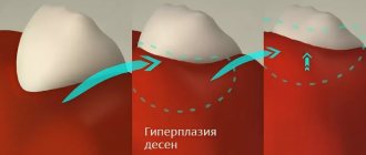
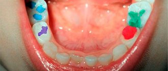
Signs of development of wedge-shaped defects
V-tooth shape
The main symptom of a wedge-shaped defect is the appearance of a V-shaped depression on the surface of the tooth. There are no tissue changes characteristic of caries in the lesion, and tooth enamel does not lose its natural shade (changes in its color can be observed only in the last stages of the development of the disease).
Increased tooth sensitivity
In some cases, a sign indicating the occurrence of a wedge-shaped defect may be a change in the chemical, mechanical or temperature sensitivity of the teeth. Patients complain of not being able to use hard toothbrushes or eat their usual foods.
What to do if caries appears on teeth
First of all, you need to immediately contact your dentist. When a tooth is destroyed due to caries, treatment cannot be delayed. Otherwise, the complications described above arise. It is worth noting that no available means at home will relieve the patient of this problem. Only a dentist can cure this disease.
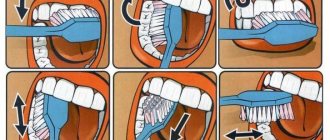
You should also pay attention to your oral hygiene:
- Brush your teeth at least twice a day.
- Use floss and mouth rinses.
- Visit your dentist for a checkup every six months.
- Come for professional hygiene every 6 months.
Dentists will help you select individual hygiene products (brushes, pastes, threads, rinses) at your appointment. The doctor will also determine why dental caries appears in your particular case and give the necessary recommendations.
Stages of development of the defect
In modern dental practice, it is customary to distinguish the following stages of development of wedge-shaped dental defects:
- initial (tissue loss is not noticeable to the naked eye, the patient may complain of a slight increase in tooth sensitivity);
- superficial (presence of visible defects 0.3 mm deep and less than 3.5 mm long);
- medium (the appearance of defects up to 4 mm long and 0.4 mm deep, formed by two planes located relative to each other at a slight angle);
- deep (the appearance of a defect reaching a length of 5 mm, non-carious damage to dentin).
According to statistics, initial and superficial wedge-shaped defects are most often found in young people, and deep and medium ones - in older people.
Formation of a hole on the side
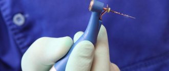
The process of formation of “side” holes in teeth is no different from the occurrence of carious cavities in other places of the tooth. The inconvenience lies in the very “lateral” location, it is simply difficult to get there and therefore such holes are discovered already at the final stage, the symptomatology of which is acute pain due to pulpitis. When treating such formations, the dentist uses a speculum and multiple instruments, and the patient must be prepared for the fact that the mouth will have to be kept wide open and for quite a long time.
Methods for eliminating wedge-shaped defects
Methods aimed at eliminating wedge-shaped dental defects include:
- applications of medications that restore the mineral composition of enamel and enhance its ability to withstand adverse factors;
- the use of toothpastes and other products that can reduce the sensitivity of dental tissues;
- closing the formed defect using filling materials;
- installation of veneers;
- covering the affected teeth with metal-free dental crowns or metal-ceramic crowns;
- carrying out orthodontic measures aimed at correcting the patient’s malocclusion.
The choice of dental treatment method depends on the stage and severity of the pathological process, as well as on the patient’s age and state of health. Only timely identification of a wedge-shaped defect, drawing up a competent treatment regimen and strict adherence to medical instructions allows:
- get rid of all manifestations of this disease,
- to keep healthy,
- beauty of teeth,
- prevent the development of other dental pathologies.
What happens if caries is not treated?
- For a long time, the disease may not bother the patient in any way. When the destruction of hard tissue has reached a significant extent, sensitivity appears during eating.
- If you do not consult a doctor at the first stage, the carious process will go further and reach the pulp (nerve). Spontaneous severe pain appears. Treatment of pulpitis will be required.
- If the patient does not seek help at this stage, the infection spreads beyond the root. Periodontitis occurs. The prognosis here is unfavorable, since such a tooth often needs to be removed to eliminate the infection.
Therefore, timely visit to the dental clinic will help to avoid acute pain and complications.
Periodontal tissue
The definitions we give are absolutely scientific and generally accepted. At least tell me during the exam. But they are also absolutely incomprehensible (Unless, of course, you are a dentist, or a 2nd year dental student). Therefore, we will add our comments to the scientific definitions.
Periodontium
- a complex of tissues surrounding the tooth and holding it in the alveolus, having a common origin and function. I'll explain. Periodontal tissue is the tissue that holds the tooth in the jaw. If you remove at least one of them, the tooth will immediately fall out.
The periodontium unites four different, but closely related tissues: the gums, periodontal ligament, alveolar bone and dental cementum. They are connected by one chain by common origin and function. Gums – protects the periodontium. The other three hold the tooth.
Gum
- this is the mucous membrane that covers the alveolar process of the upper jaw and the alveolar part of the lower jaw and covers the teeth in the cervical area and extends to the transitional fold.
Well, here everything was immediately clear. The gum is a mucous membrane. Look in the mirror, everything around your teeth is gums. If you feel the gum, for example, on the lower jaw - it is dense tissue below the teeth, soft tissue begins even lower - this is no longer gums. The place where soft tissue transitions into hard tissue is called a transitional fold.
Gingival edge
- This is the edge of the gum that lies on the necks of the teeth. If you look in the mirror, you will see the gum line just above the tooth.
Dentists divide gums into two types:
- Free gum (or marginal) is gum that is not connected to bone or tooth. It is mobile and is located in the area of the necks of the teeth. It also fills gaps between teeth.
And again to the mirror - between the teeth there are triangular shaped gum protrusions. They are called gingival papillae.
- Attached gum is a fixed gum. It is firmly connected to the cementum of the root and to the alveolar bone. (In the mirror, this is almost the entire gum except for the papillae and gingival margin).
Gingival epithelium
Epithelium is the tissue that is the top layer of the mucous membrane. And the gingival epithelium covers the gum. It is divided into three types:
- Oral epithelium - covers the largest surface of the gums. It starts from the transitional fold and ends at the gingival margin. Absolutely all of the gum that you see in the mirror is the oral epithelium.
- Sulcular epithelium – lines the gingival sulcus (see below). This epithelium is permeable. Bacterial toxins and harmful substances easily enter the blood through it. It also secretes gingival fluid (also see below).
- Attachmental epithelium is the epithelium that attaches to the tooth. If this epithelium is torn away from the tooth, a gum pocket is formed (This is no longer the norm).
Gingival sulcus
- This is a narrow gap between the tooth and the gum. It is located between the attachment epithelium (see above) and the gingival margin. Normally, the depth of the furrow is up to 3 mm. If more, this is a gum pocket.
Gingival fluid
The sulcus epithelium secretes a fluid called gingival fluid. (In fact, this is blood plasma). It interferes with placing a filling in the cervical area. It also contains immunoglobulins and leukocytes, and protects the gums from bacteria. And from this liquid subgingival tartar is formed. But it’s still a necessary thing.
Dental alveolus
- a hole in the alveolar bone in which the root of the tooth is located. If you had a tooth removed today, you can see it in the mirror. If not, here's a photo.
Periodontal ligament
It is located in the space between the root of the tooth and the wall of the dental alveolus. It binds them together and holds the tooth in the bone. The ligament contains collagen fibers - its main component. They perform the main function of the periodontium.
Also, cells (fibroblasts, osteoblasts, osteoclasts, cementoblasts, etc.) that synthesize collagen, build (and destroy - this is the norm!) tooth root and alveolar bone, regulate periodontal physiology. Vessels – nourish the periodontium and tooth root. And nerves are periodontal sensors. For example, they prevent you from clenching your teeth too hard. You can try it – it’s unpleasant.
Alveolar bone
It's just a bone. The same as the others. The only thing is that teeth grow in it. In special holes - dental alveoli. The rest is an ordinary bone.
This concludes the story about anatomy. You can remove the mirror. Next, we'll talk about what you shouldn't have in your mouth.
Selection of material for restoration
For direct tooth restoration, special composite materials are used, which have high strength, ductility, and also help achieve the effect of tooth transparency.
Most popular materials:
- Filtek (USA) - considered a universal material. It is suitable for restoring anterior teeth and chewing teeth. The main thing is to choose the right shade.
- Venus (Germany) is a more expensive option for the restoration of anterior teeth. The material consists of nanoparticles, which helps to achieve maximum aesthetic effect.
- Enamel Plus (Italy) - sometimes it is called a “chameleon” for its ability to adapt to its native enamel color.
- During indirect restoration, ceramics, zirconium dioxide, and metal ceramics are used.