1.General information
It would seem that any person who has had a runny nose at least once is familiar with this uncomfortable sensation: somewhere in the depths of the nasopharynx, something has accumulated and interferes with every swallow, and it is necessary to get rid of it urgently, by any means... Considering that a runny nose is the most common from painful human conditions, we can safely assume: this feeling of “internal runny nose” is known to everyone, and it is known so well that there seems to be nothing to talk about here. In the end, the mucous membranes of the nasopharynx normally produce large volumes of secretion, some of which flows down the back wall, is swallowed with saliva and food, sometimes accumulates - for example, after a night's sleep - but, in general, does not create any special problems.
However, the flow of moisturizing and bactericidal mucus along the posterior nasopharyngeal wall into the laryngopharynx is not always “normal” and problem-free. It's all about the causes of hypersecretion, the composition and properties of mucus, and its quantity.
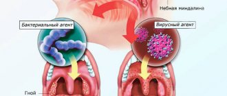
Rhinitis (runny nose) and accompanying rhinorrhea (profuse discharge of mucous discharge from the nasal passages) is, by and large, not a disease. This is a symptom, a sign of a particular pathology. Sometimes this symptom is quite expected and really does not have any particular clinical significance, but in some cases it requires special attention and examination. For such cases, a special term has been introduced, which is found in the literature in several synonymous spellings: postnasal drip syndrome (English: “postnasal drip syndrom”), retronasal drip (sometimes written “drip”), retronasal discharge, internal runny nose, postnasal syndrome. It was first described at the end of the 18th century under the name “chronic pharyngeal catarrh.”
A must read! Help with treatment and hospitalization!
Breathing with turbulence and friction
Chief, I can hear you
Bronchial breathing is a sound phenomenon that occurs when air turbulence passes through the larynx, and is also the site of the division of the trachea into two main bronchi (tracheal bifurcation). Since zones of turbulence occur both during inhalation and exhalation, bronchial breathing is clearly audible both in the first phase of breathing and in the second.
You've probably noticed that the doctor doesn't listen to the lungs at one point, but strives to poke the stethoscope all over the back, and in young beautiful girls he listens especially intently to the lungs in front. In general, he is not doing this out of a good life, believe me. Our lungs are rather large, and the doctor needs to determine whether the noise is heard over their entire surface or whether it is localized. Is one type of breathing heard everywhere, or is there an alternative somewhere?
A striking example of such an alternative is bronchial breathing. As we have already said, normally vesicular breathing should be heard over the entire surface of the lungs. Let me clarify: almost all of it. Above the larynx, in the projection of the trachea and especially in the projection of its bifurcation - the level of the IV-V thoracic vertebrae (in young beautiful girls - this is right at the very top of the cleavage;)) bronchial breathing is normally heard. Sometimes, to distinguish it from pathological bronchial breathing, it is called laryngo-tracheal breathing. That is, the laryngo-tracheal is somehow the norm, but the bronchial is no longer the norm.
Let me emphasize once again that normally bronchial breathing should not be heard over other parts of the lungs. If it appears somewhere else, it means there is a problem in the lungs. Most often, associated with compaction of the lung tissue (under such conditions, sounds are conducted much better than through air lungs).
Depending on the severity of the process, bronchial breathing may overlap with vesicular breathing, or may completely displace it. What can cause hardening of lung tissue? Pneumonia, first of all. Especially massive, lobar, for example.
In addition to pneumonia, compaction of lung tissue can cause tuberculosis, pulmonary infarction, compression atelectasis of the lung (collapse of lung tissue and compression of the lung to its root), pneumosclerosis (when lung tissue is replaced by connective tissue).
To be fair, it should be noted that bronchial breathing can rarely be heard over the entire surface of the lungs. Most often, it is perfectly audible above the source of compaction, and if you move to the side, its volume drops exponentially. This fact helps the doctor to say with a certain degree of probability where exactly the problem is located.
Amphoric breathing is also a variant of pathological bronchial breathing. It can be heard if a cavity in the lung (5-6 cm in diameter, usually smooth-walled) connects to a bronchus. A cavity can appear, for example, with a lung abscess.
Essentially, we get the Pan Flute element. The air passes over the opening of the cavity - and we hear a fairly loud and somewhat musical sound.
The old professors mentioned more than once suggested taking an empty champagne bottle and blowing over its neck. The sound is almost the same as with amphoric breathing.
It's cold!
Fortunately, quite rarely, there are disturbances in the innervation of the respiratory muscles. With such a pathology, they do not all work at the same time, but one by one, step by step. As a result, we get a stepped inhalation. Our exhalation is passive, muscles are not particularly needed there, so the second phase of breathing is usually not changed.
This type of breathing is called saccoded.
But in the overwhelming majority of cases, saccoded breathing is explained by completely normal and not at all pathological reasons. First of all, the cold. It’s good for the doctor, under his coat he may have not only a fiery heart, but also, for example, a woolen vest. But the patient has no choice, he has to be naked to the waist (from above), even if it is not hot in the office or ward, to put it mildly. The muscles begin to engage in tremulous thermogenesis, resulting in saccoded breathing upon auscultation.
Therefore, it is recommended to examine patients in a more or less warm room. And warm the head of the phonendoscope in your hands before applying it to the patient’s skin. For experienced therapists, this is already at the level of automation: you interview a person, and the stethoscope warms up in your hand.
Another option is people who, when they see a doctor, begin to get terribly worried. And again we hear saccoded breathing. Again, if the patient sobs before the doctor arrives, then for some time he will again experience saccoded breathing. And again, the reason is not illness at all, it’s just the consequences of crying.
Well, to figure out whether it is a violation of the innervation of the respiratory muscles or recent crying - this is precisely the art of the diagnostician.
Noise maker mouse
And now we move on to the most notable noisemaker in the lung area. As you know, our lungs are covered with a kind of elastic film - pleura. And the chest cavity is lined from the inside with the same pleura. Between these two layers there is a small cavity, which is called the pleural cavity. Both leaves are coated with a small amount of lubricant to facilitate breathing movements.
If the pathological process is localized not in the depths of the lungs, but on the surface, the pleura covering the lungs can also become inflamed. In this case, the lubrication can either disappear (as with the rapid loss of a noticeable amount of fluid by the body - cholera, blood loss, indomitable
vomiting and other delights), or fibrin may be deposited on the surface of the pleura, adhesions and scars may occur (tuberculosis, cancer, etc.).
As a result, the sliding of the pleura becomes no longer so easy and carefree. And not so silent.
Pleural friction noise is one of the loudest sound phenomena. Already annoying, the old professors compared it to the crunch of snow on a frosty day. Amazingly accurate definition.
Distinctive features - does not disappear when coughing, is clearly audible both on inhalation and exhalation, if you press with the head of the phonendoscope, the pleural friction noise will intensify. Thus, it can be distinguished from fine moist rales and crepitus. There is another sadistic technique: cover the patient’s mouth and nose and ask him to imitate breathing movements with his stomach, perhaps accompanied by music. The pleural friction noise will remain, but there will be no wheezing or crepitus. The main thing with this technique is to quickly run away after it is completed.
Most often, pleural friction noise is heard in places of greatest amplitude of respiratory movements, that is, from below on the sides. Much less often (with tuberculosis) it can be detected in the area of the apexes of the lungs
2. Reasons
Possible causes of hypersecretion of nasal mucus are very numerous and varied:
- acute respiratory viral infections (ARVI);
- rhinitis of allergic etiology;
- sinusitis (inflammation of the paranasal sinuses);
- foreign bodies in the nasal cavity (more common in childhood);
- long-term use of hormone-containing contraceptives, antihypertensive drugs and drugs of some other groups;
- congenital or acquired anomalies of the anatomical structure (most often a deviated nasal septum);
- adenoiditis (inflammation of hyperplastic growths of lymphoid glandular tissue);
- vasomotor rhinitis (hypersensitivity to changes in temperature, humidity, etc.);
- food allergies or ingestion of spicy, sour or other irritating foods;
- air pollution with irritants (substances or particles that irritate the mucous membrane).
In the absence of the above reasons, postnasal drip can be observed in women during gestation (so-called rhinitis of pregnant women) and in patients with gastroesophageal reflux.
Visit our Otolaryngology (ENT) page
3. Symptoms and diagnosis
In addition to the interfering sensation described above, a feeling of congestion and an increased swallowing (and sometimes gag) reflex designed to “clear the throat,” retronasal drip is one of the causes of chronic cough, since excess amounts of mucus, flowing into the laryngopharynx, mechanically irritate the cough receptors. Often there is also pain in the tonsils and/or pharynx, a “lump in the throat” due to reflex swelling, and in more severe cases – dysphonia, hoarseness, and symptoms of salpingootitis.
As a rule, with an upright position of the body, the symptoms are significantly alleviated, and when lying down, especially for a long time, they worsen.
Diagnostics includes the study of complaints and medical history, a detailed examination of the ENT organs, as well as, as necessary, the use of additional research methods (endoscopic, tomographic, laboratory, etc.). In some cases, consultation with specialized specialists is necessary, for example, an allergist, an infectious disease specialist, etc.
Differential diagnosis is carried out with chronic bronchitis and other clinically similar conditions.
About our clinic Chistye Prudy metro station Medintercom page!
Doctor, listen to me! or what do you hear there?
How often do patients complain after being examined by a doctor: “He didn’t even listen to me…”.
And it’s sad that you still need to look for a doctor who is ready to listen and has time for this during the appointment. But auscultation or listening to the chest organs (lungs and heart) does not take much time and is a mandatory manipulation for a general practitioner. Are you wondering what the doctor hears in his “ears,” called a stethoscope, and why does he need all this?
Breathing and wheezing
Everything that the doctor listens to above the surface of the lungs, he divides into the sounds of breathing and the sounds of wheezing. Breathing can be normal (vesicular) and pathological (hard, bronchial, weakened, etc.). Normally there should be no wheezing in the lungs.
Hard and not very
Let's start with breathing. Without it, it is quite difficult to switch to wheezing. Normally, no sub-sounds or special effects should be heard in the lungs. The so-called vesicular breathing is considered “healthy”. It is so called because it occurs as a result of vibrations of the walls of the pulmonary alveoli (vesicles) during inspiration. Naturally, not the entire volume of the lungs is filled at once, so we have a sound extended in time and increasing in intensity.
The old professors explained what vesicular breathing sounded like this: try saying the letter "f" as you inhale - you will get the sound of vesicular breathing. Another option is to drink tea from a saucer and sip.
Normally, in the presence of vesicular breathing, the entire inhalation and about a third of the exhalation should be audible. This is where the norm ends.
There are a lot of variations - weakening or strengthening of vesicular respiration, but this is already a propaedeutic jungle and we will not go there.
As soon as any troubles begin in the lungs or bronchi, this immediately affects the breathing pattern. For example, when bronchitis (inflammation of the mucous membrane + bronchospasm) makes it difficult for air to pass through the small bronchi, you can hear so-called hard breathing through a stethoscope, when both inhalation and exhalation are heard very well, completely.
There are many more different pathological breathings, but more about them next time. You just need to understand that there is ALWAYS some kind of main breathing, and various additional sound effects are layered on top of it, even if in intensity they cover the main breathing like the Krasnoyarsk Territory of Switzerland.
We wheeze little by little
Wheezing occurs against the background of a pathological process in the trachea, bronchi or lungs. They are divided into dry and wet, and this division is not arbitrary; the mechanisms of their occurrence differ significantly.
Let's start with dry wheezing.
The main condition for the occurrence of dry wheezing is that there must be a narrowing of the lumen of the bronchi. It is not so important what it will be - total, as in bronchial asthma, or focal, as in tuberculosis.
As a result, we get three mechanisms for the occurrence of dry wheezing:
- Bronchospasm
- Swelling of the bronchial mucosa during inflammation
- Accumulation of viscous sputum in the lumen of the bronchus
Then everything is simple: the larger the bronchus is involved, the lower the sound of wheezing will be. If the small bronchi have narrowed, we will hear wheezing; if the “strings” of viscous sputum are in the large-caliber bronchus, we will hear bass buzzing wheezing. Dry wheezing is usually very well audible not only to the patient himself, but also to the people around him, even without any stethoscopes.
From all that has been said, another conclusion follows: dry wheezing will be clearly audible both during inhalation and exhalation.
Let's move on to wet rales.
The main condition for the occurrence of moist rales is the presence of fluid in the lumen of the bronchi (sputum, blood, edematous fluid, etc.). When air passes through this secretion, many bubbles of varying diameters are formed. Overcoming the layer of liquid and entering the bronchus, the bubbles burst, making a characteristic sound. Take a thin straw and blow into the water through it - here is a model for the occurrence of moist rales.
Moist rales are better heard during inhalation, since during inhalation the speed of air movement through the bronchi will be greater.
Moist rales are divided by blistering. Depending on the caliber of the bronchi, you can hear small-bubble, medium-bubble and large-bubble moist rales. They arise, respectively, in the bronchioles and small bronchi, medium-caliber bronchi and main-caliber bronchi. The larger the blistering, the louder the wheezing. Large bubbles, for example, can be heard very well at a distance.
For example, with pneumonia, you can hear fine moist rales.
It is very difficult for a novice doctor to distinguish fine-bubbly moist rales from another sound effect - crepitus. Crepitation is not wheezing. It comes from the alveoli. When a certain amount of secretion accumulates in them, the alveoli stick together. And when you inhale, they fall apart. And this is the sound we hear.
Why is it important to find out whether it is crepitus or fine wheezing? Let me explain using the example of pneumonia: if we hear crepitus, this is bad, pneumonia is in full swing, and if fine wheezing is already good, the resolution phase has begun, or it is actually bronchitis, not pneumonia.
When you hear this damn crepitation ten times, you begin to wonder - how can it be confused with wheezing? Crepitation occurs only at the height of inspiration, it does not disappear after clearing the throat and the sound is very sharp. The same old professors advised rubbing the hair at the temple with your fingers - the resulting sound is very reminiscent of crepitus.
Here she is, my dear, against the background of bronchial breathing that I did not mention. But you will hear its peculiarity anyway.
But with bronchiectasis or if there is a cavity with fluid in the lung (abscess, cavity) that connects to the bronchus, you can hear medium- and large-bubble rales. Bubbles with such wheezing burst noticeably less often, but noticeably louder and more impressive.
4.Treatment
Depending on the diagnostic results obtained, etiotropic therapy is prescribed, i.e. measures are taken to eliminate the root cause of retronasal drip. In some cases, the primary goal is desensitization and relief of allergic inflammation (solved with medication and by minimizing contact with allergens), in others it is impossible to do without surgical restoration of normal anatomy, patency, ventilation and drainage of the nasal passages, in others, one or another strategy for the treatment of sinusitis is used , gastroenterological treatment, a course of physiotherapy, etc. are prescribed.
It is important to understand that constant and abundant flow of mucus down the back wall of the throat is not normal; this condition requires specialist consultation, examination and treatment. Effective assistance is provided in all cases.
Why is the formation of fungus in the maxillary sinus dangerous?
How can the growth of a fungal colony be dangerous? In any case, it’s not very pleasant when some kind of parasite, mold, or fungus lives in you. This is really a parasite that lives in the cavity of the maxillary sinus and feels great there. In addition, mycetoma is dangerous because the blood supply and oxygen supply to the brain deteriorates, since the function of nasal breathing is disrupted. A person simply begins to experience partial oxygen starvation due to mycetoma.
Plus, the waste products of the fungus in the maxillary sinus flow into the nasopharynx, which can additionally lead to additional complications, including the development of allergies and provoking respiratory diseases. And, of course, chronic sinusitis.
The role of CT in the study of mycetoma
Of course, a good computed tomogram gives a complete picture of the maxillary sinuses and is the main tool in diagnosing “mycetoma” when examining a patient. A CT scan of the maxillary sinus shows the location, size, and volume of mycetoma damage (local volume or total fungal infection of the cavity).
In fact, computed tomography is the gold standard for diagnosing mycetoma today.


