Manifestations Causes Classification Diagnostics Treatment Prevention
Each of us wears out our teeth with age. Hard tissues shrink due to friction between surfaces and food. In most cases, this process is uniform, takes a lot of time and does not affect chewing function. This is a natural (physiological) loss of hard tissue. But sometimes the loss of enamel and dentin (the layer under the enamel) is so intense that they talk about pathological abrasion of teeth (abrasion). This process leads to physiological disorders and affects all aspects of life.
Symptoms
At first, the disease manifests itself asymptomatically, so it is diagnosed in an advanced state. When tooth enamel is worn away and dentin is involved in the process, patients begin to complain of increased sensitivity. Teeth react to temperature (hot, cold), chemical (sour, sweet), mechanical (touch of a toothbrush) stimuli, causing inconvenience in everyday life.
Dentin destruction leads to root inflammation (periodontitis) and bone tissue atrophy.
The height of the face gradually decreases, pain in the muscles and temporomandibular joint begins. The lower jaw moves upward and backward, the volume of the oropharynx decreases, and less air enters. Problems with the respiratory system begin. The center of gravity shifts, pathological disorders occur in the musculoskeletal system. The quality of life is falling.
Prevention of enamel destruction
Dentists' recommendations:
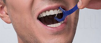
- brush your teeth twice a day, additionally use irrigators;
- use toothbrushes with medium-hard bristles, replace them with new ones every 3-4 months;
- rinse your mouth after eating;
- give up bad habits (smoking, chewing pens, opening bottles with your teeth);
- include dairy products and sea fish in your diet;
- massage the gums;
- do not abuse whitening procedures;
- treat dental diseases in a timely manner;
- Once a year, or better yet every six months, undergo a preventive examination with a dentist.
Following simple rules will significantly reduce the risk of damaging the protective layer of the tooth.
Increased tooth abrasion - causes
Factors that lead to pathological loss of hard tissue are divided into several groups:
- Inferiority of enamel and dentin
. As a result of genetic disorders, harmful effects on the fetus during pregnancy or metabolic disorders, the quality of dentin and enamel is reduced. If the balance of minerals is disturbed, the hard tissues of the tooth begin to wear away at an accelerated rate. - Uneven chewing load
. Incorrect bite, dental defects, bruxism, hypertonicity of the masticatory muscles lead to the fact that some teeth receive increased load. The ligamentous apparatus of the teeth stretches and weakens. It cannot provide height support to the lower part of the face. This leads to TMJ problems. - External factors
. Working with chemical elements and abrasive substances reduces the quality of enamel and dentin.
How to care for teeth enamel after polishing and whitening
The thickness of the protective cover varies from 2 to 2.5 mm. With thinning, increased sensitivity appears. Taking care of your teeth after professional cleaning or polishing will not only prolong the effect, but also allow you to experience less discomfort. During such procedures, the protective layer becomes very thin and, if not properly cared for, its structure may be damaged.
Dentists recommend using toothpastes to reduce tooth sensitivity and regularly using special rinses and dental floss. Immediately after the procedure, it is strictly forbidden to smoke or drink drinks that are too hot or too cold. At night, they may recommend wearing a mouth guard with a special gel. Increased sensitivity in the first days after the procedure is a standard side effect.
Care after teeth whitening includes a “white diet”. It is prohibited to consume foods with high concentrations of coloring substances. This includes wine, coffee, berries, colored vegetables and fruits, soda, spices, and products with a high cocoa content. Sticking to a strict diet is especially important in the first two weeks after whitening.
It is recommended to consume more fish, white meat, egg whites, dairy products and rice. All foods that can damage the thinned surface are excluded from the diet. Strictly monitor the temperature, as changes will negatively affect the appearance and health of the oral cavity. All restrictions are introduced only for the first 14 days after the dental procedure.
Hyperesthesia interferes with the usual way of life. It is painful to eat and drink, and discomfort occurs when the temperature changes. Fluoridation may be needed. The disease must be treated only under the supervision of a specialist. It is important to exclude products that are harmful to enamel and follow hygiene rules.
Classification
Teeth abrasion is divided according to:
- Stages
During the active stage, increased abrasion of hard tissue occurs. At the stabilization stage, their condition can be maintained at an acceptable level.
- Distribution
With localized abrasion, the front teeth are most often worn down. In generalized cases, all crowns suffer and the height of the bite decreases. In severe cases, the crown may end right at the gum.
- Shape
With a mixed form, the enamel wears off not only on top, it decreases on all sides. With horizontal, the occlusal surfaces suffer, with vertical, the lateral surfaces.
- Weights:
- Light or 1st degree (within the enamel)
- Transitional or 2nd degree (involving the superficial layers of dentin)
- Pathological or 3rd degree (with exposure of deep layers of dentin)
In grade 1, speech and chewing function are not affected. On the 2nd stage, hypersensitivity and chewing dysfunction appear. On the 3rd, pain in the temporomandibular joint begins. They radiate to the neck and head and can lead to hearing loss and decreased vision.
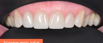
Function of tooth enamel
Enamel (lat. Enamelum) protects dentin on the entire surface of the tooth crown from damaging factors. The thickness of the enamel layer varies within the crown. The thickest layer covers dentin on the cusps of the occlusal surfaces, the thinnest on the cutting edges.
The thickness of the enamel decreases towards the neck of the tooth until it connects with the cementum of the root. The surface of the tooth enamel is smooth, but sometimes has smaller or larger depressions.
Enamel consists of inorganic substances: calcium phosphate, calcium carbonate, magnesium phosphate in the form of hydroxyapatites, which is 96.5%, and organic substances: protein, water in a ratio of 3.5%.
The enamel of temporary teeth is less mineralized than that of permanent teeth. Its layer is half as thin and less transparent. This results in baby teeth being whiter and more susceptible to tooth decay.
Diagnostics
Taking an anamnesis is of great importance in diagnosis. The patient is asked whether he has had complaints before. If yes, what treatment was given. The presence of general, especially endocrine diseases is clarified. Find out under what conditions he works.
Then an external examination is carried out. Pay attention to the symmetry of the face and the severity of the folds. Check the functioning of the TMJ. The height of the lower part of the face is assessed by taking special measurements.
When examining the oral cavity, pay attention to the bite, overlap of the incisors, and wear of the teeth. The contacts of the teeth are assessed when the jaws are closed.
To get an accurate picture of the condition of the crowns and bone tissue, an orthopantomogram (panoramic image) and targeted images are taken. According to indications, tomography of the TMJ is prescribed.
Types and features of dental hypoplasia
Based on clinical manifestations, several forms of pathology are distinguished:
- Erosive . Oval or round defects appear on the surface of the teeth. Damage comes in different sizes. They are often covered with a thin layer of enamel, and dentin can be seen inside the recesses.
- Wavy . The appearance of multiple horizontal lines on the outer surface of the teeth.
- Spotted . The structure of the enamel itself does not change, but white or yellowish spots appear on it.
- Sulcata . Grooves of varying depths parallel to the cutting edge of the tooth.
- Aplastic . Almost complete absence of tooth enamel - only small areas of the teeth are covered with it.
- Mixed . A combination of several forms, for example, erosive and wavy.
Depending on the severity, the disease can be systemic or local. In the first case, erosive pathology affects many teeth in the upper and lower jaw. In the second, the lesions are point-like and appear on one or two teeth.
Systemic hypoplasia
Pathological changes of a systemic nature are reflected in the shape of the dental units. They are classified into several types:
- Hutchinson's teeth . The anomaly affects the central upper incisors - with pathology, they have a barrel shape. There is a semicircular notch on the cutting edge.
- Pflueger's teeth . As a rule, permanent molars are subject to changes. They are barrel-shaped and have a poorly developed chewing surface. The shape of Pflueger's molars is very similar to cones.
- Fournier's teeth . The dental units look like Hutchinson's sign, but there are no characteristic grooves below.
Local hypoplasia
Local hypoplasia of tooth enamel occurs due to problems with baby teeth, inflammatory processes during the formation of the rudiments of permanent teeth, mechanical injuries to the jaw, and infections. Pathology often manifests itself in the form of shallow stripes or spots.
Treatment of tooth wear
The main task of medical intervention is to establish and eliminate the cause of the pathology. Treatment methods largely depend on this. They include stabilization of the patient’s general condition in case of endocrine and other disorders, change of place of work if we are talking about hazardous work.
In cases where the enamel on the teeth has worn off, use:
- Medication methods
These are gels, solutions, toothpastes that reduce hypersensitivity and saturate the enamel with minerals.
- Restoration methods
Restoration of teeth using composite materials (direct restoration). This technique has proven itself well on the front teeth, but is less effective on the chewing teeth.
- Orthopedic treatment
is added if the problem affects dentin.
Teeth are restored using inlays, crowns, fixed and removable dentures. The main condition for prosthetics is the use of cast crowns. Stamped ones, especially on chewing teeth, fail very quickly. The cladding is used plastic or ceramic. Recently, restoration with veneers has begun to be used.
Clasp dentures with occlusal overlays are widely used as a treatment method for increased tooth wear. The advantage of the method is that it does not require preparing the teeth (removing the pulp). Depulpation makes teeth fragile, which is already a problem with this pathology.
- Orthodontic treatment
is used for malocclusion and misalignment of teeth.
Medicinal bite plates are used. They prevent the teeth from touching to form a new bite height. For the same purpose, mouth guards are put on. Unlike plates, mouth guards can immediately separate the bite by 4 mm, which means there will be fewer intermediate stages. Mouthguards are used at 2-3 degrees of abrasion. After correcting the bite height, prosthetics are performed.
Dysfunction of the temporomandibular joint begins to be treated by restoring the height of the bite. Upon completion, signs of dysfunction disappear in most patients. Then special, inclined plane mouth guards are used to return the lower teeth and jaw to their physiological position.
- Surgery
As a result of the operation, the dentist lengthens the crown part of the tooth by removing the bone tissue of the alveolar process.
Enamel has worn off, teeth hurt – what to do?
Seek help from a dentist and begin treatment. Various methods and drugs are used to eliminate the problem. It is impossible to restore the enamel, but the use of drugs will make it harder and protect it from further destruction. Scientists are working to create drugs that can restore worn enamel. The implementation of this task will be a truly great discovery.
Rehabilitation therapy
Treatment for erosion of tooth enamel involves the appointment of local and general procedures in order to transition the disease from the active phase to the stable one. At the same time, the loss of hard dental tissues will be stopped.
If the enamel has already been worn away, then the following are prescribed:
- Local therapy includes remineralization - daily procedures of application of fluoride and calcium-containing products. The course lasts from two to three weeks. The final stage will be coating the surface of the teeth with fluoridated varnish. These procedures eliminate the increased susceptibility of enamel to irritants.
- Mineralization is carried out using calcium electrophoresis.
- If there is a high level of damage to the hard tissues of the tooth and an obvious erosive defect, a tooth restoration procedure is carried out using a light-curing composite, veneer or artificial crown.
- General therapy involves the prescription of medications with calcium and fluoride, as well as a complex of vitamins and minerals.
- Treatment of erosion in the stabilized phase involves continued use of the vitamin complex. Also, therapeutic methods at this stage will be aimed at eliminating discoloration of the areas affected by erosion. To do this, methods of polishing teeth with a special paste, moderate whitening and applying fluoride varnish and gel to erosive areas are used.
In case of erosion, filling a tooth will not always be effective, since a violation of the adherence of the filling material may occur, which can lead to the formation of a defect around the filling itself.
Treatment will be considered effective if the erosions smooth out and become invisible, pain when eating and getting air into the mouth disappears.
Stages and phases of destruction development
The disorder is classified not only by stages of development, but also by phases:
- The active phase is characterized by a progressive process of loss of tooth tissue, which causes excessive susceptibility of teeth to the action of various irritants. The enamel is destroyed very quickly, the size of the erosion increases every month.
- The stabilized phase occurs very slowly. Characterized by less severe pain. This is due to the fact that as the defect forms, the formation of tertiary dentin is observed, which is a product of the vital activity of the pulp and acts as a kind of protective barrier.
It is very important that in some cases the disease may transition from one phase to another.
There are 3 stages of disease development:
- initial, there is damage to the purely upper layer of enamel;
- medium, the enamel is affected so deeply that erosion reaches the dentin;
- deep, the enamel is completely affected, and the upper layer of dentin is also affected.
Manifestations of erosion depending on the stage
The process of development of the disease is very rapid and dangerous, since along with erosion, pathological abrasion of the hard tissues of the tooth develops.
The disease is chronic, which progresses more and more over time and affects healthy teeth.
Symptoms as the destruction progresses:
- At the primary stage, there is a loss of shine of tooth enamel in the area of a certain area of the tooth surface. At this time, it is almost impossible to identify the process of erosion development. This can only be done by drying the tooth surface with an air stream or by applying iodine to the affected area, in which case the erosive area will turn brown. Initially, the erosive defect will have an oval or round shape with a smooth bottom. The erosion is whitish in color. There is no pain.
- At the second stage, discomfort and a change in color of the affected area begin to appear.
- At the last stage, noticeable pain appears during eating and brushing teeth. The pigmentation of the affected areas changes. Brown spots on the surface of the tooth become noticeable.
The consequences of erosion are dire
Erosion affects mainly middle-aged people. This process is characterized by a long-term course and can last from 10 to 15 years. The following consequences of damage to tooth enamel can be identified:
- Tooth wear accelerates.
- As dentin is exposed, the color of the teeth changes and they become darker.
- As the enamel wears away, the sensitivity of the teeth on the inner and outer surfaces worsens, that is, touching the tongue and lips brings discomfort. Dentin is a soft tissue, so pain will inevitably occur when air enters, chewing and exposure to food acid contained in food.
- The edges of the front teeth may appear more translucent.