Ball in mouth - what is it?
When the inflammation begins to gradually progress, a blister appears on the gum. The main reason for the formation of a lump is poor oral hygiene and prevention. But the ball can also form under the influence of other factors: inflammation occurring in the roots of the teeth, gums, mucous membranes, and periosteum.
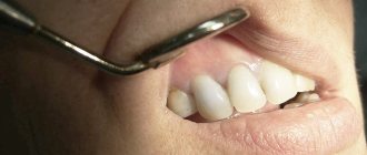
The problem is that the disease can be diagnosed in the later stages, with the development of a purulent process. This happens because the patient does not visit the dentist. For patients who experience fear while in a chair, the doctor can give sedative anesthesia, in which the receptors are turned off and the patient falls into a shallow sleep.
Attention! It is better to diagnose the disease early in order to prevent more dangerous consequences, including blood infection and inflammation of the entire jaw.
Subcutaneous acne on the chin: causes
Acne is a multifactorial disease that can occur for many reasons4,50:
- hormonal disorders;
- stress;
- genetic predisposition;
- problems with the gastrointestinal tract;
- taking certain medications (GCS, anabolic steroids, etc.);
- excess in the diet of sweets, fatty foods, dairy products, foods rich in iodine, bromine, gluten;
- use of cosmetics with comedogenic ingredients, etc.
Subcutaneous acne on the chin in women may appear before menstruation, which is associated with changes in hormonal levels.
Why does a lump appear on the gum?
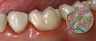
The accumulation of conditionally pathogenic and pathogenic microflora due to improper oral care leads to inflammation of the mucous membrane. Other reasons why a ball appears on the gum include:
- chemical, thermal or mechanical trauma to the periodontium;
- carious formations and their complications: periodontitis, pulpitis;
- inflammation of the wisdom tooth;
- eruption of molars;
- jawbone overgrowth;
- weak immune system;
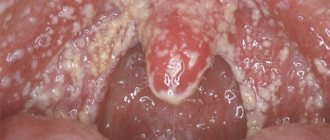
- infectious pathologies of the oral cavity: herpes, candidiasis, stomatitis.
A lump in the mouth also occurs under other conditions: epulis, exostosis, fibroma, focal fibromatosis, malignancy, cyst and flux. The main thing is to promptly establish the causes of the pathology and begin treatment.
Treatment
Most lumps are removed through surgery. However, there are formations that are treatable.
For example, boils are treated with antimicrobial and disinfectant drugs.
If the doctor expresses concerns about the identified lump, then it is worth starting treatment as quickly as possible.
After all, benign tumors eventually develop into malignant tumors, the treatment of which takes much more effort, time and money.
If the ball in your mouth does not cause discomfort
The color of the bubble will help the doctor make a diagnosis. A white lump on the gum indicates the presence of exostosis or purulent exudate. A red or bloody ball indicates the development of inflammation. If the growth matches the shade of the tissue, it is the initial stage of epulis, flux, or a malignant tumor. When the tumor does not hurt, this indicates the presence of one of the pathologies:
A fistula is a white ball on soft tissues that appears under or on a tooth. There is a hole on the surface for the release of pus. If, when pressed, suppuration flows out of the bladder, the patient does not feel pain. If the hole is closed with pus or bloody clots, the patient will experience discomfort with any impact.
A fistula is often formed due to advanced periodontitis, accompanied by periodontal hyperplasia. Overgrown tissue is good soil for the proliferation of microorganisms. In this case, the patient urgently needs treatment for periodontitis.
In the absence of therapy, the fistula enters a chronic stage, which can only be eliminated through surgical treatment. The progression of the disease must not be allowed, otherwise you may lose healthy molars.
Hematoma is a round lump on the inner surface of the cheek. Sometimes it occurs in the form of a dark bluish swelling on top of the gums. Blood accumulates in or around the root of the molar. The mucous membrane grows, the patient experiences discomfort and cannot completely close the jaw. The main reasons: consequences of filling or tooth extraction, gum damage, poor blood clotting.
Hematomas are generally not dangerous. Processes take place in the body through which soft tissues are cleared of bloody clots. After some time, the bubble disappears, but if the seal remains, you need to visit a dental clinic.
Exostosis is a hard blister that is an abnormality in which the bones protrude and protrude from the jaw. Gradually, the lump increases in volume, which causes discomfort and pain. Exostosis can be provoked by various reasons:
- jaw damage;
- heredity;
- congenital disorders;
- tissue diseases after molar removal.
An examination by a dentist or an x-ray will help detect the disease. The formation will need to be removed if the development of a malignant tumor is suspected.
Epulis is a pedunculated bubble in the form of a mushroom-shaped growth on the periodontium. The tumor may be the same color as the gums or red. The reasons causing the development of pathology include:
- improper filling of the molar or too large a filling;
- dental plaque, stone;
- jaw damage;
- malocclusion;
- hormonal imbalance;
- poor prosthetic material or incorrect prosthetics.
The symptoms of epulis resemble gingivitis; for diagnosis, the dentist prescribes radiography and histology to the patient. With their help, the degree of destruction of bone tissue at the site of the epulis lesion is determined. Mostly, pathology occurs in children during the growth of primary molars and in women.
Papilloma or fibroma is a bubble on the gum, sometimes a benign formation that does not pose a threat to the health or life of the patient. They are formed in people of different genders and ages. Predisposing factors to the appearance of a lump can be: damage to the mucosa, stress, systemic pathologies, heredity.
Papilloma is an enlargement of the papillary layer of skin. The bubble grows gradually, but with reduced immunity, systemic pathology, or stress, growth accelerates, but without turning into a malignant tumor. A papilloma neoplasm looks like a smooth, soft lump on the mucous membrane of a white or pink shade on a thin stalk.
Papilloma often does not create any discomfort. But after some time it may increase in size. You should consult a dentist and get tested.
Why should you see a doctor if you have cystic formations?
Treatment of severe acne can become more complicated due to the occurrence of acne elements of complex origin60:
- Phlegmonous. Confluences of large purplish-red pustules located in the deep layers of the dermis. They are characterized by a high rate of maturation and a tendency to form an abscess.
- Indurative. The result of the formation of a large infiltrate, which is located around the inflamed follicles. They have a bluish color. Such acne elements leave the most noticeable scars.
- Conglobate. Their appearance is also associated with the formation of infiltrate. Conglobate acne lies in the deep layers of the skin and is characterized by abscess formation. In addition to comedones, papules and pustules, many large nodes are formed; they are mainly located on the back and neck. The skin over them gradually becomes thinner, holes appear in it, from which pus comes out.
It is unrealistic to select effective drugs for therapy on your own. You should also be prepared for the fact that cystic acne can leave behind scars, which will have to be removed in a cosmetologist's office.
If the formation on the gum causes discomfort
If a bubble near a molar causes pain, this indicates an infectious inflammatory process. Discomfort is also typical with gum injuries and hematomas. Painful formations can be caused by a cyst, cancer, fibroma, periostitis, periodontitis.
Gingivitis - the disease affects only one gum, and the periodontium near the molar remains uninjured. The main symptoms of the pathology are swelling, bleeding, and peeling of the epithelium. Gingivitis often occurs against the background of halitosis. Sometimes pathology develops as a result of endocrine and metabolic disorders.
When treating, it is necessary to eliminate the cause of gingivitis. Professional oral hygiene, diagnosis and treatment of metabolic disorders are performed. To eliminate inflammation and prevent further spread of bacteria, antibiotic therapy is administered. For pain and severe discomfort, analgesics are prescribed.
Periodontitis - purulent bumps on the gums appear as a result of degeneration of the alveolar process, through which the tooth root is held in the alveolus. Tissues can be destroyed due to poor oral hygiene and internal pathologies.
In the initial stages, periodontitis is treated by teeth cleaning at the dental clinic and further proper care. An advanced form of the disease, in which the teeth become loose, pus accumulates, requires surgical intervention - restoration of the alveolar processes or removal of molars.
Periostitis or flux is a dense formation near a problematic molar with carious lesions. Patients complain of pain radiating to the temple, chest, neck, ear. The condition gradually worsens and the temperature can rise to 38 degrees. In the oral cavity, lesions due to periostitis are hyperemic. A purulent fistula forms. When the discharge is removed, the discomfort decreases.
Causes of periostitis: trauma, periodontitis, osteomyelitis, weak immune system, infectious pathologies occurring in the body and vitamin deficiency.
Elimination of pain in the muscles of the neck in front
Since this condition is classified as pathological, it requires the most prompt treatment possible. The best option is to consult a doctor, since this specialist has all the necessary knowledge regarding the fight against inflammatory and other diseases that occur in the cervical region. If you have neck pain in the front under your chin, call and make an appointment with the highly qualified, experienced doctors at the Energo Clinic. The telephone number for contact is indicated on the website in the “Contacts” section; registration is carried out on any day and time of day convenient for you.
Drug treatment
After collecting anamnesis, conducting an examination and finding out why your neck hurts in front under the chin, our neurologist
will write out the most detailed treatment plan for the disease and instruct what needs to be done to prevent the disease from worsening again. The most important point will be taking medications - painkillers and anti-inflammatory drugs, drugs whose action is aimed at maintaining the optimal condition of the muscular frame and skeletal system.
Massage and physical therapy
Conducted strictly in the presence of a chiropractor
,
massage therapist
or doctor at a physical therapy room. The neck is a rather delicate organ; it should not be pressed, and often not heated intensely, as this can cause a deterioration in the condition. At the same time, a soft, relaxing massage will help relieve muscle tension and normalize blood flow.
Surgery
It is necessary in situations where the condition threatens the normal course of a person’s life. For example, if the thyroid gland is enlarged and there is no effect from medications, the affected organ is removed in a surgical hospital. After the operation, it is necessary to follow the recommendations of the attending physician, undergo regular examinations by an endocrinologist, and take hormones.
ethnoscience
Effective for inflammatory diseases, but should only be used as an additional treatment measure. A proven folk remedy is a decoction of chamomile or calendula - it is used for colds and to relieve throat spasms. In situations where a person has pain in the neck under the chin as a result of thyroid pathologies, herbal medicine brings good results: certain medicinal plants contain components that have a positive effect on the state of the hormonal system.
Treatment of neoplasms in the oral cavity
A lump under the jaw or on the gum can be detected upon examination. For an accurate diagnosis, a biopsy or x-ray is prescribed. Treatment is selected taking into account the cause of the formation of a bubble in the oral cavity. It will be necessary to stop the further spread of infection and eliminate discomfort.
The doctor will determine how to remove the lump after a complete diagnosis, but the main measures include:
- fistula - pus is removed by rinsing with a disinfectant solution;
- epulis - surgical intervention (removal using diathermocoagulation, cryodestruction or a scalpel);
- periodontitis - removing fillings, cleaning canals, removing pus, rinsing with herbal decoctions and soda solution;
- periostitis - placement of special drugs under a temporary filling (if there is no result, the tooth will have to be removed);
- gingivitis - cleaning of periodontal tubules, antiseptic and antibacterial therapy, removal of lumps.
When a formation appears on the gum, you can rinse your mouth with vodka, diluted alcohol or juice from fresh Kalanchoe leaves.
Timely diagnosis allows you to prevent further spread of infection and save teeth.
Only an experienced dentist will help you get rid of a lump in your mouth, minimizing the risk of developing serious complications. At the first signs of pathology, you should immediately consult a doctor. The doctor's consultation
For what forms of acne does a doctor prescribe topical therapy?
For severe forms of acne, doctors usually recommend systemic therapy, and for mild to moderate acne, topical therapy is more often prescribed. Clindovit® gel is a topical antibiotic that is used to treat acne (acne vulgaris)6. The main component of the drug is clindamycin6. It exhibits antibacterial activity against propionibacteria6. Clindovit® also helps reduce the level of free fatty acids on the skin6. It is recommended to be combined with the use of benzoyl peroxide or azelaic acid (Azelik® gel) to reduce the risk of developing antibiotic resistance28.
Precancerous neoplasms (precancrosis)
Precancerous neoplasms are those that, under the influence of congenital or current causes, have acquired a tendency to malignant degeneration. As a rule, these are chronic conditions that are observed in a person for a long time.
Thus, precancerous tumors are dangerous neoplasms on the skin that can lead to the development of oncological processes. These include:
- Actinic keratoma is a keratosis in which dry crusts and scales appear on the skin of older people. When they peel off, slight bleeding may occur.
- Xeroderma pigmentosum is a hereditary tumor that develops due to increased sensitivity of the skin to ultraviolet radiation. Rarely encountered, these are pigmented spots that become warty growths.
- The cutaneous horn is a cone-shaped tumor that looks like a horn. Has a yellow or brown color. occurs in exposed areas of the body that are regularly subjected to friction or pressure. Typical for older people
- Bowen's disease is an intraepidermal cancer. Without treatment, it can transform into invasive skin cancer. In the early stage, Bowen's disease appears as a small reddish-brown spot measuring 2-50 mm. has a flaky surface and raised, uneven edges. After removing the scales, a weeping but not bleeding surface remains.