Hyperplastic gum growth is a fairly pressing problem in dentistry that requires timely professional treatment. This condition not only spoils the appearance of the teeth, but also causes discomfort and can become complicated. gum hypertrophy develops, it is recommended to immediately consult a dentist. The Nurimed clinic in St. Petersburg offers its services for the diagnosis and treatment of overgrown periodontal tissue.
Causes of gum overgrowth
Establishing factors that contribute to an increase in periodontal volume makes it possible to reduce the risk of recurrence of an unpleasant situation and draw up a competent treatment plan.
The main causes of gum hyperplasia:
- inflammatory process in the gingival area;
- mechanical damage to soft tissues;
- taking certain medications (immunosuppressants, anticonvulsants, antihypertensive drugs - calcium channel blockers);
- the presence of certain systemic pathologies (hormonal imbalances, metabolic disorders, systemic connective tissue diseases, leukemia);
- oncological processes in the body;
- accumulation of tartar in the cervical part of the tooth crown;
- increased spaces between teeth or teeth growing at an angle;
- complication of gingivitis, periodontitis.
Complications in patients with hypertrophy
In addition to aesthetic problems, the disease leads to other negative consequences.
The patient experiences pain when chewing food, and the mucous membrane becomes sensitive to food irritants.
But the most important complication is the difficulty of maintaining high-quality hygiene. The accumulation of plaque under the gum leads to the formation of tartar, the mucous membrane becomes inflamed, periodontal bleeding and bad breath appear. All this can lead to the development of caries, pulpitis, and periodontitis.
In addition, periodontitis, an inflammatory gum disease, may develop. The consequence of periodontitis can be the destruction of the ligamentous apparatus with which the tooth is held in the socket.
In childhood, periodontal hypertrophy can lead to abnormal tooth growth, malocclusion, and, without proper treatment, problems with the maxillofacial apparatus. Therefore, you cannot delay treatment, and when the first signs of the disease appear, you should immediately contact a specialist.
Types of gum hyperplasia
In dentistry, the following types of fibromatosis are distinguished:
- Localized hyperplasia (tissue near one tooth or group of teeth is affected).
- Generalized (the entire dentition of one or both jaws overlaps).
- Papillary (growth is concentrated only in the area of the papillary papilla).
- Marginal (not only the papilla is affected, but also the gingival margin).
- Diffuse (increase in all layers and areas of tissue).
In some cases, hypertrophy appears as a bump or growth when the growth affects a limited area.
Types of disease
Doctors call two types of gum hyperplasia:
- Focal, which is practically not registered. It looks like single gingival growths observed in the area of the lingual gingival zone or in the area of the tubercle of the upper dentition. Moreover, single growths can cover all gums, or just one;
- Generalized gingival hyperplasia. It is registered quite often. It is visualized as granular growths, which eventually grow together into one, subsequently covering a significant area of the tooth surface.
Manifestations of the disease
The following clinical symptoms allow you to recognize gum fibromatosis:
- In the initial stages, swelling or enlargement of tissue of a tumor nature.
- On the mucous membrane you can see thickenings that gradually increase.
- Increased gum volume without signs of swelling.
- Filling interdental spaces with soft tissue.
- Overlapping of the teeth with the edges of the gums.
- Injury to overgrown areas during cleaning (this can cause bleeding and inflammation).
- Increasing intensity of tissue color (the gums become bright pink).
If you notice any signs of fibromatosis, you should immediately contact your dentist. If you ignore the problem, unpleasant complications may develop:
- decreased quality of daily hygiene with an increased risk of developing infections;
- pain when eating;
- redistribution of chewing load;
- development of periodontal diseases.
Forms of the disease and stages of development
There are two forms of pathology: generalized, that is, affecting one or even both jaws, and limited, which appears in a small specific area.
In the generalized form of the disease, there may initially be several lesions in different places in the oral cavity, which later merge into one. A limited form of hyperplasia may not manifest itself at all for a long time.
Stages of disease development:
Stage 1 – the gingival papillae increase in size, the periodontium is slightly enlarged and extends onto the tooth. Stage 2 – the tissue grows over half of the crown part of the tooth. Stage 3 – most of the tooth is hidden under the gum. In this case, the mucous membrane is regularly injured, which causes bleeding, swelling and general inflammation of the oral cavity.
Also, periodontal hypertrophy is divided into fibrous and medicinal forms. Fibrous is extremely rare. Due to similar symptoms - the appearance of periodontal pockets that are filled with plaque, the pathology is often confused with periodontitis. The most common form of the disease is the drug form.
Mechanism of development of hyperplasia
The mechanism of development of the hypertrophic process remains not fully understood. However, scientists have discovered that the pathogenesis of the disease is influenced by impaired calcium transport. This leads to increased production of collagenase, which causes the gums to grow. Hyperplasia develops gradually, so patients do not pay attention to it right away.
Stages of the disease:
- First: the tissue covers a third of the tooth, there are no signs of an inflammatory process.
- Second: covers half of the tooth, bleeding may occur, especially with mechanical impact.
- Third: overlap from two-thirds to the entire crown, sometimes even the cutting edge and chewing surface are covered with soft tissue, granulations are visible over the entire surface.
What causes gum hyperplasia?
Overgrowth of gum tissue is also known as hyperplasia. The main reason is genetic predisposition. Dentists also include the following as provoking factors:
- serious jaw injuries,
- malocclusion,
- chronic inflammation of the gums,
- hormonal imbalances,
- taking certain medications,
- accumulation of soft plaque and tartar at the edge of the gum.
The disease is diagnosed in children and adults. If it is not treated, the child may have problems with the loosening of baby teeth and the eruption of permanent teeth. People with a mature bite have an increased risk of developing various dental diseases.
How to diagnose and how to treat gum hyperplasia?
The diagnostic algorithm is focused on not confusing the disease with other pathologies. Therefore, the examination should be trusted only to a qualified dentist. The Nurimed Clinic offers a modern algorithm for confirming hypertrophy:
- examination of the oral cavity (allows the doctor to suspect a problem);
- X-ray examination to exclude similar pathologies;
- histological analysis of tissues taken for biopsy (a conclusion on the benign quality of the process and cellular composition is required).
After confirmation of the diagnosis, which is impossible without histology, treatment of gingival hyperplasia is carried out. Modern dentistry offers safe, painless surgical removal of overgrown gum tissue. The dentist peels off and removes excess tissue, after which the mucous membrane is sutured. This procedure is considered minimally invasive and does not require special recovery. In some cases, the dentist prescribes a number of medications to prevent complications.
An additional stage of treatment is sanitation of the oral cavity. Manipulation allows you to eliminate infectious processes that could cause hyperplasia. Also, professional cleansing can reduce the risk of inflammation and relapse of hypertrophy.
In the case of drug-induced fibromatosis, it is recommended to consult with your doctor and, if possible, change medications. To prevent re-growth, the cause of hypertrophy must be eliminated: the bite is corrected, tartar is removed, inflammation is treated, the balance of hormones is controlled, and so on.
Treatment
The first thing a doctor does is diagnose the pathology. The disease is easily confused with other gum diseases such as gingivitis and periodontitis.
If drug-induced hyperplasia is diagnosed, they try to eliminate the disease by discontinuing medications. If this is not enough, surgery is performed to remove excess gum.
That is, if therapeutic methods are not enough, the pathology is eliminated using gingivectomy. The essence of the operation is to excise the overhanging edge of the gum. After gingivectomy, when the gums have healed, damaged teeth are treated. Plaque is removed using professional oral hygiene, and the enamel is strengthened with fluoride-containing products.
The sooner treatment begins, the easier it will be to eliminate the pathology and return the gums to a healthy and attractive appearance.
The structure of enamel growths
Hyperplasia also develops after changing teeth. Excess enamel appears in the form of drops measuring 1 mm. In the severe stage, pearl drops can reach 5 mm.
Initially, the development of hyperplasia is asymptomatic. The reason why the patient goes to the doctor is to complain about the unaesthetic appearance of his teeth.
Places where the disease spreads:
- at the root of the tooth;
- on the neck of the tooth;
- on the crown of the tooth.
The classification of hyperplasia occurs depending on their structure:
- True enamel.
- Enamel dentin.
- Enamel dentin, filled with pulp inside.
- Enamel drops in the form of nodules, the so-called Rodriguez-Ponty drops.
- Growths that form on the dentin of the crown or inside the tooth itself.
During a visual examination of the teeth, excess enamel may not be detected. When using a drill, you can only accidentally stumble upon a hard area in the dentin.
Nadent – Natalia Khvorostinova’s Universal Dentistry in Moscow
general information
Gingival hyperplasia is a pathological growth of gum tissue until it covers a significant surface of the tooth crowns.
As a rule, hypertrophic growth of the gums is not associated with inflammatory processes and does not affect other tissues of the oral cavity (cheeks, tongue, etc.).
This anomaly is often accompanied by hyperemia - increased blood supply to the gum tissue, which entails pain and a high risk of bleeding when brushing teeth or eating rough food.
If gum hyperplasia is not treated in time, it can prevent the replacement of baby teeth with permanent ones, as well as lead to osteoporosis and destruction of interdental septa.
In addition, gum hyperplasia significantly complicates oral hygiene and indirectly provokes the development of various types of oral diseases.
Causes of gum hyperplasia
Despite the fact that gingival hyperplasia is relatively common, the reasons that cause its development have not yet been clearly established.
Very often (in 25-40% of cases), gum hyperplasia develops with the active use of drugs such as phenytoin (prevents epileptic seizures), cyclosporine (taken after organ transplantation, preventing organ rejection).
Up to 10% of patients taking various drugs that block calcium channels are also at risk of this disease: nifedipine, diltiazem, felodipine, verapamil, etc.
Other factors predisposing to this disease include:
- Pregnancy
- Certain blood diseases (such as leukemia)
- Puberty
- Genetic predisposition
- Some malocclusions
- Abundance of tartar
Gum hypertrophy most often occurs in adults. In children, it is observed only in the case of genetic causes of the disease.
Drug-induced hyperplasia is possible at any age.
Types of gum hyperplasia
Experts distinguish two types of hyperplasia:
- Generalized hyperplasia - occurs most often and consists of several foci of granular growths of the gums merging into one, covering a fairly large surface of the teeth
- Limited (focal) hyperplasia is relatively rare and represents single growths of the gums in the area of the tubercle of the upper jaw or in the area of the lingual surface of the gums in the lower jaw. Single growths can be localized either on one side of the gum or have a bilateral localization.
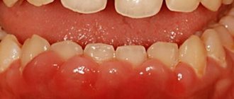
Symptoms of gum hyperplasia
The main manifestations of gum hyperplasia:
- Swelling of the gums (gingival margin and interdental papillae) and increase in their mass
- Filling the interdental spaces with gum tissue
- Covering of a significant part of the surface of the crown of the teeth with gum tissue
- Thickening of hypertrophied gum tissue
- Homogeneous pink gum color
Development of gingival hyperplasia
Despite the fact that the mechanism of development of gingival hyperplasia has not been fully studied, it has been established that most often this disease is caused by a violation of the movement of calcium through the membranes of gum fibroblasts, which entails a disruption of homeostasis in the cells of the gingival tissue and the active production of collagenase, which promotes gum growth.
Gum hyperplasia develops quite slowly.
First, small dense formations in the form of a roller appear on the gums (usually in the area of the front teeth), which do not differ in color from the rest of the surface of the gums and do not cause pain when pressed. The crowns of the teeth are covered with gum tissue by about 1/3.
Next, hypertrophy of the gingival margin and gingival papillae begins to progress, the shape of the gingival papillae becomes deformed, and the growing gum covers up to half the height of the tooth crown.
The extreme degree of gum hyperplasia is characterized by maximum growth of the gingival margin, covering more than 2/3 of the height of the crowns and sometimes reaching the cutting edge or chewing surface of the teeth. Swollen gingival papillae are covered with bleeding granules of various sizes.
Diagnosis and treatment of gum hyperplasia
The main task when diagnosing gum hyperplasia is not to confuse it with other gum diseases. Therefore, when diagnosing here, in addition to visual examination, radiographs and histological analysis are often used.
Treatment of gum hyperplasia is currently only possible surgically by excision of excess hypertrophic gum tissue. At the same time, a complete sanitation of the oral cavity is performed.
If gum hyperplasia is caused by taking certain medications, you should replace them with other drugs. In this case, discontinuation of the drug can contribute to the natural return of the gums to their normal state. If this does not happen, the excess gum tissue is removed surgically.
If the patient again starts taking the drugs that caused gum hyperplasia, then the likelihood of a relapse of the disease is high.