Tooth canal treatment: when is it necessary and how is it carried out?
Tooth canal treatment is a procedure that is carried out for certain indications and allows you to stop active inflammation in the internal tissues of the tooth and avoid the removal of a dental unit. Treatment of tooth canals has its own specifics and is complex; only an experienced and competent specialist can carry out the procedure efficiently. In the article we will look in detail at the process of root canal treatment: we will find out under what circumstances the procedure is carried out, how it goes, what methods are used for treating tooth canals.
Where is the tooth canal located, how is it structured
Human teeth have almost the same anatomical structure, including three main parts: crown, neck and root. Inside the tooth there is a pulp chamber, from which canals originate and extend all the way to the root. The pulp chamber contains the pulp or nerve of the tooth, which is a bundle of nerve fibers and blood vessels. Nerve fibers and vessels also pass through the entire internal space of the dental canal. The shape of the tooth canals can be either straight or curved, and their number varies depending on the type of tooth. Thus, the canines and incisors of the upper jaw have one canal, the canines and incisors of the lower jaw have two. There may be two or three canals in molars, and the maximum number of canals is observed in wisdom teeth - from three to five. The exact number of dental canals is determined using radiographic examination.
As mentioned above, the shape of the canals can have bends and turns, which complicates the process of their high-quality cleaning and treatment. Meanwhile, when treating canals, it is important to thoroughly clean their internal space - otherwise it will not be possible to stop the inflammatory process and save the diseased tooth.
Obliteration of root canals. Clinical case.
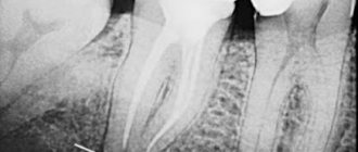
A patient contacted us. The orthopantomogram image clearly shows complete obliteration of the lumen of the root canal of the 21st tooth.
Fig. 1 Complete obliteration of the root canal of the 21st tooth on an orthopantomogram
Diagnosis was made using cone beam computed tomography (CBCT).
Rice. 2 Sagittal section of the area of the 21st tooth on a CBCT image
Taking into account the skeletal asymmetry of the lower jaw in the image, the main cause of obliteration of the root canal in this case can be assumed to be trauma.
Rice. 3 Axial section of the upper jaw area with obliteration of the root canal of tooth 21
Indications for dental canal treatment
Tooth canal treatment is a specific dental procedure that is advisable to perform in the following clinical cases:
When an active inflammatory process is detected inside the root of a dental unit. The inflammatory process in this part of the tooth can lead to gradual tissue necrosis. If tissue necrosis begins, the diseased tooth will have to be removed.
The condition is diagnosed by radiography;
Treatment of tooth canals is required for pulpitis - inflammation affecting the pulp bundle;
Endodontic treatment is carried out for periodontitis, a disease affecting the apical part of the tooth root;
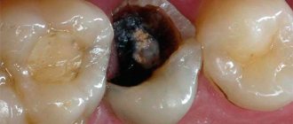
In some cases, cleaning and treatment of dental canals is required for advanced forms of caries.
Dental canal treatment is also indicated for abscesses, the onset of an inflammatory process under an old filling, or a fractured tooth root. The need for measures to treat tooth canals may be indicated by such symptoms as: severe, excruciating pain in the tooth, occurring mainly at night, swelling of soft tissues, discoloration of the gums and tooth enamel.
In some cases, tooth canal treatment is carried out before prosthetics. The nerve is removed from the tooth, and the canal cavities are cleaned, treated with antiseptic drugs and filled.
Main stages of root canal treatment
Treatment of tooth canals is a complex, specific process that includes several successive stages. Below we will consider all stages of the treatment process in detail.
Diagnostics
During the initial visit to the dentist, the doctor will conduct a thorough examination of the patient’s oral cavity and will also prescribe x-rays. An image of the tooth will allow you to assess the condition of the dental canals, see their number, determine the stage of development of the inflammatory process, and also select adequate treatment for a specific clinical case.
Anesthesia
To gain access to the canals of a tooth, the dentist will drill out the coronal part of the tooth to reach the pulp chamber, behind which the canals are located. Both the pulp and the canals are penetrated by nerve fibers and therefore the specialist’s actions can cause some discomfort to the patient. To relieve a person of discomfort, local anesthesia is performed before starting root canal treatment. The drug is selected individually for each patient.
Moisture insulation of the area of the diseased tooth
To prevent pathogenic microorganisms that may be contained in saliva from entering the internal space of the tooth, the area of the diseased tooth is first protected from moisture by covering with a special latex bandage - a rubber dam.
Reaming a tooth
To gain access to the dental canals, the doctor will drill the tooth, removing all tissue affected by caries. The access hole is made either on the chewing surface of the tooth (when treating molars) or on the inside (when treating incisors and canines). After drilling, the doctor will remove either the entire pulp bundle or only part of the tissue affected by the inflammatory process.
Treatment of dental canal cavities
For high-quality treatment of dental canals, a specialized tool is used - files. Before the cleaning process, it is important to obtain detailed information on the structure of the dental canals, as well as their length. To do this, a photograph of the diseased tooth is taken, and a special type of device is used - an apex locator. The dentist will use the files to clean the canal cavities, and then rinse them with an antiseptic solution and treat them with antibacterial agents. During cleaning and antiseptic treatment, the dentist will expand the cavity of the canals, clean and smooth their internal surfaces. Then the cavities of the dental canals are thoroughly dried with paper points.
Next, the doctor puts medicine into the canal cavity, places a temporary filling on the tooth and sends the patient home. After a few days, you should come back to the dentist to evaluate the results of the treatment. If the image shows that the inflammation has been stopped, the canals are filled.
Sealing
Gutta-percha pins are used to fill dental canals. It is important that the dental canal is sealed correctly and efficiently, otherwise a recurrence of inflammation is possible and the tooth cannot be saved. After filling the canals, the crown part of the tooth is restored with a photopolymer filling or orthopedic structures. It will be useful to know that mild pain in the tooth within 14 days after the procedure is considered normal by dentists. Taking painkillers prescribed by your doctor will help eliminate discomfort. However, if the pain does not go away, its character is sharp, and does not subside even after taking anesthetics, you should immediately contact the dentist!
Search for the mouths of heavily sclerotic canals
Clinical case of endodontic treatment of upper incisors with heavily sclerotic canals. How to navigate a situation where there are “no mouths.”
Today I would like to share a clinical case that will demonstrate how a seemingly trivial situation can lead to serious complications, as well as how to avoid these complications.
The reference patient was referred by a colleague with a request to help carry out endodontic treatment before prosthetics in teeth 12 and 21. The doctor made an independent attempt at treatment, but unexpectedly the problem of severe obliteration of the orifices in both teeth was revealed, which did not allow the doctor to find the entrances to the root canals. The referring dentist only had binoculars in his arsenal, which, alas, did not allow him to confidently navigate inside the tooth.
This is how these teeth came to me. What do we see?
Firstly, we see that the doctor is great for realizing in time the risk of searching for the mouths “blindly”, without magnification. Since in both cases there were a couple of strokes of the tip left before the root wall was perforated.
Secondly, the search vector was shifted to the palatal side in both cases.
Thirdly, the obliteration really did not leave even a hint of the mouth where it should have already appeared. The situation in tooth 21 turned out to be especially difficult.
The green line shows the outline of the actual root canal. And here you can clearly see how the search for the mouth was carried out away from the true course of the root canal, threatening to end in perforation.
How to find the entrance to the canal when even the thinnest file (No. 6, No. 8) does not “catch” anything? It is necessary to remove sclerotic dentin. This can be done with a rotating instrument on a long stem or with an ultrasonic nozzle. But the main thing to remember is 2 important rules:
1. Any actions with aggressive instruments inside the tooth (canal) can ONLY be carried out under visual control. Ideally, under the control of armed vision, i.e. good lighting and magnification. We predictably can only do what we see with our eyes. If we work virtually, by touch, “by experience” - predictably, we can only do perforation, which will significantly worsen the future prospects of the tooth and the confidence of your patients.
2. Use practical laws of orientation in the pulp chamber when searching for orifices. One of the most important is the color of the fabrics...
This helps to understand where to “dig” to find the entrance. By removing a certain amount of sclerotic dentin under a microscope, we obtained a point that was barely noticeable at high magnification.
However, this turned out to be quite enough to make sure that this was the entrance to the root canal. Further work on processing and expanding channels in this situation was quite simple. The working length was determined using manual K-files No. 10, No. 15. Further instrumentation of the channels was carried out with the BioRaCe system to sizes No. 50.04.
Filling of the canal in the 12th tooth was carried out only in the apical third using hot gutta-percha (for subsequent restoration of the tooth using a pin structure). In 21 teeth, cold hydraulic obturation of gutta-percha with a bioceramic coating and a bioceramic sealer was used.
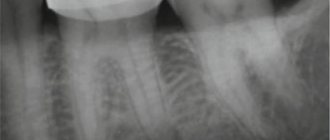
Progress of treatment of both teeth according to radiographs:
The patient was returned to the referring doctor for continued treatment and prosthetics.
Conclusions from this case, especially for young doctors:
- Inside the channels, do only what you see, do not work by touch!
- If you encounter a problem, stop on time! There is no need to be a hero if you do not have enough equipment, skills, or experience to cope with the situation. Refer the patient to more experienced and equipped colleagues, or at least warn the patient of the significant risk to him if he continues. This will be better for both of you and will save you from unnecessary conflict situations.
- use the rules of orientation in the pulp chamber! They are very effective, there are only a few of them, we talked about one of them today.
In subsequent articles and cases, I will tell you about other rules of orientation inside the tooth, which will help you not to make any mistakes. Stay tuned!
How long does root canal treatment take?
The duration of tooth canal treatment depends on the complexity and characteristics of the clinical case, as well as the number of canals that the specialist will have to treat. On average, the duration of the procedure can range from 20 minutes to 1.5 hours.
It is also worth considering that for high-quality root canal treatment, you will have to visit the dentist’s office several times.
Depophoresis
All modern methods of filling root canals are imperfect - they do not guarantee complete tightness of the filling. This is why the depophoresis method is so useful - it allows, using a special device, to completely sterilize the canal to get rid of infection.
In fact, this method cannot be fully called filling, since it is used even on already filled teeth. Copper and calcium hydroxide is injected into the tooth canals, sterilizing everything, even the most inaccessible areas, and also leaving a medicinal depot there that prevents the occurrence of infections. Depophoresis can be performed several times every week. Its cost is quite high, but it allows you to keep the tooth sufficiently hard for many years, even with the pulp removed, and to avoid infections.
Is it painful to treat root canals?
Thanks to the use of modern anesthetics, root canal treatment has become a painless procedure. The patient may feel mild discomfort only during the injection of the anesthetic.
Therefore, there is no need to be afraid of root canal treatment; moreover, you must remember that you should never postpone the procedure due to fear of the dentist! Inflammation in a tooth that has progressed to a certain stage of development cannot be cured, which means that the diseased tooth will have to be removed and a prosthesis put in its place. In addition, more serious complications may occur.
Obturation with Thermofil system
Thermophile are special plastic carriers with gutta-percha. This method allows you to seal not only the main root canal, but also the side canals. First, a small amount of sealer is injected into the canal to ensure better glide and contact with the surface. Then the plastic rod is heated, inserted into the channel and cut. Gutta-percha penetrates into all cavities of the canal, which ensures sealing. This system is simple and reliable, and filling with its help occurs quite quickly, however, the filling material often extends beyond the canal, which can cause irritation.
Modern method of treating tooth canals: treatment of the canal cavity under a microscope
The quality of dental canal treatment depends on the correctness and accuracy of cleaning and processing of their internal space. This is a very painstaking work, since the channels themselves are quite narrow (no more than one millimeter), and in addition, they can have bends and turns. If a specialist makes a mistake when treating the canals and does not remove particles of the affected dentin, a relapse of the inflammatory process is guaranteed. The use of a special tool in treatment - a dental microscope - will help eliminate possible errors and inaccuracies in the treatment of dental canals.
Using a microscope, the dentist will perform a high-quality removal of all infected tissues, while preventing injury to healthy tooth tissues. The use of the device simplifies the process of cleaning and processing channels with significant lengths, anomalous structure,
with curvatures and branches. In addition, the use of a dental microscope during the treatment of tooth canals improves the quality of tooth restoration as a whole, and also contributes to the timely detection of hidden caries.
At our dental clinic “Uni Dent” in St. Petersburg, you can receive services for dental canal treatment under a microscope. The procedure is carried out by highly qualified doctors using modern instruments and materials, and therefore when you contact us, you can rest assured that the treatment result will be of high quality! Dentistry "Uni Dent" - come to us for a beautiful smile and painless dental treatment!
Diagnostics
Treatment of impassable root canals begins with determining their length and anatomical features. The most informative diagnostic method is radiography. To obtain accurate data on the condition of the tissues, a photograph of the tooth root is taken with the instrument inserted into it. This allows you to see:
- length of root canals,
- the presence of curvatures and perforations,
- impassable areas.
The condition of the periodontium is also assessed. This allows you to determine further treatment tactics.