715
Endodontic treatment of a tooth involves invading its internal cavities - the pulp chamber and root canals for treatment (vitalization) or removal of the pulp.
The location of the opening may be the bottom of the carious cavity or the undamaged, intact surface of the tooth.
The essence and purpose of manipulation
Tooth trephination (Greek Trypanon - trephine, drill) is the initial stage of endodontic treatment, which involves opening the top of the pulpous chamber, removing the pulp located in the coronal part, and creating access to the mouths of the tooth root canals.
This procedure is also called “crown expansion” or “endodontic coronal preparation.”
It should be noted that trephination does not involve expanding or changing the shape of the dental cavity and penetrating the tooth canals. Its more modest task is to open the crown cavity and provide access to the mouth of the root canals. The operation can be performed on both natural and artificial crowns.
According to the general medical classification, trephination is indicated by code A 16. 07. 092, where “A” means medical, indicating that this is a surgical intervention, “07” - that it is carried out in the oral cavity, and “092” indicates that this the procedure, in fact, is trepanation.
In recent years, metal-ceramic structures have occupied a major place in prosthetics for partial loss of teeth and crown defects, since they allow not only to optimally reproduce the anatomical shape of teeth and completely restore chewing function, but also meet the increased aesthetic requirements of patients, allowing them to achieve a functional and aesthetic optimum.
Odontopreparation for metal-ceramic structures involves grinding down tooth tissue up to 2 mm, which can adversely affect the condition of the dental pulp. To avoid complications, it is necessary to strictly adhere to the regimes for preparing hard dental tissues, know the safety zones, and master the methods of local anesthesia. One of the main conditions is also to take measures to protect the prepared tooth from temperature and chemical influences and the penetration of infection through open dentinal tubules. All this requires highly qualified orthopedic dentists, additional time costs, the availability of high-tech equipment and modern materials.
In some cases, as part of special therapeutic preparation of teeth, it becomes necessary to depulp them before prosthetics. Indications for such preparation of teeth have long been formulated and defined. According to various authors [1, 3, 5, 7], therapeutic preparation in the form of tooth depulpation is a last resort measure that should be taken in the following cases:
- if a wide tooth cavity is determined radiologically, during preparation the tooth may be opened or after preparation there will be a thin layer of dentin that is not able to protect the pulp;
— with dentoalveolar elongation or protrusion of teeth >5 mm;
- with anomalies of position, inclination of the teeth >15°, rotation of the central incisors of the upper jaw >30° and lateral incisors >50°;
— in patients with periodontitis, when splinting the teeth of the anterior group, if a decision is made to significantly shorten the crowns of the teeth;
- with pathological abrasion of teeth II and III degrees;
- if persistent hyperesthesia occurs after tooth preparation, which does not go away after conservative treatment;
— in cases of significant destruction of the crowns of natural supporting teeth, when a decision is made to restore with core inlays [1, 3, 5, 7].
According to the literature [2, 4, 6] and our own observations, despite the existence of strict indications for depulpation of teeth before prosthetics with metal-ceramic crowns, as part of orthopedic practice, most intact abutment teeth are depulped before prosthetics to reduce the incidence of complications and ease of preparation.
Unreasonable depulpation - deprivation of an organ's viability - cannot be lawful from both an ethical and a legal point of view.
The purpose of the study is to determine the prevalence and validity of tooth depulpation during therapeutic preparation before prosthetics with metal-ceramic crowns.
Material and methods
Outpatient records of patients from several dental clinics of different forms of ownership were retrospectively analyzed. A total of 873 outpatient records of patients whose prosthetics were performed with metal-ceramic crowns and bridges were analyzed.
The protocol for analyzing case histories included the following points:
1. Diagnosis.
2. The total number of teeth used to support metal-ceramic crowns and dentures (previously pulpless and intact).
3. The number of teeth subjected to endodontic revision (from those previously depulped), the reasons for depulpation.
4. Number of teeth with satisfactorily filled root canals.
5. The number of intact teeth subject to prosthetics and depulpation in the direction of orthopedic doctors.
6. Justification for referral for pulp removal (availability of indications).
7. The number of teeth depulped by general practitioners in the direction of orthopedic doctors.
Results and discussion
Analysis of the 1st point showed that out of 873 patients, 267 (30.58%) were diagnosed with partial tooth loss; the diagnosis of “crown defect” was recorded in 141 (16.15%) medical records; a combination of these 2 diagnoses occurred in 465 (53.27%) case histories.
According to paragraph 2 of the protocol, 3174 teeth were used as supports for metal-ceramic structures, of which 1539 (48.53%) were previously pulpless and 1635 (51.47) were intact.
By determining the number of teeth subjected to endodontic revision (from previously pulpless teeth), we found that out of previously pulpless teeth (1539), 606 (39.38%) underwent endodontic revision; Thus, the number of qualitatively filled teeth was 933 (60.62%).
Repeated endodontic treatment was carried out on 606 (39.38%) teeth, including due to inflammatory changes in the periapical tissues - in 225 (14.62%), due to poor-quality root canal filling - in 381 (24.76%) case.
The number of intact teeth subject to prosthetics and depulpation by orthopedic surgeons was 1377 (84.22%) of the total number of intact teeth - 1635. Moreover, in 1371 (83.85%) cases, the reason for referral for depulpation was listed in outpatient records It was stated: “for orthopedic indications.” In 6 (0.37%) cases, the reason for referral for depulpation was the presence of changes in the periapical tissues on radiographs. Finding out the reasons for referral for tooth depulpation if there was an entry in the outpatient card “for orthopedic indications”, neither in the “objective examination” section nor in the diagnosis we found any grounds for depulpation.
Of 1635 teeth, 264 (15.78%) were replaced with metal-ceramic crowns while maintaining pulp vitality.
From the records in outpatient medical histories it is clear that dental therapists unconditionally depulped intact teeth in 100% of cases after referral from orthopedic doctors.
An analysis of 873 outpatient case histories showed that during prosthetics, out of 3174 teeth used as supports for metal-ceramic prostheses, 1635 (51.47%) initially had living pulp. Of the 1635 intact teeth subject to prosthetics with metal-ceramic crowns, 1377 (84.22%) were sent for pulp removal. Changes in the periapical tissues detected on diagnostic radiographs in 0.37% of cases were indicated in outpatient records as indications for depulpation of teeth before prosthetics. The remaining 1371 (83.85%) teeth underwent pulp removal “for orthopedic indications.”
While maintaining the vitality of the pulp, 264 teeth (15.78%) out of 1635 initially intact were replaced with metal-ceramic crowns. In not a single case of referral of teeth for pulp removal “for orthopedic indications” was there an objective basis for this type of special preparation before prosthetics.
Thus, our study revealed a significant expansion of indications for depulpation of teeth under metal-ceramic crowns and, in the vast majority of cases, completely unfounded.
From a legal point of view, this type of special preparation of teeth before prosthetics is in the vast majority of cases illegal.
Indications
Trephination is the first stage of endodontic treatment; it is indicated in all cases where it is necessary to penetrate inside the tooth for its treatment (vitalization or pulp removal, obturation of dental canals, installation of a pin or other operations).
Most often, trepanation is performed in the following cases.
- The presence of a deep carious cavity in the area of the clinical crown of the tooth with a thin or completely absent septum between it and the pulpous chamber.
Even if the thin wall is preserved, it is capable of allowing pathogenic microorganisms to pass through, resulting in possible infection of the pulp. Therefore, if appropriate symptoms are present, the doctor may consider trepanation of the tooth necessary. - Chronic or acute pulpitis.
- Any form of periodontitis.
A contraindication to trepanation is the impossibility of restoring the functionality of the tooth and the need for its removal:
- impassable, curved root canals;
- severe periodontal damage;
- tooth root fracture.
In all these cases, the tooth is simply removed.
Why does a tooth hurt when you press it, and what to do in such a situation.
Visit here to learn more about the signs of dental pulp necrosis.
At this address we will tell you whether garlic works against toothache.
Causes of toothache under a crown
The list of factors that can provoke an inflammatory process even under a recently installed crown is:
- Hypothermia of the body.
- Decreased immunity; previously suffered acute respiratory diseases; viral infections.
- Failure of the crown (i.e. the crown has expired its service life) each orthopedic design has its own service life, therefore, in order to avoid pain in the tooth under the crown, it is necessary to undergo a professional examination. dental examination every 6 months.
- Chronic dental injuries. (if the filling on the antagonist tooth is too high)
- Acute injury (fall and bruise or damage in a fight).
Preparatory stage
Trepanation should be preceded by a diagnosis, a complete assessment of the condition of the crown of the tooth, its roots, and periodontal tissues.
To achieve this, the following activities are carried out:
- Questioning the patient regarding complaints. Inspection and probing of the tooth to determine the presence and depth of carious cavities in the clinical crown. Assessing the condition of the gums in the area of the problem tooth. If there are fillings, their condition is assessed.
- Electroodontometry. Determination of the condition of the pulp by its reaction to electric current.
- X-ray of the tooth. This is a mandatory and very important stage in endodontic treatment.
Using fluoroscopy, the number and condition of root canals, their length, and anatomical shape are determined. The shape and dimensions of the pulp chamber, the thickness of its walls and lid are established. The presence of lateral canals, pathological changes in the tooth ligament.
By applying the bur to the radiograph, the required size of the instrument and its correct direction when opening the tooth are determined.
Before treatment, professional cleaning of the entire dental cavity or just a segment of the jaw arch with the element being prepared may be necessary.
The procedure for gaining access to dental canals
To ensure convenience and high quality of root orifice treatment, the dentist needs to ensure a wide opening of the tooth cavity. To do this, he needs to perform the following list of operations:
- Formation of an intradental cavity in full accordance with its anatomical dimensions and shape, as well as the number, location and curvature of the canals.
- Adaptation of the shape of the prepared cavity to make subsequent manipulations as convenient as possible by: creating free access to the root orifices;
- providing a direct approach to the apical foramen;
- preparation for a specific filling technique;
- creating conditions for complete control over the movement of the tool.
Sequence of doctor's actions
Trepanation involves three main stages:
- local anesthesia;
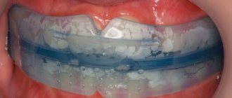
- tooth isolation by applying a rubber dam;
- directly opening the crown.
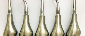
After anesthesia and the application of a rubber dam, the doctor proceeds directly to trepanation. The preparation is performed using a turbine drill with a diamond or carbide bur with a supply of coolant.
First, the crown is opened - installed by drilling a point connection between the bottom of the carious cavity or the intact surface of the tooth with its internal cavity.
Then the coronal cavity is opened - the edges of the enamel-dentin cap overhanging it are removed. Dentin sawdust and necrotic tissue are washed off with liquid.
The coronal part of the pulp is removed with a sharp excavator and EDTA (sodium salt dihydrate, which softens surface dentin) and disinfectant sodium hypochlorite are supplied into the cavity.
The walls of the cavity and the mouth of the dental canals are not additionally prepared. The main thing is that they are clearly visible and access to the dental canals is provided.
At this point, the trephination of the tooth ends and the next stage of endodontic treatment begins according to indications.
In the video, watch the process of tooth trephination.
Main types of tooth preparation
In modern dental practice, five main methods of tooth preparation are used:
- tunnel;
- laser;
- ultrasonic;
- chemical;
- air-abrasive.
Tunnel preparation
For tunnel turning of abutment teeth, turbine units are used, equipped with speed regulators, as well as metal or diamond tips.
Advantages of the tunnel view
The main advantage of this technique is the ability to control the thickness of the removed layer of dental tissue.
Disadvantages of the tunnel view
The disadvantages of tunnel grinding of teeth include:
- severe overheating of the enamel;
- increased risk of injury to the soft tissues of the mouth if the technique of performing the procedure is violated;
- a high probability of cracks and microscopic chips appearing on hard dental tissues when using worn out or low-quality instruments.
Laser preparation
For laser preparation, pulsed lasers are used to heat the water contained in the hard dental tissues and destroy the integrity of the enamel and dentin. Pieces of sanded fabric are cooled and removed using a special air-water mixture.
Advantages of the laser type
The advantages of laser tooth preparation include:
- high turning speed;
- safety (no equipment elements rotating at high speed);
- absence of noise from the operation of laser equipment;
- low heating temperature of the treated tissues;
- no cracks or chips on the edges of the pin.
Ultrasonic preparation
With ultrasonic preparation, the grinding of teeth is carried out due to high-frequency vibration of the dental instrument under the influence of ultrasound.
Advantages of Ultrasonic View
The main advantages of this method are:
- no negative effect of ultrasonic waves on pulp tissue;
- release of a small amount of thermal energy that is not capable of causing significant overheating of dentin and enamel;
- painlessness;
- no chips or cracks on the pin stacks.
Air abrasive preparation
When using air-abrasive tooth preparation, instead of a drill with a rotating drill, an abrasive-air mixture is used, supplied to the desired area under high pressure. When abrasive powder gets on enamel or dentin, tissue destruction occurs.
Advantages of the air-abrasive type
The advantages of this turning method are:
- absence of tissue overheating and excessive vibration;
- high speed of the procedure and its relative simplicity;
- maintaining the maximum volume of dental tissue;
- painlessness of the performed manipulations.
Chemical preparation
The chemical preparation method of teeth involves softening enamel and dentin using acids and other active chemicals.
Advantages of the chemical type
The advantages of this technique include:
- absence of microcracks and chips on the treated surface;
- painlessness;
- absence of thermal damage to dental tissues.
Disadvantages of the chemical type
The only disadvantage of chemical preparation is the long duration of the procedure.
Some nuances
The starting point for opening the pulpous chamber should be located at the bottom of the caries lesion or on the intact surface of the tooth closest to the pulp, that is, at the site of the pulp horn. The moment the drill enters the cavity manifests itself as a feeling of “sinking” of the instrument.
When is a rubber dam applied?
There is no consensus. Some experts recommend doing this before opening the cavity, others - after opening. The latter explain their choice by the fact that the absence of a rubber dam at the stage of opening the cavity helps to more accurately guide the tool.
Trepanation technique
Trephination methods vary somewhat depending on the group of teeth and their location (upper or lower jaw).
Technique for preparing incisors and canines
The carious cavity on the cutting surfaces of the incisors and canines is transferred to the lingual/palatal surface, and only then the pulpous chamber is opened.
Preparation of intact incisors and canines begins from their lingual/palatal surface - from its central point. Preparation from the cutting edge can lead to destruction (breakage) of the lateral surface of the incisor or canine. The intact incisors are exposed in the area of the blind fossa.
During trephination, the instrument is first directed perpendicular to the palatal or lingual surface of the canine or incisor. After the hole is formed, the drilling direction is changed to parallel to the longitudinal axis of the tooth.
Trepanation of premolars
In premolars, carious cavities begin to be opened from their bottom in the place closest to the pulp. In this case, they try to transfer the preparation to the chewing surface.
The opening point for intact premolars should be located on the occlusal surface in the middle of the fissures. When drilling, the bur is directed towards the most pronounced tubercle.
The dental cavity should be opened in the palatal-buccal direction. Doing this in a mesial or distal direction is not recommended, since this quite often leads to perforation of the walls of the coronal cavity.
The opened cavity should have a round or oval shape.
Molar preparation
Carious cavities in the molars of the upper jaw are trepanned in the same way as premolars. During opening, the bur is directed in the palatal-buccal direction.
Lower intact molars have their own opening characteristics. The bur entry point should be located in the middle of the longitudinal fissure. The instrument is directed towards the anterior buccal cusp. Unlike maxillary painters, the cavities open in the mesial-distal direction. Buccolingual opening is considered faulty.
The second and third maxillary molars are the most difficult for trephination due to the different structures of their crowns.
Difficulty identifying canal mouths
If the orifices of the root canals are poorly identified, their location is established using a dye that colors the entrances to the canal dark blue.
What is the danger of chronic caries and its treatment tactics.
In this publication you will find instructions for using a caries marker.
Here is all the most important information about fissure caries.
How is tooth preparation done?
The choice of preparation method directly depends on:
- clinical situation,
- technical equipment of the dental clinic,
- client's wishes.
Grinding of pulpless teeth is carried out without anesthesia (except for those cases when the doctor has to use special threads to move the gums back). If the procedure is performed on living (vital) teeth, the patient may require local anesthesia.
Grinded teeth are necessarily protected with temporary crowns. This allows you to keep damaged dental tissues in an acceptable condition until they are covered with a permanent prosthesis.
Teeth preparation should be carried out only in specialized medical institutions that have positively proven themselves in the dental services market. Only a professional and competent approach to the procedure allows not only to achieve the desired result, but also to avoid the development of complications.
Possible complications
Coronal preparation is less risky in terms of complications than root preparation. However, they still happen:
- Perforation of the wall of the pulpous chamber. The reason is the carelessness of the dentist, the wrong choice of instrument, the lack of an x-ray, which determines the shape of the cavity.
If the hole is small and the perforation site is located above the periodontal groove, the defect can be easily eliminated - with the help of cement.If the hole is larger than 1.5 mm, and the perforation is located below the periodontal groove, there is a risk of filling material leaking into the periodontium, which can lead to inflammation.
- Broken tool. Occurs due to excessive application of force, the use of low-quality instruments, poor condition of the crown, and the doctor ignoring the shape of the cavity.
- Change in color (darkening) of the crown . The reasons are a violation of the operating mode of the tool (high speed, lack of cooling), improper processing of the cavity walls.
- Pain. They may appear after the instrument enters the periodontium, when working without cooling, or due to poor-quality antiseptic treatment (re-infection).
In general, complications are caused by preparation errors, lack of cooling, incorrect choice of instrument, and lack of high-quality x-rays. To avoid them, you need to strictly follow the operation protocol.
Expert opinion
Igor Yurievich Malinovsky
Maxillofacial surgeon, implantologist
Experience: more than 11 years
Transplanting teeth from one area to another is not widespread. The risks of complications are quite high; the tooth may not take root. And it is morally difficult for the patient to experience psychological stress again. Especially considering that these are often children - teenagers, boys and girls. Among dentists there is no clear opinion on this method of restoration. And I am also inclined towards temporary prosthetics, until the time when it will be possible to put reliable and comfortable crowns on the implants. Of course, it is worth mentioning that autologous dental transplantation should not even be considered for older patients.
Reviews
The vast majority of visitors to dental offices are familiar with tooth trephination, which everyone simply calls “drilling.”
If you are one of them, and some special incident happened to you during tooth trephination, share your experience with other visitors to our site. You can do this by filling out the form at the bottom of this page.
If you find an error, please select a piece of text and press Ctrl+Enter.
Tags toothache
Did you like the article? stay tuned
Previous article
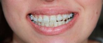
In what cases are braces + springs used and how effective?
Next article
What you need to know about supraocclusion
Grigoriev Alexander.
SCAD member, academic director. International lecturer on adhesion and interdisciplinary restoration.
Highly aesthetic works from metal ceramics, press ceramics and frame ceramics on zirconium and aluminum oxide are a reality today. The skills and craftsmanship of the medical technician team create masterpieces that stun patients with their results. Today, aesthetic rehabilitation services can improve the patient’s personal status and sometimes change their fate.
But there is also a downside to this happiness: a chip of a fragment of ceramics, a chip of a veneer, a disintegration of an orthopedic crown, and sometimes even a defect in the tooth stump. Moments of happiness stop at this moment for our patient. The beloved restoration is no longer pleasing to the eye.
Using the example of one clinical case, I will show how to effectively and accurately carry out the inversion (reverse) prosthetics method.
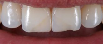
Patient M., 35 years old, came to the clinic with complaints of decementing of an aluminum oxide crown by 2.1.
Objectively: the crown had a movable position on the stump of tooth 2.1 and was removed without effort. A chipping of 1/3 of the tooth stump was noted (the inner part of the crown was filled with the separated fragment).
Defect of stump of 2.1 tooth
In the past, the tooth was subjected to endotherapy followed by restoration using a polymer post. There was complete preservation of the polymer pin throughout. Only the inner surface of the crown with a fragment of core material broke off.