The remedy for Fordyce granules is an effective treatment option for neoplasms that appear on the skin of the human body. The phenomenon of seborrheic cysts is quite common, and for the most part does not cause concern among its owners. Nevertheless, for some people they become an acute problem, and all possible methods are selected to get rid of this phenomenon. Patients are looking for a method that will help get rid of Fordyce granules without causing harm to health.
Features of Fordyce granules
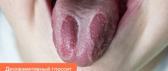
Fordyce granules look like small compactions - nodules, the size of which does not exceed 2 millimeters and a height of 1 millimeter. They have a light, flesh-colored or white color. They are located mainly in groups of several dozen specimens. Formations can be seen on the nipples of women, in the genital and anal areas, in the armpits and on the face. This anomaly affects about 35% of people, mostly women. When such a granule is squeezed, a curd mass may come out of it, white or yellow in color with a specific odor.
The presence of granules is almost unnoticeable. They do not cause pain, and the discomfort of their presence is completely eliminated.
Many experts consider such neoplasms to be an absolutely normal phenomenon. The reasons for their occurrence are associated with the abnormal location of the sebaceous glands, which is considered completely acceptable or normal.
The growths do not cause physical discomfort to a person. The desire to get rid of granules is associated with psychological discomfort due to the ugliness of the skin. The formations are not prone to complications, transmission to another person is excluded.
The appearance of granules can occur due to:
- stress;
- poor nutrition;
- bad habits;
- environmental impact.
The appearance of granules is observed during the period of hormonal changes in the body. Vulnerable periods are puberty and pregnancy. Often the granules dissolve on their own, without any intervention.
Cost of removal of Fordyce's angiokeratoma
| Cost of laser removal | price, rub. |
| Removal of Fordyce's angiokeratoma with CO2 laser (taking into account application and infiltrative anesthesia) | 7 900 |
| Removal of Fordyce granules with CO2 laser (taking into account application and infiltrative anesthesia) | 7 900 |
| Removal of an epidermal cyst | 5 000 |
Make an appointment for Fordyce angiokeratoma removal
- Full name
- Telephone
Description and symptoms of angiokeratoma
Angiokeratomas have many varieties and are divided into groups of diseases. One of them is Fordyce's disease, described by a researcher 120 years ago. The genetics of the formations have not been clarified, although it is known that they are caused by a developmental defect.
Most often, men, not women, face the problem. According to statistics, angiokeratomas of the labia are less common than angiokeratomas of the scrotum or penis. The first manifestations of the disease usually occur in adulthood, although there are exceptions.
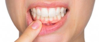
What are the symptoms and features of the disease? The latter manifests itself in the form of small brown-red nodules. The skin underneath remains smooth. When rubbed, angiokeratomas of the thighs, vulva, penis or scrotum may bleed. Possible damage to the sebaceous glands, nipples, and oral mucosa.
The disease is not dangerous to the patient's health. Typically, benign formations that arise are removed only for aesthetic purposes. Therefore, this disease should not be confused with seborrhea, focal eczema, neurodermatitis, or, for example, molluscum contagiosum.
Treatment of Fordyce's angiokeratomas
Currently, various methods for removing such pathologies are used, but none of the methods has proven to be 100% effective. This is mainly due to the fact that the clinical causes of the disease have not been clarified.
To remove angiokeratomas of the thighs, vulva, penis, scrotum, the following can be used:
- Retin-A;
- Jojoba oil;
- Electrocoagulation (removal of formations by electric current),
- Cryotherapy;
- Laser application;
- Use of liquid nitrogen (cryodestruction).
Under no circumstances should you squeeze out angiokeratomas or treat them yourself! Any procedures can be carried out only after consultation with an experienced dermatologist and tests.
Laser therapy for Fordyce's angiokeratosis
For limited (mild) forms of the disease, laser treatment is the best treatment. Photons not only remove Fordyce angiokeratomas, but also trigger many important processes. At the same time, the elasticity of the skin membrane is maintained, microcirculation of the blood flow is normalized, and tissues are regenerated.
Usually, laser therapy allows you to get rid of angiokeratomas completely, unless, of course, the area of skin damage is too extensive and the disease is not chronic and hereditary. The duration of laser treatment is minimal, and the results are immediate.
If you are interested in removal of Fordyce angiokeratoma, you can make an appointment with one of the Center’s specialists by phone
Types of treatments
Treatment is not a necessary measure in this matter. Nevertheless, for many people it becomes relevant, as it causes many complexes about the appearance of the body.
Ointments
The most popular remedy for combating Fordyce's disease is Retin A ointment. The drug affects the secretion of the sebaceous glands and dissolves the formations that have arisen. Trans-retinoic acid, which is part of the ointment, neutralizes the contents of sebaceous plugs and regenerates skin tissue. You need to apply the cream once a day, preferably in the evening, before bed. It is important to avoid contact of the substance with the eyes and mucous membranes of the body. After 6 hours, it must be washed off with running water. The first effects are observed after 2-3 weeks. The full course of treatment lasts up to 2 months. Analogs of the cream drug Airol and Lokacid.
Removal by devices
New growths can be removed by performing an instrument procedure. Modern medicine offers many options for removing skin defects in a hospital setting:
- Cryodestruction - granules are frozen with liquid nitrogen. A special device is applied to the affected area for a few seconds. After this, the growth begins to collapse and dies.
- Laser removal is the most popular. It occurs under the influence of a special laser beam, which burns out the growth layer by layer. Removing Fordyce granules with this laser takes several minutes.
- Electrocoagulation – neutralization of the cyst occurs using high frequency electric current. The current energy accumulates in the tip, after which it evaporates the liquid from the tissues of the growth. Thanks to the method, elimination takes place without blood loss.
- The radio wave method is the most gentle. Excision is carried out by exposure to high-frequency waves that cut off the formation. Moreover, at this moment there is no contact between the device and the tissues.
We recommend reading:
- How to get rid of warts on the neck
- Is it possible to cauterize condylomas?
- Features of the appearance of a wen on the scrotum
Medications
Treatment of cysts is carried out without the use of various medications. In special cases, your doctor may recommend taking anti-inflammatory drugs or antibiotics to prevent bacterial infection. The selection of medications is solely at the discretion of the doctor, depending on the symptoms of the disease and the characteristics of the patient.
How does a cosmetologist remove it?
In addition to standard clinical treatment methods, a person with rashes can be treated at home, or seek the help of a cosmetologist.
After a diagnostic examination, the cosmetologist can select one of the most suitable methods. Most often, for such a problem, treatment with the Darsonval apparatus is used or permanent makeup is applied.
The use of Darsonval allows you to cleanse the skin, saturate it with oxygen and restore natural metabolic processes in tissues. The sebaceous department is normalized. The skin becomes more pleasant and smooth to the touch. In this case, the course of treatment will be about 10 sessions. During the procedure, the specialist installs the necessary attachment – an electrode – into the device holder. After this, light movements massage the skin. Unfortunately, cosmetologists do not give a 100% guarantee of an effective outcome. A similar procedure can also be carried out at home.
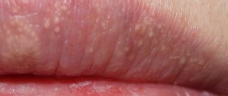
Permanent makeup is used for minor tissue damage. Especially if the growths are on the lips. To do this, a special pigment is introduced into the required area, which colors the granules that stand out in color. Tissue tattooing takes place. The result of the procedure lasts for 1-2 years, after which the event must be repeated.
One of the rare skin diseases of interest to dermatologists is Fordyce angiokeratomas (Fordyce angiokeratomas of the scrotum/vulva).
Angiokeratomas are vascular tumors formed by persistent subepidermal capillary dilations and changes in the epidermis with symptoms of hyperkeratosis, papillomatosis and acanthosis.
There are several clinical forms of angiokeratoma: limited angiokeratoma, nevoid angiokeratoma of the Mibelli fingers, solitary papulous angiokeratoma of the scrotum/vulva of Fordyce, diffuse angiokeratoma of the Fabry trunk.
Fordyce's angiokeratomas (Fordyce's angiokeratomas of the scrotum/vulva) were described in detail in 1895 by researcher Fordyce. This type of angiokeratoma is a developmental defect. The disease is benign. To date, it has not been possible to identify the genetic cause of Fordyce disease [1, 2].
Clinical picture.
This type of disease is manifested by the formation of multiple brown-red nodules located on the skin of the scrotum, less often on the penis and inner thighs. In women, rashes appear on the thighs and labia. Vascular nodules are bright red in color, gradually increase in size, and their color becomes darker.
Nodules with Fordyce angiokeratoma are small - 1-5 mm in diameter, the surface of the skin over them is smooth, less often - hyperkeratotic. Sometimes patients complain of itching; most patients have no subjective sensations [1, 2].
When scratched, pressed, or rubbed with linen, angiokeratomas begin to bleed. If bleeding occurs frequently, then Fordyce's disease can cause the development of mild iron deficiency anemia.
Scrotal angiokeratomas may be associated with varicocele, inguinal hernia, or thrombophlebitis. Angiokeratomas of the vulva are much less common than angiokeratomas of the scrotum, mainly in older women. In young patients, vulvar angiokeratomas may develop due to increased venous pressure during pregnancy or the use of oral contraceptives.
Diagnostic methods.
The diagnosis is relatively simple and is based on characteristic clinical data. In some doubtful cases (severe hyperkeratosis) is confirmed by the results of histological examination.
Histological picture
. The phenomena of hyperkeratosis, acanthosis, and papillomatosis are observed. The capillaries are sharply expanded and located close to the upper layer of the epidermis or in its outgrowths. Some capillaries contain lymph [3].
The prognosis is favorable
. Complications are rare and uncharacteristic. Only in rare cases (when scratched or injured by clothing) angiokeratomas of the scrotum and vulva can cause minor bleeding [4, 5].
Differential diagnosis
carried out with melanoma, lymphangioma, hemangiomas, Kaposi's sarcoma.
Considering that the localization of Fordyce angiokeratomas in the area of the glans penis is extremely rare, we present the results of our clinical observation.
Patient X
., 21 years old, applied to the center for viral pathology of the skin on January 26, 2015 with complaints of a rash in the area of the glans penis, the inner layer of the foreskin (see picture).
Clinical manifestations of Fordyce angiokeratoma in patient X.
History of the disease.
He considers himself sick for about 3 months, when he first noticed the appearance of rashes in the area of the glans penis, which gradually increased in size, spread to the inner layer of the foreskin and did not cause subjective sensations. When contacting a commercial medical center, the patient was diagnosed with “Anogenital warts.” To clarify the diagnosis, the patient was sent to the center for viral pathology of the skin of the Babushkinsky branch of the Moscow Scientific and Practical Center of Culture and Culture of the Department of Health.
Local status
: the process is widespread, asymmetrical, sub-inflammatory in nature, localized on the skin in the area of the glans penis and the inner layer of the foreskin, represented by multiple papules of a bluish-burgundy color, tending to group, round in shape, with clear boundaries, with a diameter of 0.3-0 .8 cm.
Microflora smear
from 09/16/14 - norm;
blood for HIV, syphilis, hepatitis B and C
dated 09.18.14 - negative.
The patient was sent for a diagnostic biopsy on January 26, 2015, where several lesions were collected for pathomorphological examination with a preliminary diagnosis of Fordyce hemangiomas (?). Kaposi's sarcoma (?)".
Results of histological examination:
in the preparations there are fragments of skin formations with dilated blood vessels with flattened endothelium and the presence of blood clots in their lumens. The epidermis is flattened in places, with shallow acanthosis and focal hyperkeratosis.
Histological diagnosis
from 01/30/15: Fordyce angiokeratomas.
Thus, according to the above clinical observation, Fordyce angiokeratomas can be localized in places atypical for a given nosology, which can lead to diagnostic errors and require a histological examination to clarify the clinical diagnosis.
No conflict of interest
.