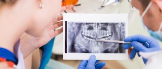
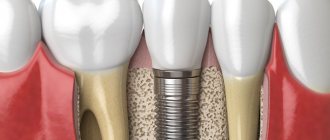
Dental implants are one of the most widely used methods to replace missing teeth. To do this, you will visit your dentist, and after an examination, your dentist may inform you that a bone graft is required before the implant can be placed. And this is often scary for patients, especially when they are not familiar with bone grafts for dental implants. But don't worry because the dental bone grafting procedure is simple and painless. If you are reading this, you are probably a candidate for bone graft surgery and want to learn more about it. You have come to the right place because this article will explain everything you need to know about bone grafting, including the technique, different types of grafts, surgery cost, success rate, risks and side effects.
What is a Dental Bone Graft?
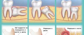
Part of the jawbone may degenerate over time due to the loss of one or more adult teeth. In this case, you may need a bone graft before getting a dental implant to increase the amount of bone in the affected jaw area. Overall, dental bone graft surgery is a painless and simple procedure in which bone taken from any part of the body is fused to the existing jaw bone. These bone grafts are taken from the patient's bones, such as the hip, thigh, or back of the jaw. It may also be artificial. You may ask, “Are dental bone grafts necessary?”
In answer to this question, we must state that this surgery is necessary if the patient does not have enough bone in the jaw to support the implant. Factors such as gum disease, facial injury or trauma, and developmental problems can also damage part of the jawbone, in which case bone grafting is necessary.
Also visit our bone graft products: Monib Bone Graft
Types of grafts for bone grafting
Natural bone material for bone grafting in modern medicine is not used as often as it seems. Innovative technologies for the production of artificial transplants make it possible to avoid many of the disadvantages and risks of using natural materials.
There are four types of grafts for bone grafting:
- Autogenous material. Transplants that are taken from different parts of the jaw, chin or hard palate from the patient himself. It takes root the fastest and most successfully, since it does not have antigens to which the human immune system will react. However, the installation of such grafts requires additional intervention. During the recovery period, multiple surgical approaches will need to be monitored and cared for.
- Allogeneic material. Natural bone material that is taken from another person. As a rule, corpses are used, that is, the use of such transplants requires special laws that allow the removal of any body parts from deceased people. Allogeneic material takes root less well, but is also absolutely safe, as it undergoes a number of treatments and quality controls.
- Xenogeneic transplant. Materials of animal origin are enjoying great success in modern technologies. They are made from bovine tissue and carefully modified into a human-fit structural design or shavings. By filling the space with such material, the doctor increases the likelihood of increased regeneration of the patient’s own bone.
- Synthetic material. Artificial structures are widely used for several reasons. The main one is the low cost of the material. In addition, the patient can have 100% no doubt about the safety of a synthetically created graft.
The patient is involved in the selection of material for bone grafting, since the financial side of the issue is not in last place. The dentist is obliged to explain to the person in detail all the features of natural and synthetic transplants, as well as recommend the most optimal options.
How does Dental Bone Graft Work?
There are several methods for performing bone graft surgery. However, the general process involves the dentist making a small incision in the jaw and inserting a bone graft into the jaw. So how do these bone grafts or powders work? Bone matrix is found in the skeletal system. It is a hard material that helps strengthen bones. The matrix is formed and maintained by the living bone cells within it. Doctors connect bone grafts containing matrix and living cells to the jaw. The cells inside the new bone can then attach to the old bone. As a result, the living bone cells in the bone grafts form a matrix and heal the damaged bone. As a result, your jawbone will eventually become strong enough to support the implant.
Introduction
Elimination of jaw defects plays one of the leading roles in craniomaxillofacial surgery. Bone grafts are used to fill bone deformities and defects in the craniofacial skeleton. Bone tissue is the only tissue in the body capable of reparative osteogenesis without scar formation and organotypic regeneration, subject to a number of conditions. Free bone grafts are bone blocks obtained from donor areas. In maxillofacial surgery they are usually divided into intra- and extraoral [7, 8]. Extraoral zones include both parietal bone and fibula grafts. It should be recognized that such a division is somewhat arbitrary, since in terms of their structure and properties all types of grafts, including intraoral ones, have a sufficient number of differences among themselves, which makes such a division convenient only when planning surgical treatment, but not from the point of view of predicting its effectiveness [9 , 11, 12].
The method of autotransplantation of bone blocks is widely used in such areas as traumatology and orthopedics, maxillofacial surgery, reconstructive surgery, oncology, neurosurgery, etc. In literature sources it appears as the “gold standard” in the treatment of many diseases due to the possibility of obtaining a predictable clinical result [3, 17]. Most publications on the high effectiveness of bone block transplantation are based on clinical and aesthetic results. The few works containing the results of clinical and histological studies are, as a rule, descriptive in nature [4, 19]. The main mechanism for integrating the transplanted block is considered to be the osteoconductive properties of cancellous bone, which maintains viability. As for the cortical plate, its role has not been fully determined [2, 14]. However, clinical data indicate signs of dystrophic changes in the transplanted free bone beams, which is manifested in a decrease in mineralization over time [15]. Since replacement of extensive bone tissue defects with free grafts is associated with a risk of complications, the next generation of autografts became vascularized grafts, or grafts with a vascular pedicle. They are part of an organ with a preserved vascular network, take root in the bed and undergo integration and restructuring under load much faster than non-vascularized ones, which is explained by the same structural features of bone tissue [18]. The few scientific publications containing the results of histological studies do not shed light on the mechanisms of graft engraftment, regeneration, or their future fate [6, 10]. A comparative analysis of vascularized and nonvascularized grafts using radiation and morphological research methods showed that a graft on a vascular pedicle retains the ability to retain the mineral component and reconstruct the graft according to the type of cortical bone in the anastomotic area [5, 13, 16].
Thus, at present, favorable conditions have developed for the dynamic study of reparative osteogenesis during autologous bone tissue transplantation.
Types of Dental Bone Grafts
Another concern for patients is the type of dental bone graft. Because the quality and material of the grafts, as well as the procedure, are important to them. Dental bone grafts are divided into four types based on their origin and components:
Allografts
Allografts are obtained from the bones of another person, usually a corpse. Patients generally do not prefer to use these types of grafts, but dentists use them widely today. Allografts themselves come in various forms, such as the socket graft and the lateral ridge sparing graft . The main goal of socket grafting is to avoid alveolar bone atrophy. A lateral ridge preservation graft is used to expand the jawbone to accommodate a dental implant.
Autografts
These types of grafts contain bones from your own body, such as your hip or jaw. Block bone graft is a well-known example of an autograft. Dentists use this type of graft when there are significant defects in the jawbone.
Xenografts
These grafts are made from bones from other species, such as cows, pigs or coral. Dentists often use these grafts to treat jaw bone defects.
Alloplasts
The newest type of dental bone graft is completely artificial and made from compounds such as calcium phosphate or calcium phosphosilicate.
Bone reconstruction: materials and possibilities
The history of bone augmentation with materials of various origins reaches almost a century ago. Significant problems associated with transplantation materials and growth enhancers relate to their origin, rationale for use, method of collection, taking into account the volume of so-called biological “costs”. Another key issue with bone grafts concerns the potential “repair” or regeneration aspect of the intervention site. Reparation is the process of replacing a defective area with something physically similar in structure, but different biologically and physiologically. Regeneration, on the contrary, involves the use of different materials and methods that make it possible to restore the biological structure of the defective area with completely identical tissue. Surgeons planning to perform augmentation need to clearly understand what outcome they can predict at the end of rehabilitation, and how successful the techniques they use will be. By clearly understanding all aspects, the physician can reach a consensus on the restoration of vital bone, the amount of augmentation performed, the rate of healing and the optimization of the iatrogenic intervention procedure.
Directed tissue regeneration involves the implementation of a number of stages to achieve a certain volume of bone tissue formation in the area of interest. This approach involves the formation of a scaffold on which new tissue will form. Blood vessels must grow into the developing area to provide it with a nutrient medium. Calcium is necessary for the mineralization of the organic matrix, and osteoblasts will ensure the deposition of collagen, which will subsequently turn into bone tissue. Signaling molecules promote the attraction of progenitor cells (osteoblasts, endothelial cells), which ensure the formation of a specific type of tissue in the area of interest. Achieving a successful result of tissue regeneration in the maxillofacial area is a rather complex process, taking into account the constant activity of muscles, the presence of variable forms of bacteria and the liquid environment of saliva. Understanding the aspects of using various bone grafting materials is an important step in achieving the required successful outcome during the complex rehabilitation of a dental patient.
Recommendations regarding bone reconstruction
The choices faced by the surgeon during an extraction, augmentation or sinus lift procedure primarily concern the following aspects:
- access to the defect area. The access can be created without flap separation - minimally invasively, or classically - by separating the volume of surrounding tissue.
- source of bone material. Autogenous bone, allogeneic analogs, alloplasts, xenografts, or other materials are all suitable for reconstructing residual ridge parameters.
- the type of graft used. The graft can be presented in the form of particles, paste or block, of different sizes and granularity, and can also be collected from sites of different origin (cortical or cancellous bone).
- specific characteristics of each graft. Bone substitutes are characterized by varying levels of mineralization and varying resorption tendencies. consideration should be given to whether the material is osteoconductive or osteoinductive and how quickly or slowly, or if at all, it biodegrades.
- the type of biological barrier used. The membranes used can be synthetic or collagen; they can also be resorbable or non-resorbable, biologically active or inert.
Early studies indicate that vital bone formation still occurs in the extraction socket, even without the appropriate use of graft or membrane. However, you should not rely only on such an outcome. In 2008, Fickl et al examined the differences between different extraction approaches (with and without flap separation) with subsequent filling of the socket with augmentation or without performing a reconstruction procedure. They found that the separation of mucosal and periosteal tissues resulted in greater loss of alveolar ridge width and height compared with the results obtained after implementing the minimally invasive approach. They also noted the fact that the primary bone dimensions were preserved when filling the socket with an inorganic graft of bovine origin, which was not observed without the augmentation procedure. Iasella et al, however, noted that more vital bone was formed in the postextraction socket area without any type of graft used, although the loss of initial hard tissue volume was approximately 30%. Similar consequences were avoided by filling the socket with a mineralized allograft covered with a resorbable collagen membrane.
Another study compared two different augmentation methods. A xenograft obtained from cattle was installed in the sockets and covered with a resorbable membrane - in this way it was possible to achieve a slight increase in tissue height, but the volume of the formed vital bone did not exceed 26%. When a demineralized allograft with calcium sulfate was used as an analogue, which was covered with a barrier of calcium sulfate, a similar result was noted in changes in the geometric parameters of the bone crest, but the amount of vital bone was almost twice as high as what was noted when using the allograft.
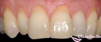
Augmentation and insulation materials
Calcium sulfate
Calcium sulfate is a synthetic analog that is used in many configurations as both a graft and a growth promoter (Figure 1). A 2004 study demonstrated that this material, in a hemihydrate form without a membrane, provides excellent tissue volume retention and approximately 60% vital bone formation. The new bone graft material described in a 2012 report is biphasic and contains both hemi- and dihydrate calcium sulfate. This material is self-precipitating in the presence of blood or saliva, and can be used with or without a barrier membrane. The report presented histological results of its use in several types of defects that indicate bone growth. In addition, it was also possible to achieve retention and increase the geometric parameters of the alveolar ridge. The patient, shown in photo 1, sought help after a significant abscess of endodontic etiology, which provoked a loss of both the vestibular and lingual bone plates. Covering the biphasic calcium sulfate with a dense polytetrafluoroethylene (PTFE) membrane for 3 weeks promoted healing of the defect site. It was clinically evident that the volume of the residual ridge was maintained for over 5 years, which in turn supported the overlying keratinized soft tissue (Figure 2). Most importantly, histological evaluation revealed 58% vital bone with no remnants of bone graft granules (Figure 3).
Photo 1. Augmentation procedure using BPCS and PTFE membrane coating.
Photo 2. View of the patient 5 years after treatment.
Figure 3. Histological examination results show 58% vital bone 4 months after augmentation.
Beta-tricalcium phosphate
Previously, there were concerns that transplant materials, which are completely reserved in a short period of time, could provoke collapse of the augmented area. Pure beta-tricalcium phosphate (ß-TCP) (photo 4) was one of the materials developed to solve this problem. Clinical and histological studies have shown that it can provide retention of 91% of the bone crest width by filling the defect area with Cerasorb (Curasan, Inc.) and covering the intervention site with collagen or dense PTFE membranes. 4-6 months after augmentation, dental implants were installed in the reconstruction area, which also had a high success rate. The patient in photo 4 had only three teeth left in the upper jaw, which supported a removable denture. Taking into account the specific conditions of the clinical situation, it was decided to reconstruct the residual ridge to create conditions for the installation of dental implants. 7 months after healing of the augmentation site, the flap was separated (photo 5) and the implant was installed. It was found that the augmentation allowed restoration of a significant part of the previously registered defect. Analysis of the resulting specimen revealed that it was composed of 85% vital bone in the apical portion and 90% in the midline portion, with β-TCP present only in a thin stripe at the coronal portion (Figure 6).
Photo 4. Beta-tricalcium phosphate coated with a collagen membrane.
Photo 5. View 7 months after treatment during re-exposure of the ridge.
Photo 6. Histological examination results demonstrate significant volume of vital bone.
TCP (tricalcium phosphate) with PLGA coating
To facilitate use, tricalcium phosphate packaging has been developed in the form of syringes, the material of which can be directly used for augmentation. Such a system consists of a single syringe containing 99% β-TCP beads coated with polylactide-co-glycolide (PLGA). The contents of this syringe are mixed with an ampoule containing BioLinker (N-methyl-2-pyrrolidone and water) (GUIDOR easy-graft, Sunstar) (Photo 7). The study demonstrated the biocompatibility and good resorption of this material in examples of reconstruction of tooth sockets after tooth extraction. This augmentation was slowly resorbed and maintained the required shape for a long time, thus currently representing a suitable alternative for bone reconstruction with or without the use of membranes. Photo 7 shows the process of using this material to augment the socket in the area of the lower molar. The intervention area was covered with a collagen membrane without primary suturing of the wound. After 8 months, a dental implant was installed in this area (photo 8). From photo 8 you can see that a significant amount of the material used was resorbed and replaced by vital bone tissue, while at the same time it was possible to maintain the original parameters of the width of the bone crest.
Photo 7. Packing PLGA-TCP into the socket of an extracted tooth.
Photo 8. View 8 months after socket augmentation.
Membranes
Although the issue of infection and exposure of PTFE membranes with wide pores remains poorly understood, dense, non-porous analogues have proven their clinical effectiveness for many years. The use of dense types of PTFE membranes (Cytoplast TXT, Osteogenics) to protect the blood clot, according to numerous studies, allows one to largely maintain the original width of the alveolar ridge. At the same time, replacement of the socket space with vital bone tissue, according to radiographic and histological studies, was noted already 3 months after covering the intervention areas with membranes. Membranes can be used similarly effectively to cover the augmentation (photo 1-3). Resorbable types of membranes have been developed to support the required volume of space, taking into account their stiffness parameters and the distance between the inner and outer layers (GUIDOR Matrix Barrier, Sunstar). The porous nature of the surfaces, consisting of a combination of citric acid ester and polylactic acid, allows tissues to grow somewhat into the membrane, thus providing a higher level of membrane stabilization. At the same time, the frequency of infection or inflammation of the tissues around the membranes is observed quite rarely, which argues for the advisability of their use in regenerative dentistry, especially in cases of restoration of bone defects around implants or even before their installation. In the case presented in photos 9-11, a synthetic membrane was installed immediately after extraction of a tooth with bone deficiency on the palatal side. The rigidity of the membrane ensured the formation of such a volume of space that was necessary to restore a certain configuration of the bone crest. After six weeks, the intervention site showed signs of complete healing without any signs of inflammation or infection. In this way, it was also possible to achieve the formation of a wider zone of keratinized tissue.
Photo 9. Subperiosteal membrane placement for significant bone loss on the buccal side of the socket.
Photo 10. View of the final ridge shape.
Photo 11. View 6 weeks after healing.
Fillers
Considering the disadvantages of powder forms of augmentations, some of their analogues have been developed in the form of pastes for multifunctional use: reconstruction of the socket area, sinus lift and bone crest augmentation. The previously mentioned BPCS and PLGA/TCP are just such materials. They are essentially a combination of demineralized/mineralized allograft and collagen gelatin (Optecure, Exactech), thus representing a semi-rigid material for bone tissue reconstruction. According to the manufacturers, these materials are also characterized by osteoinductivity with the possibility of forming significant volumes of vital tissue. The patient in photo 12 sought help with a bone width of approximately 1 mm in the frontal area of the lower jaw. The allograft was mixed with sterile saline and covered with calcium sulfate (3D Bond, Augma Biomaterials) and Ossix Plus membrane (Datum Dental Ltd). Narrow diameter implants (ANEW, Dentatus) were installed 4 months later. The clinical result of the treatment is depicted in photo 13, representing the view 3 years after loading of the installed implants.
Photo 12. View of the area of bone tissue deficiency in the frontal area of the lower jaw.
Photo 13. View after 3 years of the prosthetic field.
Inorganic graft of bovine origin
Early studies of the sinus lift procedure used bovine inorganic bone graft. Many publications have described the use of this xenograft either alone or together with autogenous bone tissue, or even with certain growth factors. When the xenograft was used in isolation but covered with a membrane, the percentage of vital bone formation ranged from 12% to 17% at 6 to 10 months postoperatively. In another study, a xenograft was used together with growth factors to raise the level of Schneiderian membrane. The results of this multicentric study involving 25 patients demonstrated that bone formation after 6 months was about 50%, which is more effective not only in terms of tissue growth, but also in terms of time spent on rehabilitation.
conclusions
In discussing the results of a histological study of the area of tooth socket reconstruction using calcium sulfate hemihydrate, Guarneri and colleagues stated the following: “The goal of any augmentation procedure is to achieve the formation of 100% living bone tissue surrounding the implants.” Depending on the size and location of the defect, the specifics of the relationship with surrounding tissues, this goal can be achieved, or, conversely, impossible to achieve. According to long-term studies of the success of implants installed in the area of augmentation, the presence of non-vital graft particles in the intervention area does not in any way compromise the success of treatment. Studies describing the experience of using resorbable membranes indicate the possibility of immediate installation of implants with a reasoned choice of augmentation method. Materials for restoring the volume of the bone crest, as well as for isolating the reconstruction area, provide the physician with various options for adapting the surgical approach during the complex rehabilitation of the patient. Having determined the final goal of the procedure, and having predicted its possible outcome after 3, 6, 9 or more months, the doctor can more convincingly approach the choice of intervention algorithm, based on preliminary data described in respectable scientific sources.
Authors: Robert A. Horowitz, DDS Minas D. Leventis, DDS, MS, PhD Michael D. Rohrer, DDS, MS Hari S. Prasad, BS, MDT
Best Candidate for Dental Bone Implantation
Is it possible to install a dental implant without a bone graft? Good question and the answer is yes. Dental implants do not always require bone graft surgery. This is possible when you go to the dentist immediately after losing a tooth. However, if you wait a few months, the jawbone in the place of the missing tooth will begin to deteriorate. In these conditions, you will need a bone graft before getting a dental implant. Dental bone grafts are not just for implants. This surgery is necessary in circumstances such as gum disease, trauma and trauma to the jawbone, and developmental abnormalities. As a result, choosing an experienced dentist and using high-quality bone grafts allows the operation to be performed without complications.
Bone Marrow Transplant Center at Anadolu Clinic
A specialized bone marrow transplant center has been operating at our medical center since 2010. On average, Anadolu doctors perform 250 transplants per year on patients 16 years of age and older.
The center includes an outpatient clinic, a clinical department for bone marrow transplantation and three laboratories: genetic typing, hemophoresis, cryopreservation and stem cell processing. Our specialists are highly qualified transplantologists and hematologists who are proficient in all modern techniques of bone marrow transplantation. In their work, they strictly follow international protocols for the treatment of blood cancer and observe all precautions and safety measures.
The Anadolu Bone Marrow Transplantation Department works not only with the international, but also with the Turkish bone marrow bank, which allows you to select a suitable donor as quickly as possible - in just a few weeks. In a global registry, this process can take several months.
The material was prepared in agreement with the doctor, medical professor Zafer Gülbash.
Dental Bone Graft Rejection
Another concern for patients is the failure of dental bone grafts. You may ask, is this possible? Although dental bone implants have a success rate of approximately 99.3%, failure can occur. But what causes this failure? There are several factors involved, but the leading cause of bone graft failure is bone graft surgery performed by an inexperienced surgeon. But this can happen to professional surgeons too. Bone graft rejection or failure can occur for the following reasons:
- If the bone grafting material is contaminated with bacteria, the procedure will fail;
- Infection of surgical instruments can lead to the transfer of bacteria to the surgical site and graft failure.;
- Failure is possible if the patient does not properly follow the postoperative care prescribed by the surgeon,
- If you do not maintain proper oral hygiene after surgery, you may experience problems.
Symptoms of Single Dental Graft Rejection
Bone graft rejection can occur in early and late stages, each with its own symptoms. You may experience early problems within three to four months after surgery. In this case, the following symptoms may occur and you should visit your dentist immediately:
- Swelling of the dental bone graft: Swelling in the surgical area and part of the face is common a few days after surgery. However, if the swelling lasts more than a few weeks or gets worse, it is a sign that your body is rejecting the bone graft.
- Acute pain: A few days after surgery, pain and swelling are normal because bone implant pain is one of the most common side effects of this surgery. However, if the pain persists and worsens over time, it may indicate that the bone graft has failed.
- Large amounts of leakage: As with other surgeries, some drainage from the treated area is expected. However, if the leak persists and a large amount of drainage occurs, this is one of the most important symptoms of graft failure and you should see a doctor immediately.
- Gum disease: If you have a persistent gum infection for three or four months after surgery, this may indicate a problem with your treatment.
These were signs of early bone graft failure, but sometimes this failure can occur 6-12 months after surgery. Symptoms of late stage failure vary, and you should pay attention to whether it is due to graft rejection or another medical condition. These symptoms include the following:
- A bacterial infection in the mouth that affects the gums and other teeth;
- If your oral health worsens despite serious and regular care;
- You notice that you are clenching your teeth much harder than usual;
- If you notice that your gum tissue is receding or that the amount of bone around the surgical site is decreasing over time, contact your dentist;
- You may feel pressure on the implant when you eat or chew
- You suffer from neck or head pain.
As a result, constant vigilance for many years is recommended after any surgery. If you are aware of these symptoms and see your doctor promptly, you can prevent many oral diseases that may be bothering you. By contacting a specialist as soon as possible, your surgeon will be able to offer a suitable solution to the problem. However, even a small delay can have irreversible offsetting consequences.
Alternative to natural material
Modern medicine is constantly creating synthetic structures for various applications. In dentistry, synthetic grafts are being actively modernized, which are much cheaper in cost, but are not inferior in performance to natural materials. It is recommended to use the most modern and proven types of synthetic grafts, because they take root best.
Some dentists completely abandon natural bone material for bone grafting and purchase artificial grafts. Most often they have a granular form, which allows you to quickly adjust the amount of bone restoration. The absence of an organic component in the material reduces immune incompatibility and minimizes rejection processes. Often, it is the use of synthetic grafts that allows immediate implantation and firmly fixation of artificial roots for future crowns.
Risks of Bone Tissue Implantation
Bone graft for dental implants is a simple and generally low-risk procedure. However, patients are always concerned about the risks and side effects of this surgery and are willing to learn more about it. Generally, surgeries may have some complications, some mild and some very severe. The severity of side effects depends on various factors such as the experience of the surgeon, the use of quality materials and the physiology of the patient. Although bone graft surgery is a low-risk procedure, you may experience some of the following side effects, which are normal:
- Light bleeding from the gums that may last for several days;
- Swelling of the gums and parts of the face, which recovers after a few days;
- Difficulty eating, speaking and chewing,
- Pian
These are common side effects that all patients experience three to four days after surgery. However, in rare cases, serious complications may occur during bone implantation, which can have dangerous consequences for the patient. Possible complications include:
- Severe bleeding and infection in the surgical area;
- Inflammation, swelling and excruciating discomfort;
- Damage to nerves of parts of the face or gums during surgery;
- An adverse reaction to anesthesia that may occur during or after surgery;
- Your body may reject the grafted bone;
But remember that these complications are rare and may occur in less than 1% of patients. Additionally, by choosing your surgeon carefully and taking the necessary precautions after surgery, you can greatly reduce the likelihood of serious side effects.
How is the operation performed?
Bone block transplantation, as a rule, occurs not in a hospital, but in a dental office. All manipulations are performed under local anesthesia - the patient does not feel pain during the operation.
A bone block of the required size and volume is installed in the area of the jaw where there is a deficiency of bone tissue. The graft is fixed using screws made of titanium or zirconium oxide - biologically neutral materials. The transplanted fragment is covered with a special protective membrane, a flap of soft tissue is placed on top and sutures are applied.
Small holes are made in the native bone with which the graft is in contact, through which new blood vessels grow. Within six months they penetrate the bone block, and it becomes part of the body. After strong fusion of the grafted bone with the native bone, implantation is performed. An implant embedded in repaired bone can last as long as an implant placed in natural bone.
During bone grafting, the patient does not experience any discomfort - he is under sedation. In the postoperative period, pain may occur, which can be relieved with painkillers. The appearance of swelling and bruising is prevented by applying ice to the surgical site.
Recovery Time After Bone Graft Surgery
The recovery period after bone graft surgery can range from two weeks to three months, depending on factors such as the type of surgery and the patient's physical strength. However, the bone graft will take three months to heal. Some patients resume their normal activities within two weeks after surgery, while others may require 6-12 months to recover. Post-operative care is essential, and if you do it carefully, you will have a shorter recovery period. Here are some steps of bone graft healing that will help you recover quickly:
- You will have light bleeding 12 to 24 hours after surgery. To stop this, place a gauze pad on the surgical area and bite down lightly; keep it in this position for an hour.
- Swelling around the mouth, chin, eyes and some parts of the face is normal. Use an ice pack to reduce swelling for 48 hours at 30-minute intervals.
- Complete medications prescribed by your doctor, such as pain relievers and antibiotics.
- Avoid vigorous activity for two weeks after surgery as this may cause bleeding.
- Smoking should be avoided for one month before surgery and during the healing period as smoking delays the healing process and the development of new bone tissue.
- For the first few days after surgery, eat a soft, moderate-temperature diet to avoid damaging the surgical site.
results
Histological structure of bone regenerates after transplantation of mandibular branch blocks
Histological examination of bone trephine biopsy samples from the area of transplantation with a free graft revealed that the bone tissue consists of a well-defined cortical plate and cancellous bone. The intertrabecular space is filled with reticular stroma of the bone marrow without foci of hematopoiesis and with areas of coarse fibrous unformed connective tissue. The bone mass of the cortical bone had a lamellar structure with well-defined Haversian canals. Part of the trabeculae consisted of a heterogeneous bone matrix—the internal structure of the trabeculae was represented by a bone matrix with empty lacunae with slits and stratified areas of bone substance (see Fig. 1, a). On the surface of the beams there was bone tissue with osteocytes, sometimes immature. The inner surface of the Haversian canals and the outer surface of part of the trabeculae showed signs of smooth resorption - disintegration of the bone substance with a change in the tinctorial properties of the dye. The interbeam space was filled with unformed loose fibrous connective tissue of the regenerative type. In turn, the trabeculae of the cancellous bone of the graft with preserved viability had a mature structure; in places, osteoid formation and proliferating osteoblasts were detected on the surface. Osteoclastic activity was evident by the presence of erosive surfaces on the surface of the bone beams. In contrast to the cortical plate, trabeculae carried osteoclasts in moderate quantities. Haversian canals were often without vessels. In some cases, the entire column of trephine biopsy specimen was represented by necrotic bone.
During the morphometric study, attention was drawn to the presence of two variants of bone structures in the following proportions: necrotic bone (BDV/CV=9.76±1.04%) and viable, including newly formed bone tissue (BTV/CV=45.07± 3.88%). The tissue of the intertrabecular space was represented by yellow bone marrow (MaV/CV=40.08±5.84%) and unformed dense fibrous connective tissue (Fb.V/CV=3.67±0.57%).
Osteogenesis processes occurred both on the surface of the graft, which retained its viability, and, to a greater extent, on the surface of bone structures with signs of necrosis. The phenomena of osteoresorption took place according to the mechanism of smooth and osteoclastic resorption, and on the surface of necrotic areas of transplantation, osteoclasia prevailed (Ra.Oc = 15.6 ± 1.5%). Assessment of bone balance showed a predominant contribution to the regenerative osteogenesis of the graft from newly formed bone tissue with preserved viability (BB-BT=5.6±1.8) compared to necrotic areas (BB-BD=2.54±0.75) at this time observations.
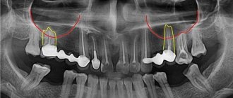
According to multislice computed tomography (MSCT), performed 4 months after transplantation of a free bone block of the mandibular ramus, when examining transverse and axial sections, a moderate decrease in the bone density of the graft compared to the cortical plate of the alveolar process of the mandibular ramus was noteworthy. There was no pronounced fusion of the receptive bed with the graft (see Fig. 1, b).
Histological structure of bone regenerates after transplantation of iliac bone blocks on a vascular pedicle
Histological examination of trephine biopsy specimens obtained from a bone graft, 12 and 24 months after autotransplantation of a fragment of the iliac bone, determined that the bone tissue of the column at the initial stage consisted of a cortical plate and underlying cancellous bone. The cortical plate was of moderate thickness, the trabeculae of the cancellous bone were large and wide. The intertrabecular space was filled with red bone marrow, sometimes with signs of replacement by yellow bone marrow. The cortical plate often had large Haversian canals that were lined with endosteum. In them, reticular stroma of the bone marrow was observed, turning into yellow bone marrow. The trabeculae of the cancellous bone were also covered with endosteum, had signs of osteoclastic resorption with the formation of characteristic lacunar grooves, and osteoblastic activity was moderate (Fig. 2, a).
Rice. 2. Disorganization of the bone matrix of the transplanted bone block of the lower jaw branch 4 months after surgery. Papanicolaou staining. ×400 (a). Decrease in bone mineral density of the graft according to MSCT data after 4 months (b). After 24 months, the intertrabecular space was only partially filled with red bone marrow. The trabeculae of the graft were thickened due to the layering of newly formed bone tissue. Signs of osteoclasia occurred both on the bone substance of the graft and on the surface of the newly formed bone.
A morphometric study also observed the presence of two variants of bone structures: necrotic bone (BDV/CV=5.7±1.5%) and viable bone, including in newly formed bone tissue (BTV/CV=48±3.75%). The tissue of the intertrabecular space was represented by yellow bone marrow (MaV/CV=38.8±7.4%). Assessment of bone balance (BB) demonstrated a predominant contribution to regenerative osteogenesis of newly formed bone tissue (BB=44.2±5.8) compared to the graft and its necrotic areas at this observation period. The phenomena of osteoresorption were determined by the mechanism of both smooth and osteoclastic resorption (Ra.Oc=36%). The processes of osteogenesis and osteoclasia on the surface of the graft and necrotic bone resulted in the formation of a lamellar bone matrix around them.
In an MSCT study of transverse and axial sections of the lower jaw after plastic surgery with a bone block on a vascular pedicle from a branch of the iliac crest, 12 months after transplantation, the formation of an artificial lower jaw was observed through integration and restructuring of the bone graft with initial signs of the formation of the cortical plate and well-defined spongy substance. The bone density of the artificial jaw was comparable to the contralateral half (see Fig. 3, b).
Rice. 3. Bone regenerate 12 months after transplantation of the iliac bone block on a vascular pedicle. Masson staining. ×200 (a). MSCT of the transplant area: after 6 months, the density of bone tissue in the graft is comparable to the skeletal bones not involved in the process (b).
Histological structure of bone regenerates after transplantation of free parietal bone blocks with bone chips
According to a histological study of trephine biopsy specimens obtained from patients after osteoplasty with autologous parietal bone grafts, the bone tissue was represented by a loose cortical plate, under which a layer of spongy bone was located. The cortical plate is narrow, surrounded by Volkmann canals. The bony trabeculae of the cancellous bone were narrow in places and of normal thickness in others. Bone tissue of trabeculae with small areas of incompletely mineralized bone tissue (Fig. 4, a).
Rice. 4. Trephine biopsy from bone regenerate 12 months after transplantation of a parietal bone block in combination with bone chips from the parietal bone. Papanicolaou staining. ×200 (a). Formation of bone structures of the artificial jaw from the graft according to MSCT data 12 months after surgery (b). Osteoblasts of a round or cubic shape, with a basophilic nucleus and nucleolus, surrounded by osteoid or lying freely on the surface, were found on the surface of the trabeculae. For the most part, the trabeculae were focally lined with flattened cells, the precursors of resting osteoblasts. In some places, osteoclastic activity was recorded, accompanied by smooth resorption of the surface of the bone beam. The interbeam space was filled with reticular soft fibrous connective tissue and was moderately vascularized. The reticular stroma did not contain adipose and blood tissue. Interbar space in the cortical layer with some fibrosis.
A morphometric study revealed the presence of two variants of bone structures in the following proportions in trephine biopsy specimens 6 months after surgery: necrotic bone (BDV/CV=5.7±1.5%) and viable, including newly formed bone tissue (BTV/CV =48±3.75%). The tissue of the intertrabecular space was represented by yellow bone marrow (MaV/CV=38.8±7.4%). After 12 months, the necrotic part of the graft was determined by a few islands of bone tissue located in the center of the bone structures. It was not possible to identify the graft tissue. The relative bone volume (BV/CV) was 72.4±5.4%. The reticular stroma did not contain hematopoietic elements. Osteoresorption 6 months after transplantation was carried out according to the mechanism of osteoclastic resorption (Ra.Oc=14±3.8%). After 12 months, a decrease in resorption activity was observed (Ra.Oc=12±5%). Assessment of bone balance (BB) showed that a greater contribution to regenerative osteogenesis was made by newly formed bone tissue (BB=44.5±8.6) than by the graft and its necrotic areas. Reparative osteogenesis remained active after 12 months (BB=25.7±7.6).
MSCT results 6 months after transplantation of bone blocks of the parietal bone and bone chips: when studying transverse and axial sections of the lower jaw, it was recorded that the structure of the lower jaw at the transplant site anatomically corresponds to the bone structure on the contralateral side, the cortical plate and spongy substance are pronounced. Bone density was comparable to the opposite side (see Fig. 4, b).
Histological structure of bone regenerates after transplantation of vascularized fibular bone blocks
Histological examination of trephine biopsy samples obtained from a fibular bone graft 6, 12 and 24 months after transplantation revealed that the trephine column mainly consisted of a dense mass of bone tissue, which could correspond to the cortical plate of the tubular bone. The Haversian canals were almond-shaped and tended to widen over time. In the early stages, only vessels and reticular stroma of the bone marrow were observed in the lumen of the Haversian canals, which over time was supplemented with newly formed bone tissue; by 24 months, the lumens were partially filled with newly formed bone tissue. The process of graft resorption began as a smooth resorption through swelling and disintegration of the matrix, which changed its tectorial properties with polychrome stains. Osteoclasts in the early stages were rare. Osteoblastic activity is minimal and is represented by single osteoblasts. 12 months after transplantation, the Haversian canals were widened, often irregularly shaped, and branched, resembling cancellous bone. In the area of physiological bone restructuring, single osteoclasts were found. Osteoblastic activity was minimal and was represented by a few osteoblasts in the areas of reversion (Fig. 5a).
Rice. 5. Trephine biopsy from bone regenerate 12 months after transplantation of a fibular bone block on a vascular pedicle. Papanicolaou staining. ×200 (a). Moderate decrease in bone mineral density of the graft according to MSCT data 12 months after transplantation. After 24 months, reticular bone marrow stroma and vessels containing formed elements, as well as bone structures resembling spongy bone tissue, were detected in the lumen of the Haversian canals. In some cases, accumulations of hematopoietic tissue cells were encountered. The trabeculae were unevenly distributed in space, their thickness varied significantly both in the thickness of the column and in its length. The inner surface of the Haversian canal with endosteal lining extending to the bone structures within the canals often showed signs of osteoclastic resorption. Single osteoclasts were detected in the area of physiological bone restructuring. Osteoblastic activity was minimal and was represented by a few osteoblasts in the areas of reversion on the surface of both the graft and the newly formed bone inside the Haversian canals. The bone substance contained osteocytes with a basophilic nucleus; some of the osteocyte lacunae did not have cells, especially at a distance from the blood vessels. The bone plates in the bone substance predominantly retained a characteristic pattern corresponding to the lamellar bone tissue of the cortical plate of the tubular bone - osteonic structures with circular plates and a few intercalated plates (Fig. 6, a).
Rice. 6. Trephine biopsy from bone regenerate 24 months after transplantation of a fibular bone block on a vascular pedicle. Papanicolaou staining. ×200 (a). Formation of cortical plates and spongy substance in the graft according to MSCT data 24 months after transplantation (b).
Thus, the microscopic picture in dynamics corresponded to a surviving graft, partially necrotic, with signs of bone tissue restructuring at the beginning according to the smooth type, and later 1 year according to the osteoclastic type, as well as signs of reparative osteogenesis inside the lumens of the Haversian canals with the formation of unorganized bone substance and sometimes with foci of hematopoiesis.
The morphometric study also determined two types of bone structures in the following proportions in samples 6 months after transplantation: partially necrotic bone (BDV/CV=42±7.5%) and viable, including newly formed bone tissue (BTV/CV=22 ±9.75%). The tissue of the intertrabecular space was represented by yellow bone marrow (MaV/CV=33.4±8.2%). 12 months after transplantation, the relative volume fraction of bone tissue decreased mainly due to resorption of necrotic bone (BDV/CV=18±4.1%, BTV/CV=27±3.7%). 24 months after transplantation, the relative volume fraction of bone tissue tended to increase mainly due to graft resorption and bone tissue formation (BDV/CV=23.7±4.4%, BTV/CV=37±5.9%).
The dynamics of reparative osteogenesis in the surviving graft consisted of a six-month period of relative stagnation (BB=2.25±0.6, Ra.Oc=5.7%±0.3%), characterized by some moderate surge in regeneration by 12 months against the background of pronounced osteoresorption (BB=3±0.3, Ra. Oc=7.2±0.6%) and subsequent slowdown of osteogenesis by 24 months against the background of ongoing resorption (BB=1.5±0.03, Ra. Oc=9, 4%±0.8%).
A CT study of transverse and axial sections of the lower jaw of patients after plastic surgery with fibular grafts revealed that the structure of the graft changed starting from 12 months (see Fig. 5, b). If during this period the structure of the graft completely repeated the fibula in terms of anatomical structure and bone density, then by 24 months there was a thinning of the cortical plate, an increase in the size of the artificial jaw, an expansion of the size of the graft with a change in the ratio of the cortical plate and cancellous bone in favor of the latter. Bone density was comparable to the opposite side (see Fig. 6, b).
How Much Does a Dental Bone Graft Cost?
The cost of dental bone graft surgery varies depending on various factors, including the patient's geographic location, the type of graft used, and other medical procedures. The average cost is between $200 and $1,300 per graft. This cost is for artificial bone grafts only. Thus, patients who require an autologous bone graft will have to pay more because an additional procedure will be performed in these circumstances. Overall, the cost of dental bone grafts is very reasonable and therefore we advise patients not to delay undergoing this procedure.
Disadvantages of bone block transplantation
The main disadvantage of the procedure is that it is a surgical operation with all the ensuing consequences: additional expenses, long-term rehabilitation and delay in the actual installation of implants and prostheses, since it must take 2 or more months for the complete restoration of the built-up bone tissue.
Is it possible to perform implantation without block transplantation?
The procedure has a small number of alternatives. The first is a splitting of the jaw ridge. The bone is cut horizontally, its edges are moved apart, and the gap is filled with material from the donor's own bone or artificial bone. This is a more difficult operation to perform - transplantation of blocks is much easier to tolerate.
There are also implantation methods without bone augmentation. But they also have their limitations and contraindications, especially for very narrow bone tissue - it is simply not enough to fix the implants. Of course, there are thin models specifically designed for narrow bones. But they are selected strictly individually.
Bone block transplantation operations are performed by experienced maxillofacial surgeons. The absence of complications after the procedure and its effectiveness largely depend on the professionalism of the doctor. We guarantee our clients a professional approach and caring attitude.
1 Perova M.D. Complications of dental implantation, their treatment and prevention. New in dentistry. 2002.
summary
A dental bone graft is a simple procedure used to replace missing jawbone. In this method, surgeons use different types of bone grafts according to the patient's needs and the severity of the problem. When is bone graft surgery necessary? If you know how dental implants are made, you will understand the importance of bone graft surgery. There must be enough bone in the jaw to hold the implant posts in place so that the tooth is securely anchored. Therefore, the use of bone grafts is essential for bone growth in the target area. If you are also a candidate for bone graft surgery and have questions about a specific topic, please write to us so we can guide you.