Types of foreign bodies that can damage gums
A splinter in the oral cavity is caused by:
- habit of chewing pencils and pens;
- inaccurate eating of seeds;
- picking teeth with wooden toothpicks.
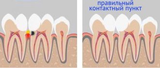
A foreign body in the gum can cause both minor discomfort and serious signs of inflammation.
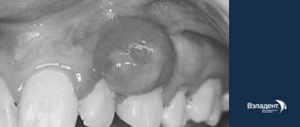
Splinter in the gum
When the gums are damaged by a splinter, sharp pain occurs. When examining the mouth, a foreign body is visible, half of which is located inside the periodontium, and the other half lies on the surface of the mucous tissue. You can get a splinter in your gums in everyday life:
- if you use a toothpick carelessly;
- when eating popcorn, sunflower seeds and unboned fish;
- with severe destruction of a carious tooth;
- if an instrument breaks during dental treatment.
After a person picks the gum with a toothpick, the tissue around the mole turns red, swells and bleeds. Unpleasant symptoms of damage are a good reason to visit a doctor.
Fish bone stuck in gum
The most common cause of a splinter in the mouth is chewing fish with bones. Thin small bones extend deep under the gum and cause sharp pain when pressed.
In 78% of cases, this type of splinter in the gum occurs in childhood. Children cannot remove all the bones from fish on their own, and to prevent unpleasant consequences, adults need to take care of the safety of the prepared dishes before eating.
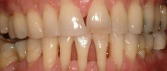
Symptoms of a foreign body in the gum
The moment a bone gets stuck in the gum is not always noticeable to a person. The first signs of damage appear upon repeated consumption of food or immediately after the development of the inflammatory process.
Symptoms of a splinter in the gum near a tooth include:
- redness of the mucous membrane around a foreign body;
- swelling of the gums;
- the formation of a translucent growth with purulent contents;
- the appearance of an abscess at the site of injury;
- itching in the inflamed area;
- sharp pain, aggravated by mechanical stimuli.
The mouth contains a large amount of pathogenic and conditionally pathogenic microflora. When there is wound damage, bacteria quickly penetrate the wound and begin active reproduction. In this case, inflammatory diseases of the oral cavity develop.
To prevent unwanted consequences, if you find a foreign body in your mouth, you should immediately consult a dentist for medical help.
How to diagnose a splinter in the gum or a bone from a fish
Splinters that appear in the gums from a toothpick, fish bones or an old filling force their owner to stick his tongue into the damaged area. You can see the smallest particles in your mouth at home. For this purpose, you will need a clean mirror and light. The examination should be carried out in clean conditions, and hands should be treated with alcohol-based antiseptics.
To prevent infection, the mouth is rinsed with Miramistin or an aqueous solution of chlorhexidine.
When the patient identifies the bone on his own, he uses all methods to remove it from the gums. Unfortunately, removing a foreign body at home is not always successful.
Incorrect bone removal technique further injures the tissue and provokes the development of purulent inflammation. In such cases, you need to trust the dentist and follow all the recommendations he gives.
If the splinter in the upper gum or lower jaw is stuck deep and cannot be seen, an x-ray examination is prescribed.
Specialist actions
It doesn’t matter whether you turned for help to a professional dentist in a clinic or to an emergency room, the doctor’s algorithm of action is always the same. First of all, an external examination of the gums and teeth is required, and an x-ray examination is prescribed. X-ray helps determine the number of fragments, their location and depth.
Only after a preliminary examination can the doctor choose a specific technique for extracting fragments. After their removal, the hole must be washed with antiseptic solutions; if necessary, a drug with a powerful antibacterial effect is added to it. If fragments were found very close to the surface, they can be removed using a tool. If they are deep enough, then dissection of the gum tissue will be required.
Today, in all these cases, anesthesia is used, so the patient does not experience any discomfort. In rare cases, general anesthesia may be prescribed. It is very important to seek help immediately after identifying the first symptoms, this will allow you to remove the particles as quickly as possible and avoid serious complications in the future. Unfortunately, even the most highly qualified specialist is not immune from such situations, because everything depends on the complexity of the specific clinical picture and the individual characteristics of the human body.
previous post
What to do if a tooth grows from the roof of your mouth?
next entry
How to remove bone from gum
As a rule, the fish bone is clearly visible in the mouth and can be removed using non-sharp tweezers. Before this, the instrument must be treated with an antiseptic, and hands must be washed with soap and wiped with ethyl alcohol.
The bone should be removed with careful and slow movements so that the fragment does not remain in the gum. After removing the splinter, the wound is treated with a 0.05% solution of chlorhexidine or hydrogen peroxide. It is not advisable to chew on the inflamed side of the mouth for several hours after the procedure.
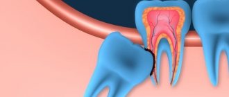
Broken instrument in the canal. Clinical tactics.
This article presents the author's view on the problem of broken instruments from a biological point of view.
The author created a clinical decision framework based on the following factors:
1. The tooth is vital or infected.
2. Initial or final stage of canal cleaning.
3. The tool is broken before or after bending.
Based on this pattern, doctors may decide not to remove the instrument, try to bypass it, or try to remove a broken instrument.
A broken instrument in the canal is, unfortunately, a complication of endodontic practice.
Broken instrument in the medial canal
Research shows that steel tools break off in 1 to 6% of cases. With the advent of nickel-titanium rotary tools, a marketing myth arose that they were unbreakable due to their flexibility...
However, the practical activities of endodontists around the world, unfortunately, have refuted this statement. Even among experienced endodontists, breakage of nickel-titanium instruments ranges from 0.5% to 5% of cases. Moreover, while the problem of steel instruments breaking off can be solved by observing signs of metal fatigue and removing such instruments from use, with nickel-titanium instruments, doctors are faced with a situation in which the instrument can break without showing signs of metal fatigue. This occurs either due to fracture due to bending fatigue of the metal or due to a metallurgical defect. Therefore, even one-time use of Ni-Ti tools does not completely prevent breakage.
Broken nickel titanium instrument behind a bend in the medial canal
There are no signs of any deformation on the surface of the nickel-titanium tool after breaking.
The only solution to prevent instrument fracture is to stop clinical work. All the tools break...
In the course of clinical work, doctors are faced with a situation where they may discover a broken instrument in the canal during previous treatments, or they themselves break the instrument in the canal.
Today, under the influence of marketing pressure and with the availability of certain tools and equipment, doctors automatically begin attempting to remove the broken instrument.
Let's look at this problem from different points of view:
1. Broken instrument and failure of endotherapy.
The cause of periapical processes is infection of the root canals.
A broken instrument in itself does not naturally cause inflammation.
Studies have shown that SI is the cause of endodontic treatment failure in only 0.96% of cases (OUTCOME OF ENDODONTIC TREATMENT AND RE-TREATMENT J. Ingle, J. Simon, P. Machtou and P. Bogaerts Endodontics - 5th Ed. (2002) Chapter 13) .
Often we see old endotherapy with broken instruments without any signs of periapical
inflammation. The influence of SI on the forecast will depend on several parameters:
— The tooth is vital or infected.
— At what stage of instrumentation and canal cleaning did the instrument break?
2. Procedure for removing a broken instrument.
The procedure involves creating direct access to the instrument using a modified Gates glidden and the use of ultrasonic tips; some technicians recommend the additional use of grasping instruments, such as IRS, Masserannkit, STN, etc.
This procedure is associated with loss of root dentin, and therefore can lead to complications such as perforation and vertical root fissure due to weakening of the root wall due to dentin loss. It is dentin that provides the strength of the tooth and its loss increases root deformation. Vertical root fracture is responsible for 11% of endodontic treatment failures.
2 broken instruments in the middle part of the canal
After removing the broken instruments, the root walls are thinned and weakened, in one of the areas the root is perforated
3. The instrument broke in the apical part of the canal behind the bend.
In such a situation, no technique is usually effective.
4. Existence of the ByPass procedure.
The procedure is carried out next to a broken instrument. This procedure allows us to clean the canal more apically and thereby solve the problem of infection...
Diagnostic image. Periapical process on the M and D roots, a broken instrument in one of the M canals.
A snapshot of determining the working length. The ByPass of the broken tool is visible.
A snapshot of determining the working length. The ByPass of the broken tool is visible.
Photo at the end of endodontic treatment. Hybrid condensation method.
Follow-up snapshot after 6 months. Almost complete disappearance of periapical processes on M and D roots.
1. Vital teeth.
Breakage of the instrument behind the bend of the canal in its apical part.
The prognosis is good. There is no point in trying to remove the tool.
This solution is applicable only if modern standards of endodontics are observed: working with dental
irrigation with sodium hypochlorite and cleaning of coronal caries before starting canal treatment.
Diagnostic image of the 48th tooth with irreversible pulpitis due to deep caries.
A snapshot of determining the working length.
Image after completion of endodontic treatment. Arrows indicate a fragment of an NT instrument.
Instrument fracture in the coronal part of the canal with direct access.
A photograph after extirpation, during which the instrument broke at the dittobuccal root.
The instrument has been removed by the IRS.
Photo after canal filling is completed.
Breakage of the instrument in the middle or apical part of the canal without bending.
Try to do ByPass (removing the tool is not necessary).
Diagnostic image of the 47th tooth. Broken instrument in one of the M channels.
A snapshot of determining the working length. Bypass a broken tool.
During the process of widening the canals, the broken instrument was removed from the canal.
Photo at the end of endodontic treatment.
2. Infected tooth.
The breakage occurred at the final stage of channel formation and cleaning.
There is no need to remove a broken tool.
Diagnostic image with gutta-percha in the fistula tract (tracing). Periapical lesions on M and D roots.
Working length shot.
Photo after canal filling is completed. The arrow indicates a broken NT instrument in the Mesiobuccal
channel.
Follow-up after 6 months. Complete disappearance of periapical processes. The normal contour of the periodontal fissure has been restored.
The tool broke off in the initial stages of canal formation and cleaning.
1. Trying to pass near a broken instrument (ByPass)
Diagnostic image. Broken and in the medial canals there is a periapical process on the M and D roots extending into the furcation area.
Working length shot. SI bypassed.
Photo at the end of endodontic treatment. The broken instrument is visible in the canal.
Follow-up after 6 months. Reduction of the periapical process.
2. If ByPass fails and the tool breaks off before bending.
Diagnostic image of the 37th tooth. The SI in the apical part of the canal partially extends into the periapical region. The patient has persistent sensitivity when chewing.
Picture after SI extraction. Gates Glidden technique and ultrasonic tips were used.
Photo after completion of filling the distal canals.
3. If the ByPass attempt was unsuccessful and an experienced doctor was unable to remove the instrument within an hour, or
the instrument broke behind the bend and attempting to remove it is dangerous from the point of view of weakening the root and perforations.
Intracanal attachment based on Ca(OH)2 for a period of 2 to 4 weeks. Canal filling and follow-up after 6 and 12 months.
If the periapical process increases, the decision is about apical surgery or tooth extraction.
When re-treating the 36th tooth with SI in the M channel behind the bend, it was not possible to make ByPass.
Canal filling after 4 weeks of using Ca(OH)2.
Follow-up after 6 months. Reduction of the periapical process.
The clinical tactics I have proposed are based on an understanding of the biological processes underlying periapical pathologies and dental biomechanics.
I believe that the mechanistic approach to solving the problem of a broken instrument - attempting to remove it in any case - is not correct. The procedure for removing a fragment of a broken instrument is associated with the loss of healthy root dentin. During this procedure, there may be perforations, or, as a delayed complication due to weakening of the tooth, a vertical root crack may occur.
Of course, in practice there may be cases that do not fall 100% under this scheme, and the doctor must make decisions based on a biological understanding of the situation, and not due to marketing pressure.
Author:
Dr. Solomonov Mikhail